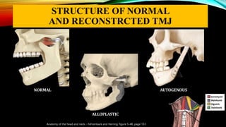
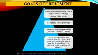
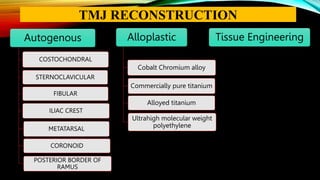
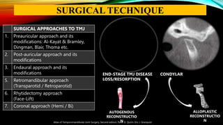
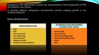
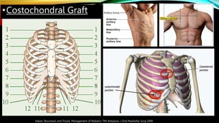
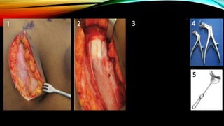
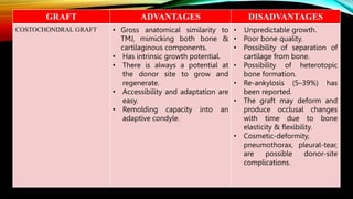
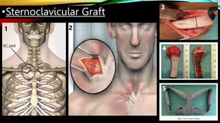
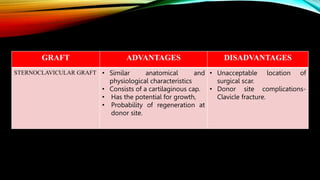
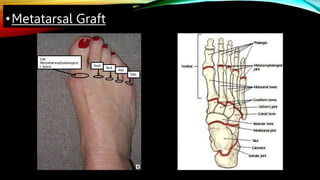
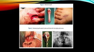
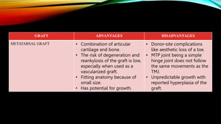
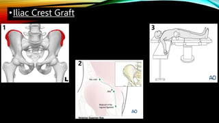
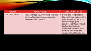
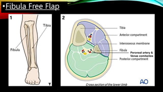
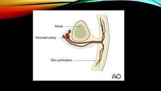
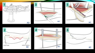
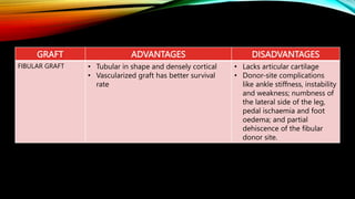
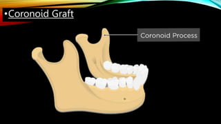
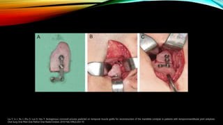
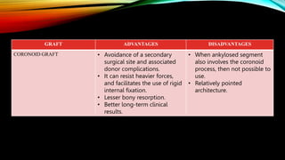
TMJ RECONSTRUCTION
PRESENTER:
Dr. Harjeet Yadav, PG III
MODERATOR:
Dr. Alok Bhatnagar, Professor
CONTENTS
• Introduction
• History
• Goals
• Indications/Contraindications
• Anatomy
• Autogenous reconstruction
• Alloplastic reconstruction
• Combined orthognathic and TMJ prosthetic
reconstruction
• Post-operative management
• Complications
• Distraction Osteogenesis/Transport Distraction
• Tissue Engineering
• Conclusion
• References
INTRODUCTION
o PLANE JOINTS – Carpal and Tarsal bones
o HINGE JOINTS – Elbow, Knee, TMJ
o CONDYLAR JOINTS – Wrist, Toes, TMJ
o PIVOT JOINTS – Neck, Wrist, Elbow
o BALL AND SOCKET JOINTS – Hip, Shoulder
o SADDLE JOINTS – Thumb, Middle Ear
INTRODUCTION
human-anatomylessons.blogspot.com
Advanced diseases or irreparable fracture,
with functional and anatomic distortion
dictates the need for TMJ reconstruction.
A successfully reconstructed TMJ should
reproduce normal joint structure, provide
functional articulation, and permit adaptive
growth or remodeling.
STRUCTURE OF NORMAL
AND RECONSTRCTED TMJ
Anatomy of the head and neck – Fehrenback and Herring, figure 5-4B, page 133
NORMAL
ALLOPLASTIC
AUTOGENOUS
HISTORY OF TMJ RECONSTRUCTION
1840 – Carnochan attempted to mobilize a patient’s ankylosed jaw by placing a small block of wood between the
raw bony surfaces after resection at the condylar neck.
1909 – Bardenheuer replaced the mandibular condyle with a patient’s 4th metatarsal.
1920 – Gillies first described the use of CCG for TMJ reconstruction.
1960 – Christensen reported resurfacing of the glenoid fossa with a thin Cast Vitallium fossa-eminence
hemiarthroplasty prosthesis for the management of TMJ ankylosis.
1970 – Homsy developed interpositional TMJ implant. It was a laminate of either Vitreous Carbon, Aluminum
Oxide, or Synthetic Hydroxyapatite and Polytetrafluoroethylene (PTFE).
1990 – Christensen developed a Cast Vitallium ramus-condyle component with a Polymethyl Methacrylate
condyle to create a total joint prosthesis.
1995 – Mercuri et al developed a custom CAD/CAM total TMJ replacement system.
2000 – Quinn introduced a stock TMJ replacement device (Zimmer Biomet).
Mercuri LG. Temporomandibular joint replacement devices - dark past to challenging future.
Restoration of mandibular ramus
length and morphology
(vertical facial height)
Acceptable range of motion
Reestablishment of premorbid
jaw relations and occlusion
In pediatric patients, the
reconstructed RCU must also
grow at a rate similar to the
opposite side and in concert
with the entire maxillofacial
skeleton
Atlas of Temporomandibular Joint Surgery, Second edition, Peter D. Quinn, Eric J. Granquist
GOALS OF TREATMENT
INDICATIONS
• TMJ ankylosis (cases requiring resection of the condylar process).
• Advanced osteoarthritis of TMJ.
• Inflammatory joint diseases (like rheumatoid and psoriastic arthritis).
• Well-progressed idiopathic mandibular condylar resorption.
• Congenital diseases.
• Post-traumatic condylar loss or damage.
• Postoperative condylar loss (including neoplastic ablation).
• Multiple invalid surgical history.
• Patients with a poor postoperative course after revision surgery with artificial joints.
• Patients with a poor postoperative course after costochondral graft.
Patients who present with above-mentioned diseases/conditions and any of the two following
main criteria are considered for surgery:
 Patients with occlusal abnormality and difficulty eating and chewing on a daily basis.
 Patients with a limited mouth opening (<35 mm).
Tetsuya Yoda et al , Clinical guidelines for total temporomandibular joint replacement
INDICATIONS
NORMAL BONE
NORMAL BONE OSTEOPENIA
LOCAL INFECTIOUS CONDITIONS EFFECTING THE TMJ
SEVERELY IMMUNO-COMPROMISED PATIENT ALLERGIC REACTIONS
CONTRAINDICATIONS
OSTEOPOROSIS SEVERE OSTEOPOROSIS
SEVERE OSTEOPOROSIS
Absolute contraindications:
• Deficient bone form (mandible, temporal bone) or poor bone quality.
• Local infectious conditions (Staph. aureus, Neisseria gonorrhoeae, and haemophilus influenzae)
• Severely immuno-compromised patient.
• History of metal allergy (CoCr, Ni, molybdenum).
• The patient who require a surgical correction of TMJ other than total replacement.
Relative contraindications:
• Progressive or chronic inflammation.
• Patients in the period of skeletally immature growth.
• Abnormal parafunctional habits, such as clenching, grinding, etc.
• Patients who cannot understand and accept medical instructions after surgery (including those with
neuropsychiatric disorders).
Tetsuya Yoda et al , Clinical guidelines for total temporomandibular joint replacement
CONTRAINDICATIONS
Autogenous
COSTOCHONDRAL
STERNOCLAVICULAR
FIBULAR
ILIAC CREST
METATARSAL
CORONOID
POSTERIOR BORDER OF
RAMUS
Tissue Engineering
Alloplastic
Cobalt Chromium alloy
Commercially pure titanium
Alloyed titanium
Ultrahigh molecular weight
polyethylene
TMJ RECONSTRUCTION
Atlas of Temporomandibular Joint Surgery, Second edition, Peter D. Quinn, Eric J. Granquist
SURGICAL APPROACHES TO TMJ
1. Preauricular approach and its
modifications: Al-Kayat & Bramley,
Dingman, Blair, Thoma etc.
2. Post-auricular approach and its
modifications
3. Endaural approach and its
modifications
5. Retromandibular approach
(Transparotid / Retroparotid)
6. Rhytidectomy approach
(Face-Lift)
7. Coronal approach (Hemi / Bi)
END-STAGE TMJ DISEASE CONDYLAR
LOSS/RESORPTION
ALLOPLASTIC
RECONSTRUCTIO
N
AUTOGENOUS
RECONSTRUCTIO
N
SURGICAL TECHNIQUE
AUTOGENOUS
RECONSTRUCTION
RAMUS CONDYLE UNIT
• CCG
• SCG
• Iliac Crest Graft
• Free Fibula Flap
• Coronoid Graft
• Metatarsal Graft
• Posterior Surface of Ramus
AUTOGENOUS DISC REPLACEMENT
(GLENOID FOSSA LINING)
• Dermis Graft
• Buccal Fat Pad
• Auricular Cartilage Graft
• Temporalis Myofascial Flap
• Fascia
• Skin
Autogenous TMJ replacement involves the reconstruction of the components of TMJ
with patient's own tissues.
In growing patients, autogenous reconstruction permits ongoing growth in the
reconstructed joint.
Donor site alternatives:
RAMUS CONDYLE UNIT
AUTOGENOUS DISC REPLACEMENT
(GLENOID FOSSA LINING)
Indications
• Absent TMJ condyle
• Zero to 1 previous TMJ surgeries (for free
grafts)
• Both hard and soft tissues are required
(vascularized fibula graft)
• Growth center transplant indicated (rib or
SCG)
• Custom-fitted total joint prosthesis are
unavailable
• Patient preference
• Allergy to metals in total joint prosthesis
Contraindications
• Multiple TMJ surgeries (2 or more previous
procedures)
• Connective tissue autoimmune disease or
inflammatory disease
• Previously failed TMJ allografts or
autogenous grafts
• Polyarthropathies
• Concomitant TMJ and orthognathic surgeries
are indicated
Wolford LM, Perez DE. Surgical management of congenital deformities with temporomandibular joint malformation. Oral Maxillofac Surg Clin
•Costochondral Graft
Kaban, Bouchard, and Troulis. Management of Pediatric TMJ Ankylosis. J Oral Maxillofac Surg 2009.
1 2 3
5
4
GRAFT ADVANTAGES DISADVANTAGES
COSTOCHONDRAL GRAFT • Gross anatomical similarity to
TMJ, mimicking both bone &
cartilaginous components.
• Has intrinsic growth potential.
• There is always a potential at
the donor site to grow and
regenerate.
• Accessibility and adaptation are
easy.
• Remolding capacity into an
adaptive condyle.
• Unpredictable growth.
• Poor bone quality.
• Possibility of separation of
cartilage from bone.
• Possibility of heterotopic
bone formation.
• Re-ankylosis (5–39%) has
been reported.
• The graft may deform and
produce occlusal changes
with time due to bone
elasticity & flexibility.
• Cosmetic-deformity,
pneumothorax, pleural-tear,
are possible donor-site
complications.
•Sternoclavicular Graft
1 2
3
4
5
GRAFT ADVANTAGES DISADVANTAGES
STERNOCLAVICULAR GRAFT • Similar anatomical and
physiological characteristics
• Consists of a cartilaginous cap.
• Has the potential for growth,
• Probability of regeneration at
donor site.
• Unacceptable location of
surgical scar.
• Donor site complications-
Clavicle fracture.
•Metatarsal Graft
D
GRAFT ADVANTAGES DISADVANTAGES
METATARSAL GRAFT • Combination of articular
cartilage and bone.
• The risk of degeneration and
reankylosis of the graft is low,
especially when used as a
vascularized graft.
• Fitting anatomy because of
small size.
• Has potential for growth.
• Donor-site complications
like aesthetic loss of a toe.
• MTP joint being a simple
hinge joint does not follow
the same movements as the
TMJ.
• Unpredictable growth with
reported hyperplasia of the
graft.
•Iliac Crest Graft
1
*
2
*Anterior Superior Iliac
Spine
*
3
L
GRAFT ADVANTAGES DISADVANTAGES
ILIAC CREST GRAFT • Has a cartilage cap, mimicking both the
bone and cartilaginous components.
• Has potential for growth.
• Donor-site complications
like prolonged postoperative
pain, altered gait, sensory
nerve damage, poor
scar/bone contour, delayed
healing, herniation of
abdominal contents, ilium
fracture, peritonitis, and
retroperitoneal hematoma.
•Fibula Free Flap
1 2
Peroneal artery &
Venae comitantes
Cross section of the lower limb
T
4
2
3 5
1
6
Fibular head Lateral malleolus
2 cm
GRAFT ADVANTAGES DISADVANTAGES
FIBULAR GRAFT • Tubular in shape and densely cortical
• Vascularized graft has better survival
rate
• Lacks articular cartilage
• Donor-site complications
like ankle stiffness, instability
and weakness; numbness of
the lateral side of the leg,
pedal ischaemia and foot
oedema; and partial
dehiscence of the fibular
donor site.
•Coronoid Graft
Liu Y, Li J, Hu J, Zhu S, Luo E, Hsu Y. Autogenous coronoid process pedicled on temporal muscle grafts for reconstruction of the mandible condylar in patients with temporomandibular joint ankylosis.
Oral Surg Oral Med Oral Pathol Oral Radiol Endod. 2010 Feb;109(2):203-10
GRAFT ADVANTAGES DISADVANTAGES
CORONOID GRAFT • Avoidance of a secondary
surgical site and associated
donor complications.
• It can resist heavier forces,
and facilitates the use of rigid
internal fixation.
• Lesser bony resorption.
• Better long-term clinical
results.
• When ankylosed segment
also involves the coronoid
process, then not possible to
use.
• Relatively pointed
architecture.
Method I. (a) The bony block and prominent antegonial notch. (b) The
bony block was resected and the glenoid fossa was recreated. (c)
Vertical osteotomy was performed on the entire posterior border of the
mandibular ramus and then moved up. (d) Fixation of the bone graft
with titanium miniplates and resection of prominent antegonial notch.
Method II (c) Vertical osteotomy was performed on the proximal
posterior border of the mandibular ramus till 1.0 cm above the angle of
the mandible and the osteotomized segment was moved up. (d) The
ipsilateral autogenous coronoid was implanted in the gap and the bony
graft was fixed with titanium miniplates.
Y. Liu, A. Khadka: Sliding reconstruction of the condyle using posterior border of mandibular ramus in patients with TMJ ankylosis. Int. J. Oral Maxillofac. Surg. 2011; 40.
Posterior border of ramus
GRAFT ADVANTAGES DISADVANTAGES
POSTERIOR BORDER OF RAMUS • Bone of a size and shape
adequate for new Condyle
with similar histological
characteristics.
• Damage to the contour of
the mandibular angle.
• Lack of growth.
• Two extra-oral incisions.
• Lack of long-term studies.
ALLOPLASTIC
RECONSTRUCTION
Alloplastic reconstruction is now considered as the gold standard in
adult TMJ reconstruction.
Alloplastic TMJ prosthesis can provide accurate adaptation to the
anatomical structures.
STOCK TMJ
PROSTHESIS
CUSTOM
MADE TMJ
PROSTHESIS
HEMI-JOINT
REPLACEMENT
(HJR)
TOTAL-JOINT
REPLACEMENT
(TJR)
FDA-APPROVED ALLOPLASTIC MATERIALS
FOR TMJ PROSTHESIS
1. Commercially Pure Titanium (cpTi)
2. Alloyed Titanium (Ti6Al4V)
3. Ultrahigh Molecular Weight Polyethylene (UHMWPE)
ADVANTAGES
• Simpler surgical technique
• Lack of donor site
morbidity
• Reduced intraoperative
surgical time
• Immediate functioning
• Symmetry and occlusal
stability
• Improved predictability
than autogenous grafts
LIMITATIONS
• Loss of translation
movement
• The potential for wear
debris and the associated
biologic responses
• Mechanical failure due to
component fracture,
loosening of screw
fixation, and metal fatigue
• High cost of the device
• Unpredictable need for
revision surgery
o FOSSA-EMINENCE PROSTHESIS
o CONDYLAR PROSTHESIS
HEMI-TMJ PROSTHESIS (HJR)
Replacement of glenoid fossa and articular eminence only.
INDICATIONS
• Painful or dysfunctional internal derangement after conservative and
surgical treatment. Healthy condyle on CT or MRI.
• Associated quality of life issues.
CONTRINDICATIONS
• Disrupted condylar surface
• Avascular necrosis
• Presence of osteophytes
FOSSA-EMINENCE PROSTHESIS
Technique:
• The bony fossa and eminence are debrided and recontoured for
insertion of a non-anatomic fossa-eminence prosthesis.
• The prosthesis acts as an inter-positional material.
• The condyle is reshaped to allow minimal osseous contact on the
fossa and no eminence obstruction during translational condylar
motion.
Park J, Keller EE, Reid KI. Surgical management of advanced degenerative arthritis of temporomandibular joint with metal fossa-eminence hemijoint replacement prosthesis: an 8-year
retrospective pilot study. J Oral Maxillofac Surg. 2004 Mar;62(3):320-8.
Journal of Cranio-Maxillofacial Surgery Volume 44, Issue 10, October 2016, Pages 1670-1677
CONDYLAR PROSTHESIS
FOSSA COMPONENT
TRANSLATION PLATE
NEO-DISC
CONDYLAR SPHERE
MANDIBULAR COMPONENT
TOTAL JOINT REPLACEMENT (TJR)
“Off the shelf” devices.
In order to develop some reasonable component-to-host-bone
interface or “fit”, all stock TMJ TJR devices require:
Host bone to be removed at implantation site,
One or both components must be bent to fit,
Shimming with bone or alloplastic cement.
Atlas of Temporomandibular Joint Surgery, Second edition, Peter D. Quinn, Eric J. Granquist
STOCK TJR PROSTHESIS
Kent-Vitek Christensen Biomet
• Developed in1970’s
VK-1
Fossa component - poly tetrafluoroethylene
(PTFE).
Mandibular component - Chromium Cobalt with
a layer of Proplast.
VK – 2
Fossa component – ultra high-molecular weight
polyethylene (UHM-WPE).
Complications: glenoid fossa resorption.
• Not in use.
Atlas of Temporomandibular Joint Surgery, Second edition, Peter D. Quinn, Eric J. Granquist
N. De Meurechy, A. Braem, M.Y. Mommaerts: Biomaterials in TJR: current status and future perspectives—a narrative review. Int. J. Oral Maxillofac. Surg. 2017
KENT-VITEK TJR
• Developed in 1990’s.
• Fossa component – polymethylmethacrylate
• Mandibular component - cast vitallium
• Due to wear under functional loading, this device
was soon abandoned and replaced by a metal-on-
metal surface device.
• Not in use.
Atlas of Temporomandibular Joint Surgery, Second edition, Peter D. Quinn, Eric J. Granquist
N. De Meurechy, A. Braem, M.Y. Mommaerts: Biomaterials in TJR: current status and future perspectives—a narrative review. Int. J. Oral Maxillofac. Surg. 2017
CHRISTENSEN’S TJR
• Developed in the year 2000.
• Spherical condylar head allows positional freedom of
the mandibular component in all planes.
• Fossa component - Ultrahigh molecular-weight
polyethylene (UHMWPE)
• Mandibular component - Cobalt Chromium alloy,
(medial surface of ramal part is coated with a plasma-
sprayed titanium coating).
• The ramus of the mandibular component is currently
manufactured in lengths of 45 mm, 50 mm, and 55 mm.
• The screws used for fixation are manufactured from
titanium alloy (TI-6AL-4V).
Atlas of Temporomandibular Joint Surgery, Second edition, Peter D. Quinn, Eric J. Granquist
N. De Meurechy, A. Braem, M.Y. Mommaerts: Biomaterials in TJR: current status and future perspectives—a narrative review. Int. J. Oral Maxillofac. Surg. 2017
BIOMET / W. LORENZ TJR
• The fossa has a minimum of 4 mm thickness in
the central region and has a cavity with larger
walls to prevent heterotopic bone invasion and
displacement or dislocation of condyle.
• The neck of condyle appears as swan neck,
avoiding any obstruction at the implant-bone
interface
FAILED TMJ PROSTHESIS
1) Leibinger by Stryker (Germany)
2) THORP system by Stryker (Germany)
3) AO/ASIF condylar head add-on system by
Synthes (Germany)
4) AO/ASIF condylar reconstruction plate by
Synthes (Germany)
5) Add-on system by KLS Martin (Germany)
6) Modus1 system by Medartis (Switzerland)
CONDYLAR RECONSTRUCTION PLATES
ADVANTAGES DISADVANTAGES
Immediate availability to the surgeon (e.g.,
irreparable trauma, tumor resection).
There is limited movement of the
mandible.
Lower cost. Surgeon’s experience with multiple TMJ
reconstructions is required to manage the
variability of fit.
Not appropriate for some patients with
extensive tumors or severe deformities.
More intraoperative time.
Trimming of bone is required to make the
device fit.
Atlas of Temporomandibular Joint Surgery, Second edition, Peter D. Quinn, Eric J. Granquist
• ‘Made to fit’ devices.
• Due to distorted anatomy, caused by either failed prior
surgical interventions or primary/ secondary joint
diseases, it is extremely difficult to reconstruct with
‘Stock Devices’.
• More recently, with the advent of 3-D printing,
stereolithography and virtual surgical planning, it is
possible to manufacture individualized prosthesis.
Atlas of Temporomandibular Joint Surgery, Second edition, Peter D. Quinn, Eric J. Granquist
CUSTOM MADE TMJ PROSTHESIS
Fabrication using CAD/CAM technology based
on the CT scan (0.5mm) of patient’s skull.
A stereolithographic model of the patient’s
skull is printed, from which the final
components are designed and manufactured.
Through this custom design, the prosthesis can
be altered to the patient’s specific anatomy,
including jaw abnormalities and jaw position.
Positions of the screws can be optimized,
taking inferior alveolar nerve into account.
Optimization of device positioning and contact
with the bone.
• Requires initial joint preparation, including tumor resection,
device removal, or bony resection to be done.
• The patient is then placed in IMF and a CT scan is obtained.
• The device is then fabricated using 3-D printed models.
• The advantage to this approach is that the defect and new
mandibular position is already set.
• The disadvantages include a second procedure and the need for
prolonged mandibular immobilization during device
manufacturing.
Atlas of Temporomandibular Joint Surgery, Second edition, Peter D. Quinn, Eric J. Granquist
TWO-STAGE RECONSTRUCTION
• Requires initial site preparation and device placement in a single
surgery.
• Planning and design are first replicated on the model, with
osteotomy sites and bone contouring marked and same is later
performed on the patient.
• Disadvantages include the need to recapitulate osteotomy and
mandibular repositioning precisely as they were placed on the
model.
• 3-D printed surgical guides can be used to ensure precise
osteotomy site and device positioning intra-operatively.
Atlas of Temporomandibular Joint Surgery, Second edition, Peter D. Quinn, Eric J. Granquist
SINGLE-STAGE RECONSTRUCTION
• Developed in 1989.
• In 1999, TMJ Concepts received FDA
approval for their patient-fitted TMJ implant
system.
• The fossa of the TMJ Concepts prosthesis is
made from 4 layers of commercially pure
titanium mesh backing.
• The mesh can be adapted to the patient’s
anatomy, provides stability and allows for
bony ingrowth to occur.
• A UHMWPE lining functions as the
articulating surface.
N. De Meurechy, A. Braem, M.Y. Mommaerts: Biomaterials in TMJ replacement: current status and future perspectives—a narrative review. Int. J. Oral Maxillofac. Surg. 2017
TECHMEDICA / TMJ CONCEPTS SYSTEM
MOMMAERTS
GENOVESI RAMOS
DARSN
CHAWARE MEHROTRA
DE SOUZA
ABEL: DUNDEE
RUSTEMEYER
OTHER PATIENT SPECIFIC SYSTEMS
ADVANTAGES DISADVANTAGES
Patient-matched; anatomically stable. Higher cost.
Addresses distorted anatomy. Potential for two-stage surgeries (e.g., removal of
failed previous metallic implants).
Excessive antero-inferior movements possible. Time for fabrication of custom implant
(8–12 weeks).
Highly accurate. Limited flexibility (must replicate model surgery
exactly).
Atlas of Temporomandibular Joint Surgery, Second edition, Peter D. Quinn, Eric J. Granquist
• In cases where a large mandibular advancement or rotation is planned in
combination with TMJ reconstruction, a custom device may be required.
• The alloplastic device allows for a stable platform for facial reconstruction, as well
as the ability to allow for immediate function of the mandible.
• When a custom device is used, reconstruction of TMJ should proceed first, allowing
the custom device to act as an intermediate splint.
• The maxillary osteotomy can then be performed and the maxilla repositioned to
the mandible.
• As in all total TMJ reconstruction cases, great care must be taken to avoid intraoral
contamination of the prosthetic devices.
Atlas of Temporomandibular Joint Surgery, Second edition, Peter D. Quinn, Eric J. Granquist
COMBINED OGS AND TJR
• Limiting early postoperative opening to avoid dislocation.
• The use of training elastics in the immediate postoperative period can reduce
the potential for dislocation in the first week post-op.
• Antibiotic coverage.
• The patients should be encouraged to chew a soft diet and advance their diet
as tolerated.
• Physical therapy.
• Long-term follow-up.
POSTOPERATIVE MANAGEMENT
• Removal of implant component(s) :
- Implant changes caused by loading and/or wear.
- Degenerative changes within the joint surfaces from disease or previous implants.
- Implant materials producing corrosion particles.
• Loosening or displacement of the implant because of improper fixation.
• Dislocation
• Infection
• Foreign body or allergic reaction to implant components.
• Fossa wear trough
• Facial swelling and/or pain
• Facial nerve dysfunction
• Parotid injury
• Heterotopic bone formation
• Neuroma formation
• Ear problems
Hoffman D, Puig L. Complications of TMJ surgery. Oral Maxillofac Surg Clin North Am. 2015
COMPLICATIONS
S h a r m a R , Manikandhan R, Sneha P, Parameswaran A, Kumar JN, Sailer HF. Neocondyle distraction osteogenesis in the management of temporomandibular joint ankylosis: Report of five cases with
review of literature. Indian J Dent Res 2017;28:269-74.
1 2
DISTRACTION OSTEOGENESIS
In 1997, Stucki-Mccormick was the first to apply transport distraction
osteogenesis for TMJ reconstruction.
After gap arthroplasty, reverse L-shape osteotomy extending from the sigmoid
notch to approximately 1 cm above the inferior border of the mandible, is
marked.
The vertical cut should be planned parallel to the vector, so it can direct the
transport disc towards glenoid fossa.
The superior surface of the disc is rounded to simulate the condylar surface.
The distractor device is then oriented and fixed parallel to the vertical cut.
Once the distractor device is fixed, the osteotomy can be carried out,
mobilizing the transport segment completely.
S h a r m a R , Manikandhan R, Sneha P, Parameswaran A, Kumar JN, Sailer HF. Neocondyle distraction osteogenesis in the management of temporomandibular joint ankylosis: Report of five cases with
review of literature. Indian J Dent Res 2017;28:269-74.
• After 5 days of latency period, the distraction is initiated 1 mm daily, till the
transport disc achieves close contact with the articular surface of temporal
bone
• The distractor is left in for a consolidation period of 6–8 weeks.
• Once the mature bone formation is evident at the trailing edge of the
transport disc resulting in bridging, radiographically, the distractor can be
removed.
• The leading edge of the transport disc tends to remodel and become
rounded to form a neocondyle.
• Advantages
• No need for interpositional material.
• Patients can open and close their mouth and chew during DO.
• Simultaneous correction of secondary deformities.
• Considerably shortens the admission & operation time and the
possibility of relapse.
• Disadvantages
• Lengthy procedure.
• Patient cooperation is a must.
• Might have psychological effects on patients.
S h a r m a R , Manikandhan R, Sneha P, Parameswaran A, Kumar JN, Sailer HF. Neocondyle distraction osteogenesis in the management of temporomandibular joint ankylosis: Report of five
cases with review of literature. Indian J Dent Res 2017;28:269-74.
• The principal elements of tissue engineering are:
• A scaffold into which cells are seeded.
• An applied biochemical or biomechanical stimulus to
make the cells grow and secrete substances that
eventually produce the desired tissue.
• Pluripotent stem cells have the inherent ability to be
transformed into many different types of tissues,
depending on the growth environment to which they are
subjected.
• These can be harvested from bone marrow as well as from
fat via liposuction.
• Still in infancy.
Liu C-K et al.3-D porous internal ti scaffold or allogenic bone scaffold for tissue-engineering condyle as a novel reconstruction of mandibular condylar defects, J Med Hypotheses Ideas (2014)
TISSUE ENGINEERING
CONCLUSION
Current reconstructive techniques lie in favour of autogenous
replacement in children and alloplasts in adults.
The use of distraction osteogenesis is also being explored
although long-term outcomes are not yet available.
Newer techniques of cartilage reconstruction may ultimately
surpass any form of reconstruction.
• Atlas of Temporomandibular Joint Surgery, Second edition, Peter D. Quinn, Eric J. Granquist.
• Temporomandibular Joint Total Joint Replacement – TMJ TJR, Louis G. Mercuri Editor
• Kaban, Bouchard, and Troulis. Management of Pediatric TMJ Ankylosis. J Oral Maxillofac Surg 2009.
• A. Khadka, J. Hu: Autogenous grafts for condylar reconstruction in treatment of TMJ ankylosis: current concepts and considerations for the future. Int. J. Oral Maxillofac. Surg. 2012;
41: 94–102. # 2011
• Liu C-K et al., Using three-dimensional porous internal titanium scaffold or allogenic bone scaffold for tissue-engineering condyle as a novel reconstruction of mandibular condylar
defects, J Med Hypotheses Ideas (2014).
• Kaur K, Roychoudhury A, Bhutia O, Bhalla AS, Yadav R, Pandey RM, Evaluation of success of transport disc distraction osteogenesis and costochondral graft for ramus condyle unit
reconstruction in pediatric temporomandibular joint ankylosis., Journal of Oral and Maxillofacial Surgery (2020).
• Prasad BR, Bhat S, Bhat SS. Reconstruction of condyle following surgical correction of temporomandibular joint ankylosis: current concepts and considerations for the future. Journal
of Health and Allied Sciences NU. 2014 Jun;4(02):039-46.
• Sharma R , Manikandhan R, Sneha P, Parameswaran A, Kumar JN, Sailer HF. Neocondyle distraction osteogenesis in the management of temporomandibular joint ankylosis: Report
of five cases with review of literature. Indian J Dent Res 2017;28:269-74.
• Liu Y, Li J, Hu J, Zhu S, Luo E, Hsu Y. Autogenous coronoid process pedicled on temporal muscle grafts for reconstruction of the mandible condylar in patients with
temporomandibular joint ankylosis. Oral Surg Oral Med Oral Pathol Oral Radiol Endod. 2010 Feb;109(2):203-10. doi: 10.1016/j.tripleo.2009.09.006. Epub 2009 Dec 6. PMID: 19969485.
• Mercuri LG, Wolford LM, Sanders B, White RD, Giobbie-Hurder A. Long-term follow-up of the CAD/CAM patient fitted total temporomandibular joint reconstruction system. J Oral
Maxillofac Surg. 2002 Dec;60(12):1440-8. doi: 10.1053/joms.2002.36103. PMID: 12465008.
• Seth S, Gupta H, Kumar D, Agarwal R, Gupta S, Mehra H, Natu SS, Singh J. Sternoclavicular Graft Versus Costochondral Graft In Reconstruction of Ankylosed Temporomandibular
Joint. J Maxillofac Oral Surg. 2019 Dec;18(4):559-566. doi: 10.1007/s12663-019-01276-z. Epub 2019 Aug 20. PMID: 31624437; PMCID: PMC6795663.
• Wolford LM, Perez DE. Surgical management of congenital deformities with temporomandibular joint malformation. Oral Maxillofac Surg Clin North Am. 2015 Feb;27(1):137-54.
• Park J, Keller EE, Reid KI. Surgical management of advanced degenerative arthritis of temporomandibular joint with metal fossa-eminence hemijoint replacement prosthesis: an 8-
year retrospective pilot study. J Oral Maxillofac Surg. 2004 Mar;62(3):320-8.
REFERENCES
TMJ RECONSTRUCTION

TMJ RECONSTRUCTION

  • 1.
    TMJ RECONSTRUCTION PRESENTER: Dr. HarjeetYadav, PG III MODERATOR: Dr. Alok Bhatnagar, Professor
  • 2.
    CONTENTS • Introduction • History •Goals • Indications/Contraindications • Anatomy • Autogenous reconstruction • Alloplastic reconstruction • Combined orthognathic and TMJ prosthetic reconstruction • Post-operative management • Complications • Distraction Osteogenesis/Transport Distraction • Tissue Engineering • Conclusion • References
  • 3.
    INTRODUCTION o PLANE JOINTS– Carpal and Tarsal bones o HINGE JOINTS – Elbow, Knee, TMJ o CONDYLAR JOINTS – Wrist, Toes, TMJ o PIVOT JOINTS – Neck, Wrist, Elbow o BALL AND SOCKET JOINTS – Hip, Shoulder o SADDLE JOINTS – Thumb, Middle Ear
  • 4.
    INTRODUCTION human-anatomylessons.blogspot.com Advanced diseases orirreparable fracture, with functional and anatomic distortion dictates the need for TMJ reconstruction. A successfully reconstructed TMJ should reproduce normal joint structure, provide functional articulation, and permit adaptive growth or remodeling.
  • 5.
    STRUCTURE OF NORMAL ANDRECONSTRCTED TMJ Anatomy of the head and neck – Fehrenback and Herring, figure 5-4B, page 133 NORMAL ALLOPLASTIC AUTOGENOUS
  • 6.
    HISTORY OF TMJRECONSTRUCTION 1840 – Carnochan attempted to mobilize a patient’s ankylosed jaw by placing a small block of wood between the raw bony surfaces after resection at the condylar neck. 1909 – Bardenheuer replaced the mandibular condyle with a patient’s 4th metatarsal. 1920 – Gillies first described the use of CCG for TMJ reconstruction. 1960 – Christensen reported resurfacing of the glenoid fossa with a thin Cast Vitallium fossa-eminence hemiarthroplasty prosthesis for the management of TMJ ankylosis. 1970 – Homsy developed interpositional TMJ implant. It was a laminate of either Vitreous Carbon, Aluminum Oxide, or Synthetic Hydroxyapatite and Polytetrafluoroethylene (PTFE). 1990 – Christensen developed a Cast Vitallium ramus-condyle component with a Polymethyl Methacrylate condyle to create a total joint prosthesis. 1995 – Mercuri et al developed a custom CAD/CAM total TMJ replacement system. 2000 – Quinn introduced a stock TMJ replacement device (Zimmer Biomet). Mercuri LG. Temporomandibular joint replacement devices - dark past to challenging future.
  • 7.
    Restoration of mandibularramus length and morphology (vertical facial height) Acceptable range of motion Reestablishment of premorbid jaw relations and occlusion In pediatric patients, the reconstructed RCU must also grow at a rate similar to the opposite side and in concert with the entire maxillofacial skeleton Atlas of Temporomandibular Joint Surgery, Second edition, Peter D. Quinn, Eric J. Granquist GOALS OF TREATMENT
  • 8.
  • 9.
    • TMJ ankylosis(cases requiring resection of the condylar process). • Advanced osteoarthritis of TMJ. • Inflammatory joint diseases (like rheumatoid and psoriastic arthritis). • Well-progressed idiopathic mandibular condylar resorption. • Congenital diseases. • Post-traumatic condylar loss or damage. • Postoperative condylar loss (including neoplastic ablation). • Multiple invalid surgical history. • Patients with a poor postoperative course after revision surgery with artificial joints. • Patients with a poor postoperative course after costochondral graft. Patients who present with above-mentioned diseases/conditions and any of the two following main criteria are considered for surgery:  Patients with occlusal abnormality and difficulty eating and chewing on a daily basis.  Patients with a limited mouth opening (<35 mm). Tetsuya Yoda et al , Clinical guidelines for total temporomandibular joint replacement INDICATIONS
  • 10.
    NORMAL BONE NORMAL BONEOSTEOPENIA NORMAL BONE OSTEOPENIA LOCAL INFECTIOUS CONDITIONS EFFECTING THE TMJ SEVERELY IMMUNO-COMPROMISED PATIENT ALLERGIC REACTIONS CONTRAINDICATIONS OSTEOPOROSIS SEVERE OSTEOPOROSIS SEVERE OSTEOPOROSIS
  • 11.
    Absolute contraindications: • Deficientbone form (mandible, temporal bone) or poor bone quality. • Local infectious conditions (Staph. aureus, Neisseria gonorrhoeae, and haemophilus influenzae) • Severely immuno-compromised patient. • History of metal allergy (CoCr, Ni, molybdenum). • The patient who require a surgical correction of TMJ other than total replacement. Relative contraindications: • Progressive or chronic inflammation. • Patients in the period of skeletally immature growth. • Abnormal parafunctional habits, such as clenching, grinding, etc. • Patients who cannot understand and accept medical instructions after surgery (including those with neuropsychiatric disorders). Tetsuya Yoda et al , Clinical guidelines for total temporomandibular joint replacement CONTRAINDICATIONS
  • 12.
    Autogenous COSTOCHONDRAL STERNOCLAVICULAR FIBULAR ILIAC CREST METATARSAL CORONOID POSTERIOR BORDEROF RAMUS Tissue Engineering Alloplastic Cobalt Chromium alloy Commercially pure titanium Alloyed titanium Ultrahigh molecular weight polyethylene TMJ RECONSTRUCTION
  • 13.
    Atlas of TemporomandibularJoint Surgery, Second edition, Peter D. Quinn, Eric J. Granquist SURGICAL APPROACHES TO TMJ 1. Preauricular approach and its modifications: Al-Kayat & Bramley, Dingman, Blair, Thoma etc. 2. Post-auricular approach and its modifications 3. Endaural approach and its modifications 5. Retromandibular approach (Transparotid / Retroparotid) 6. Rhytidectomy approach (Face-Lift) 7. Coronal approach (Hemi / Bi) END-STAGE TMJ DISEASE CONDYLAR LOSS/RESORPTION ALLOPLASTIC RECONSTRUCTIO N AUTOGENOUS RECONSTRUCTIO N SURGICAL TECHNIQUE
  • 14.
  • 15.
    RAMUS CONDYLE UNIT •CCG • SCG • Iliac Crest Graft • Free Fibula Flap • Coronoid Graft • Metatarsal Graft • Posterior Surface of Ramus AUTOGENOUS DISC REPLACEMENT (GLENOID FOSSA LINING) • Dermis Graft • Buccal Fat Pad • Auricular Cartilage Graft • Temporalis Myofascial Flap • Fascia • Skin Autogenous TMJ replacement involves the reconstruction of the components of TMJ with patient's own tissues. In growing patients, autogenous reconstruction permits ongoing growth in the reconstructed joint. Donor site alternatives: RAMUS CONDYLE UNIT AUTOGENOUS DISC REPLACEMENT (GLENOID FOSSA LINING)
  • 16.
    Indications • Absent TMJcondyle • Zero to 1 previous TMJ surgeries (for free grafts) • Both hard and soft tissues are required (vascularized fibula graft) • Growth center transplant indicated (rib or SCG) • Custom-fitted total joint prosthesis are unavailable • Patient preference • Allergy to metals in total joint prosthesis Contraindications • Multiple TMJ surgeries (2 or more previous procedures) • Connective tissue autoimmune disease or inflammatory disease • Previously failed TMJ allografts or autogenous grafts • Polyarthropathies • Concomitant TMJ and orthognathic surgeries are indicated Wolford LM, Perez DE. Surgical management of congenital deformities with temporomandibular joint malformation. Oral Maxillofac Surg Clin
  • 17.
    •Costochondral Graft Kaban, Bouchard,and Troulis. Management of Pediatric TMJ Ankylosis. J Oral Maxillofac Surg 2009.
  • 18.
  • 19.
    GRAFT ADVANTAGES DISADVANTAGES COSTOCHONDRALGRAFT • Gross anatomical similarity to TMJ, mimicking both bone & cartilaginous components. • Has intrinsic growth potential. • There is always a potential at the donor site to grow and regenerate. • Accessibility and adaptation are easy. • Remolding capacity into an adaptive condyle. • Unpredictable growth. • Poor bone quality. • Possibility of separation of cartilage from bone. • Possibility of heterotopic bone formation. • Re-ankylosis (5–39%) has been reported. • The graft may deform and produce occlusal changes with time due to bone elasticity & flexibility. • Cosmetic-deformity, pneumothorax, pleural-tear, are possible donor-site complications.
  • 20.
  • 21.
    GRAFT ADVANTAGES DISADVANTAGES STERNOCLAVICULARGRAFT • Similar anatomical and physiological characteristics • Consists of a cartilaginous cap. • Has the potential for growth, • Probability of regeneration at donor site. • Unacceptable location of surgical scar. • Donor site complications- Clavicle fracture.
  • 22.
  • 24.
    GRAFT ADVANTAGES DISADVANTAGES METATARSALGRAFT • Combination of articular cartilage and bone. • The risk of degeneration and reankylosis of the graft is low, especially when used as a vascularized graft. • Fitting anatomy because of small size. • Has potential for growth. • Donor-site complications like aesthetic loss of a toe. • MTP joint being a simple hinge joint does not follow the same movements as the TMJ. • Unpredictable growth with reported hyperplasia of the graft.
  • 25.
    •Iliac Crest Graft 1 * 2 *AnteriorSuperior Iliac Spine * 3 L
  • 26.
    GRAFT ADVANTAGES DISADVANTAGES ILIACCREST GRAFT • Has a cartilage cap, mimicking both the bone and cartilaginous components. • Has potential for growth. • Donor-site complications like prolonged postoperative pain, altered gait, sensory nerve damage, poor scar/bone contour, delayed healing, herniation of abdominal contents, ilium fracture, peritonitis, and retroperitoneal hematoma.
  • 27.
    •Fibula Free Flap 12 Peroneal artery & Venae comitantes Cross section of the lower limb T
  • 29.
    4 2 3 5 1 6 Fibular headLateral malleolus 2 cm
  • 30.
    GRAFT ADVANTAGES DISADVANTAGES FIBULARGRAFT • Tubular in shape and densely cortical • Vascularized graft has better survival rate • Lacks articular cartilage • Donor-site complications like ankle stiffness, instability and weakness; numbness of the lateral side of the leg, pedal ischaemia and foot oedema; and partial dehiscence of the fibular donor site.
  • 31.
  • 32.
    Liu Y, LiJ, Hu J, Zhu S, Luo E, Hsu Y. Autogenous coronoid process pedicled on temporal muscle grafts for reconstruction of the mandible condylar in patients with temporomandibular joint ankylosis. Oral Surg Oral Med Oral Pathol Oral Radiol Endod. 2010 Feb;109(2):203-10
  • 33.
    GRAFT ADVANTAGES DISADVANTAGES CORONOIDGRAFT • Avoidance of a secondary surgical site and associated donor complications. • It can resist heavier forces, and facilitates the use of rigid internal fixation. • Lesser bony resorption. • Better long-term clinical results. • When ankylosed segment also involves the coronoid process, then not possible to use. • Relatively pointed architecture.
  • 34.
    Method I. (a)The bony block and prominent antegonial notch. (b) The bony block was resected and the glenoid fossa was recreated. (c) Vertical osteotomy was performed on the entire posterior border of the mandibular ramus and then moved up. (d) Fixation of the bone graft with titanium miniplates and resection of prominent antegonial notch. Method II (c) Vertical osteotomy was performed on the proximal posterior border of the mandibular ramus till 1.0 cm above the angle of the mandible and the osteotomized segment was moved up. (d) The ipsilateral autogenous coronoid was implanted in the gap and the bony graft was fixed with titanium miniplates. Y. Liu, A. Khadka: Sliding reconstruction of the condyle using posterior border of mandibular ramus in patients with TMJ ankylosis. Int. J. Oral Maxillofac. Surg. 2011; 40. Posterior border of ramus
  • 35.
    GRAFT ADVANTAGES DISADVANTAGES POSTERIORBORDER OF RAMUS • Bone of a size and shape adequate for new Condyle with similar histological characteristics. • Damage to the contour of the mandibular angle. • Lack of growth. • Two extra-oral incisions. • Lack of long-term studies.
  • 36.
  • 37.
    Alloplastic reconstruction isnow considered as the gold standard in adult TMJ reconstruction. Alloplastic TMJ prosthesis can provide accurate adaptation to the anatomical structures. STOCK TMJ PROSTHESIS CUSTOM MADE TMJ PROSTHESIS HEMI-JOINT REPLACEMENT (HJR) TOTAL-JOINT REPLACEMENT (TJR)
  • 39.
    FDA-APPROVED ALLOPLASTIC MATERIALS FORTMJ PROSTHESIS 1. Commercially Pure Titanium (cpTi) 2. Alloyed Titanium (Ti6Al4V) 3. Ultrahigh Molecular Weight Polyethylene (UHMWPE)
  • 40.
    ADVANTAGES • Simpler surgicaltechnique • Lack of donor site morbidity • Reduced intraoperative surgical time • Immediate functioning • Symmetry and occlusal stability • Improved predictability than autogenous grafts LIMITATIONS • Loss of translation movement • The potential for wear debris and the associated biologic responses • Mechanical failure due to component fracture, loosening of screw fixation, and metal fatigue • High cost of the device • Unpredictable need for revision surgery
  • 41.
    o FOSSA-EMINENCE PROSTHESIS oCONDYLAR PROSTHESIS HEMI-TMJ PROSTHESIS (HJR)
  • 42.
    Replacement of glenoidfossa and articular eminence only. INDICATIONS • Painful or dysfunctional internal derangement after conservative and surgical treatment. Healthy condyle on CT or MRI. • Associated quality of life issues. CONTRINDICATIONS • Disrupted condylar surface • Avascular necrosis • Presence of osteophytes FOSSA-EMINENCE PROSTHESIS
  • 43.
    Technique: • The bonyfossa and eminence are debrided and recontoured for insertion of a non-anatomic fossa-eminence prosthesis. • The prosthesis acts as an inter-positional material. • The condyle is reshaped to allow minimal osseous contact on the fossa and no eminence obstruction during translational condylar motion. Park J, Keller EE, Reid KI. Surgical management of advanced degenerative arthritis of temporomandibular joint with metal fossa-eminence hemijoint replacement prosthesis: an 8-year retrospective pilot study. J Oral Maxillofac Surg. 2004 Mar;62(3):320-8.
  • 44.
    Journal of Cranio-MaxillofacialSurgery Volume 44, Issue 10, October 2016, Pages 1670-1677 CONDYLAR PROSTHESIS
  • 45.
    FOSSA COMPONENT TRANSLATION PLATE NEO-DISC CONDYLARSPHERE MANDIBULAR COMPONENT TOTAL JOINT REPLACEMENT (TJR)
  • 46.
    “Off the shelf”devices. In order to develop some reasonable component-to-host-bone interface or “fit”, all stock TMJ TJR devices require: Host bone to be removed at implantation site, One or both components must be bent to fit, Shimming with bone or alloplastic cement. Atlas of Temporomandibular Joint Surgery, Second edition, Peter D. Quinn, Eric J. Granquist STOCK TJR PROSTHESIS
  • 47.
  • 48.
    • Developed in1970’s VK-1 Fossacomponent - poly tetrafluoroethylene (PTFE). Mandibular component - Chromium Cobalt with a layer of Proplast. VK – 2 Fossa component – ultra high-molecular weight polyethylene (UHM-WPE). Complications: glenoid fossa resorption. • Not in use. Atlas of Temporomandibular Joint Surgery, Second edition, Peter D. Quinn, Eric J. Granquist N. De Meurechy, A. Braem, M.Y. Mommaerts: Biomaterials in TJR: current status and future perspectives—a narrative review. Int. J. Oral Maxillofac. Surg. 2017 KENT-VITEK TJR
  • 49.
    • Developed in1990’s. • Fossa component – polymethylmethacrylate • Mandibular component - cast vitallium • Due to wear under functional loading, this device was soon abandoned and replaced by a metal-on- metal surface device. • Not in use. Atlas of Temporomandibular Joint Surgery, Second edition, Peter D. Quinn, Eric J. Granquist N. De Meurechy, A. Braem, M.Y. Mommaerts: Biomaterials in TJR: current status and future perspectives—a narrative review. Int. J. Oral Maxillofac. Surg. 2017 CHRISTENSEN’S TJR
  • 50.
    • Developed inthe year 2000. • Spherical condylar head allows positional freedom of the mandibular component in all planes. • Fossa component - Ultrahigh molecular-weight polyethylene (UHMWPE) • Mandibular component - Cobalt Chromium alloy, (medial surface of ramal part is coated with a plasma- sprayed titanium coating). • The ramus of the mandibular component is currently manufactured in lengths of 45 mm, 50 mm, and 55 mm. • The screws used for fixation are manufactured from titanium alloy (TI-6AL-4V). Atlas of Temporomandibular Joint Surgery, Second edition, Peter D. Quinn, Eric J. Granquist N. De Meurechy, A. Braem, M.Y. Mommaerts: Biomaterials in TJR: current status and future perspectives—a narrative review. Int. J. Oral Maxillofac. Surg. 2017 BIOMET / W. LORENZ TJR
  • 51.
    • The fossahas a minimum of 4 mm thickness in the central region and has a cavity with larger walls to prevent heterotopic bone invasion and displacement or dislocation of condyle. • The neck of condyle appears as swan neck, avoiding any obstruction at the implant-bone interface
  • 53.
  • 54.
    1) Leibinger byStryker (Germany) 2) THORP system by Stryker (Germany) 3) AO/ASIF condylar head add-on system by Synthes (Germany) 4) AO/ASIF condylar reconstruction plate by Synthes (Germany) 5) Add-on system by KLS Martin (Germany) 6) Modus1 system by Medartis (Switzerland) CONDYLAR RECONSTRUCTION PLATES
  • 56.
    ADVANTAGES DISADVANTAGES Immediate availabilityto the surgeon (e.g., irreparable trauma, tumor resection). There is limited movement of the mandible. Lower cost. Surgeon’s experience with multiple TMJ reconstructions is required to manage the variability of fit. Not appropriate for some patients with extensive tumors or severe deformities. More intraoperative time. Trimming of bone is required to make the device fit. Atlas of Temporomandibular Joint Surgery, Second edition, Peter D. Quinn, Eric J. Granquist
  • 57.
    • ‘Made tofit’ devices. • Due to distorted anatomy, caused by either failed prior surgical interventions or primary/ secondary joint diseases, it is extremely difficult to reconstruct with ‘Stock Devices’. • More recently, with the advent of 3-D printing, stereolithography and virtual surgical planning, it is possible to manufacture individualized prosthesis. Atlas of Temporomandibular Joint Surgery, Second edition, Peter D. Quinn, Eric J. Granquist CUSTOM MADE TMJ PROSTHESIS
  • 58.
    Fabrication using CAD/CAMtechnology based on the CT scan (0.5mm) of patient’s skull. A stereolithographic model of the patient’s skull is printed, from which the final components are designed and manufactured. Through this custom design, the prosthesis can be altered to the patient’s specific anatomy, including jaw abnormalities and jaw position. Positions of the screws can be optimized, taking inferior alveolar nerve into account. Optimization of device positioning and contact with the bone.
  • 59.
    • Requires initialjoint preparation, including tumor resection, device removal, or bony resection to be done. • The patient is then placed in IMF and a CT scan is obtained. • The device is then fabricated using 3-D printed models. • The advantage to this approach is that the defect and new mandibular position is already set. • The disadvantages include a second procedure and the need for prolonged mandibular immobilization during device manufacturing. Atlas of Temporomandibular Joint Surgery, Second edition, Peter D. Quinn, Eric J. Granquist TWO-STAGE RECONSTRUCTION
  • 60.
    • Requires initialsite preparation and device placement in a single surgery. • Planning and design are first replicated on the model, with osteotomy sites and bone contouring marked and same is later performed on the patient. • Disadvantages include the need to recapitulate osteotomy and mandibular repositioning precisely as they were placed on the model. • 3-D printed surgical guides can be used to ensure precise osteotomy site and device positioning intra-operatively. Atlas of Temporomandibular Joint Surgery, Second edition, Peter D. Quinn, Eric J. Granquist SINGLE-STAGE RECONSTRUCTION
  • 61.
    • Developed in1989. • In 1999, TMJ Concepts received FDA approval for their patient-fitted TMJ implant system. • The fossa of the TMJ Concepts prosthesis is made from 4 layers of commercially pure titanium mesh backing. • The mesh can be adapted to the patient’s anatomy, provides stability and allows for bony ingrowth to occur. • A UHMWPE lining functions as the articulating surface. N. De Meurechy, A. Braem, M.Y. Mommaerts: Biomaterials in TMJ replacement: current status and future perspectives—a narrative review. Int. J. Oral Maxillofac. Surg. 2017 TECHMEDICA / TMJ CONCEPTS SYSTEM
  • 62.
    MOMMAERTS GENOVESI RAMOS DARSN CHAWARE MEHROTRA DESOUZA ABEL: DUNDEE RUSTEMEYER OTHER PATIENT SPECIFIC SYSTEMS
  • 63.
    ADVANTAGES DISADVANTAGES Patient-matched; anatomicallystable. Higher cost. Addresses distorted anatomy. Potential for two-stage surgeries (e.g., removal of failed previous metallic implants). Excessive antero-inferior movements possible. Time for fabrication of custom implant (8–12 weeks). Highly accurate. Limited flexibility (must replicate model surgery exactly). Atlas of Temporomandibular Joint Surgery, Second edition, Peter D. Quinn, Eric J. Granquist
  • 64.
    • In caseswhere a large mandibular advancement or rotation is planned in combination with TMJ reconstruction, a custom device may be required. • The alloplastic device allows for a stable platform for facial reconstruction, as well as the ability to allow for immediate function of the mandible. • When a custom device is used, reconstruction of TMJ should proceed first, allowing the custom device to act as an intermediate splint. • The maxillary osteotomy can then be performed and the maxilla repositioned to the mandible. • As in all total TMJ reconstruction cases, great care must be taken to avoid intraoral contamination of the prosthetic devices. Atlas of Temporomandibular Joint Surgery, Second edition, Peter D. Quinn, Eric J. Granquist COMBINED OGS AND TJR
  • 65.
    • Limiting earlypostoperative opening to avoid dislocation. • The use of training elastics in the immediate postoperative period can reduce the potential for dislocation in the first week post-op. • Antibiotic coverage. • The patients should be encouraged to chew a soft diet and advance their diet as tolerated. • Physical therapy. • Long-term follow-up. POSTOPERATIVE MANAGEMENT
  • 66.
    • Removal ofimplant component(s) : - Implant changes caused by loading and/or wear. - Degenerative changes within the joint surfaces from disease or previous implants. - Implant materials producing corrosion particles. • Loosening or displacement of the implant because of improper fixation. • Dislocation • Infection • Foreign body or allergic reaction to implant components. • Fossa wear trough • Facial swelling and/or pain • Facial nerve dysfunction • Parotid injury • Heterotopic bone formation • Neuroma formation • Ear problems Hoffman D, Puig L. Complications of TMJ surgery. Oral Maxillofac Surg Clin North Am. 2015 COMPLICATIONS
  • 67.
    S h ar m a R , Manikandhan R, Sneha P, Parameswaran A, Kumar JN, Sailer HF. Neocondyle distraction osteogenesis in the management of temporomandibular joint ankylosis: Report of five cases with review of literature. Indian J Dent Res 2017;28:269-74. 1 2 DISTRACTION OSTEOGENESIS
  • 68.
    In 1997, Stucki-Mccormickwas the first to apply transport distraction osteogenesis for TMJ reconstruction. After gap arthroplasty, reverse L-shape osteotomy extending from the sigmoid notch to approximately 1 cm above the inferior border of the mandible, is marked. The vertical cut should be planned parallel to the vector, so it can direct the transport disc towards glenoid fossa. The superior surface of the disc is rounded to simulate the condylar surface. The distractor device is then oriented and fixed parallel to the vertical cut. Once the distractor device is fixed, the osteotomy can be carried out, mobilizing the transport segment completely. S h a r m a R , Manikandhan R, Sneha P, Parameswaran A, Kumar JN, Sailer HF. Neocondyle distraction osteogenesis in the management of temporomandibular joint ankylosis: Report of five cases with review of literature. Indian J Dent Res 2017;28:269-74.
  • 69.
    • After 5days of latency period, the distraction is initiated 1 mm daily, till the transport disc achieves close contact with the articular surface of temporal bone • The distractor is left in for a consolidation period of 6–8 weeks. • Once the mature bone formation is evident at the trailing edge of the transport disc resulting in bridging, radiographically, the distractor can be removed. • The leading edge of the transport disc tends to remodel and become rounded to form a neocondyle.
  • 71.
    • Advantages • Noneed for interpositional material. • Patients can open and close their mouth and chew during DO. • Simultaneous correction of secondary deformities. • Considerably shortens the admission & operation time and the possibility of relapse. • Disadvantages • Lengthy procedure. • Patient cooperation is a must. • Might have psychological effects on patients. S h a r m a R , Manikandhan R, Sneha P, Parameswaran A, Kumar JN, Sailer HF. Neocondyle distraction osteogenesis in the management of temporomandibular joint ankylosis: Report of five cases with review of literature. Indian J Dent Res 2017;28:269-74.
  • 72.
    • The principalelements of tissue engineering are: • A scaffold into which cells are seeded. • An applied biochemical or biomechanical stimulus to make the cells grow and secrete substances that eventually produce the desired tissue. • Pluripotent stem cells have the inherent ability to be transformed into many different types of tissues, depending on the growth environment to which they are subjected. • These can be harvested from bone marrow as well as from fat via liposuction. • Still in infancy. Liu C-K et al.3-D porous internal ti scaffold or allogenic bone scaffold for tissue-engineering condyle as a novel reconstruction of mandibular condylar defects, J Med Hypotheses Ideas (2014) TISSUE ENGINEERING
  • 73.
    CONCLUSION Current reconstructive techniqueslie in favour of autogenous replacement in children and alloplasts in adults. The use of distraction osteogenesis is also being explored although long-term outcomes are not yet available. Newer techniques of cartilage reconstruction may ultimately surpass any form of reconstruction.
  • 74.
    • Atlas ofTemporomandibular Joint Surgery, Second edition, Peter D. Quinn, Eric J. Granquist. • Temporomandibular Joint Total Joint Replacement – TMJ TJR, Louis G. Mercuri Editor • Kaban, Bouchard, and Troulis. Management of Pediatric TMJ Ankylosis. J Oral Maxillofac Surg 2009. • A. Khadka, J. Hu: Autogenous grafts for condylar reconstruction in treatment of TMJ ankylosis: current concepts and considerations for the future. Int. J. Oral Maxillofac. Surg. 2012; 41: 94–102. # 2011 • Liu C-K et al., Using three-dimensional porous internal titanium scaffold or allogenic bone scaffold for tissue-engineering condyle as a novel reconstruction of mandibular condylar defects, J Med Hypotheses Ideas (2014). • Kaur K, Roychoudhury A, Bhutia O, Bhalla AS, Yadav R, Pandey RM, Evaluation of success of transport disc distraction osteogenesis and costochondral graft for ramus condyle unit reconstruction in pediatric temporomandibular joint ankylosis., Journal of Oral and Maxillofacial Surgery (2020). • Prasad BR, Bhat S, Bhat SS. Reconstruction of condyle following surgical correction of temporomandibular joint ankylosis: current concepts and considerations for the future. Journal of Health and Allied Sciences NU. 2014 Jun;4(02):039-46. • Sharma R , Manikandhan R, Sneha P, Parameswaran A, Kumar JN, Sailer HF. Neocondyle distraction osteogenesis in the management of temporomandibular joint ankylosis: Report of five cases with review of literature. Indian J Dent Res 2017;28:269-74. • Liu Y, Li J, Hu J, Zhu S, Luo E, Hsu Y. Autogenous coronoid process pedicled on temporal muscle grafts for reconstruction of the mandible condylar in patients with temporomandibular joint ankylosis. Oral Surg Oral Med Oral Pathol Oral Radiol Endod. 2010 Feb;109(2):203-10. doi: 10.1016/j.tripleo.2009.09.006. Epub 2009 Dec 6. PMID: 19969485. • Mercuri LG, Wolford LM, Sanders B, White RD, Giobbie-Hurder A. Long-term follow-up of the CAD/CAM patient fitted total temporomandibular joint reconstruction system. J Oral Maxillofac Surg. 2002 Dec;60(12):1440-8. doi: 10.1053/joms.2002.36103. PMID: 12465008. • Seth S, Gupta H, Kumar D, Agarwal R, Gupta S, Mehra H, Natu SS, Singh J. Sternoclavicular Graft Versus Costochondral Graft In Reconstruction of Ankylosed Temporomandibular Joint. J Maxillofac Oral Surg. 2019 Dec;18(4):559-566. doi: 10.1007/s12663-019-01276-z. Epub 2019 Aug 20. PMID: 31624437; PMCID: PMC6795663. • Wolford LM, Perez DE. Surgical management of congenital deformities with temporomandibular joint malformation. Oral Maxillofac Surg Clin North Am. 2015 Feb;27(1):137-54. • Park J, Keller EE, Reid KI. Surgical management of advanced degenerative arthritis of temporomandibular joint with metal fossa-eminence hemijoint replacement prosthesis: an 8- year retrospective pilot study. J Oral Maxillofac Surg. 2004 Mar;62(3):320-8. REFERENCES

Editor's Notes

  • #28 interpositional
  • #60 The obvious disadvantages include a second procedure and the need for prolonged mandibular immobilization during device manufacturing.
  • #61 Care must be taken while transferring these measurements intraoperatively, as soft tissue not present on the model may complicate measurement on the patient.
  • #66 particularly in patients who have significant soft tissue laxity due to coronoidectomies and/or extensive dissection performed to regain opening or reposition mandible