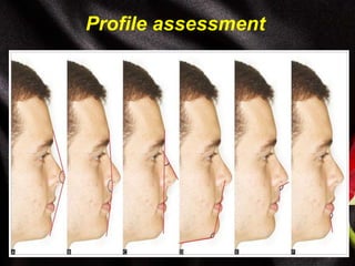
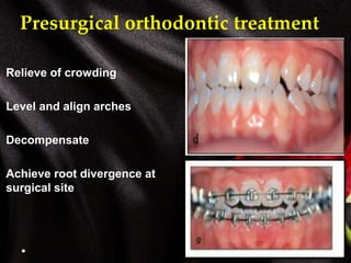
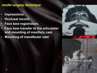
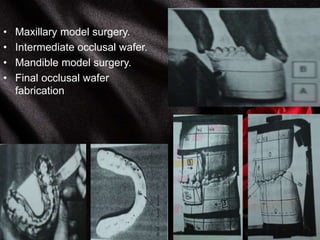
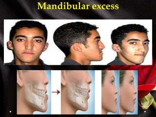
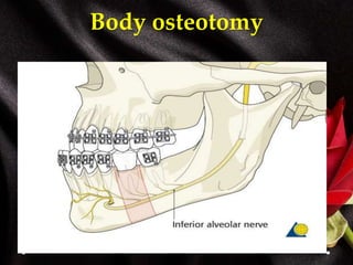
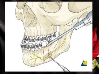
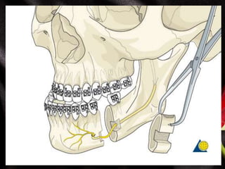
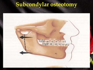
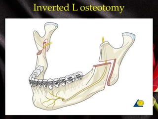
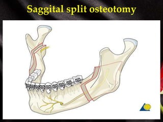
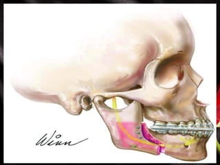
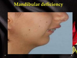
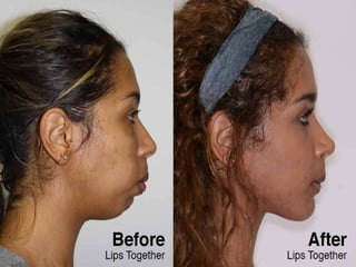
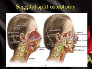
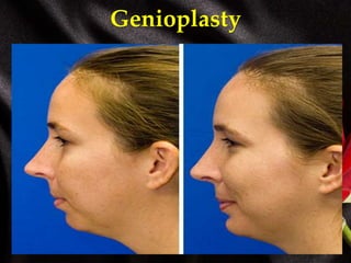
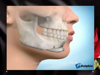
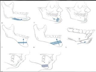
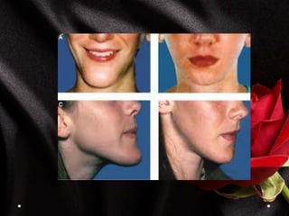
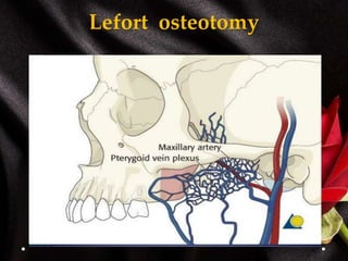
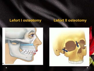
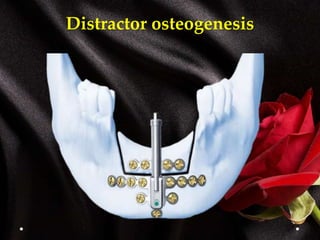
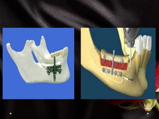
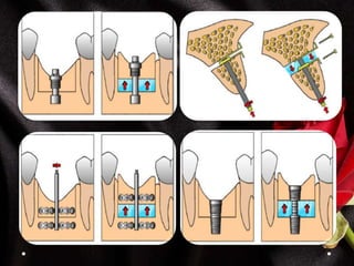
Orthognathic surgery
Dr. Mohamed Rahil
((Maxillofacial surgeon))
Tikrit dentistry college
2015 – 2016
Orthognathic surgery
 is the art and science of diagnosis, and
treatment of facial disproportion.
These disproportion may be congenital or
aquired
Basic Therapeutic Goals for
Orthognathic Surgery
• Function: obtain normal mastication, speech, ocular
function and respiratory function.
• Aesthetic: facial harmony and balance.
• Other possible benefit :
• TMJ dysfunction
• Sleep apnoea
• Traumatic occlusion and dental health
Patient Evaluation
 History
 Clinical examination
 Investigations
 Initial diagnosis
 Treatment plane
 Presurgical orthodontic
 Post –surgical orthodontic
Chief Complaints
• Understanding what patient concern, motivation,
expectation. That provide insight to psychological health of
the patient
• Patient who maintain unrealistic expectations are best not
treated
History
The purpose of history is to identify the patient’s orofacial
problems and their causes.
• Congenital deformity
• Trauma
• Medical history: bleeding disorder, autoimmune disease, any
pathological condition.
• Dental history: previous restorative, orthodontic, periodontal,
facial pain treatment.
Clinical evaluation
• Patient seated comfortably.
• Frankfort line parallel to the floor.
• Teeth in centric occlusion and lips relaxed
Frontal Assessment
Facial proportion
The full face is divided into
three equal parts
Upper part from hair line to
the glabella (or eyebrow)
Middle part from glabella to
the base of the nose
The lower part from the base
of the nose to the chin which
is subdivided into two parts,
the upper lip forms one-third
of it and the lower lip and the
chin two-thirds of it
Incisal lengths (incisal edge positions
 Is the most important
determinant in smile
creation because it serves
as a reference point to
decide the proper tooth
proportion and gingival
levels.
 Degree of tooth display:
a. 2 mm of incisor edge
show at rest
b. about 2 mm of gingival
show When smiling
vertical evaluation
• Symmetry :
1. Bizygomatic: between zygion
points the most lateral point of the
zygomatic arch.
2 . Bigonial : width of lower third
 High to width proportion are 1,3 : 1 for
female and 1,35 : 1 for males
 Bigonial width should be 30% less
than bizygomatic width
transverse evaluation
1. Role of fifth 2.intercanthal
interpapilary distance
Cant occlusion
Upper third evaluation
• Shape and symmetry: the temporal areas, frontal areas,
eyebrows, and supraorbital rims
• abnormality in this area often associated with craniofacial
syndromes, these areas usually with normal limits in
individuals with dentofacial deformaties.
Middle third evaluation
. Eyes and orbits: intercathal and
inertpupillary distance.
• The vertical symmetry of inner
and outer canthi of both eyes,(a
true horizontal line will bisect the
inner and outer canthi).
• The eyelids examination
( ptosis, entropion , ectropion)
Nose:
the symmetry of the nose,
Glabella, dorsum, tip, alar
base.
Proportion of alar base width
to the nasal length ( N-Prn)
(0.60).
Lower third evaluation
.Balance: the upper lip 1/3 of lower
lip.
.Lips:
symmetry of the lips at rest and
at smiling asymmetry exists
in
1. Intrinsic lip deformity
(patients with cleft) .
2. Facial nerve dysfunction.
3. An underlying dental-
skeletal asymmetry.
Profile assessment
Profil view
Nasolabial angle
 should be 90 -+ 10 degree
 It guid the upper lip
support by the maxillary incisors
 Influenced by decrease in
vertical maxillary dimention
 Increased in C II
 Decreased in CIII
Submental and neck examination
In esthetic examination can be
divided to
1. Mandibular angle
definition.
2. Neck-chin angle(110).
3. Neck-chin length. The
distance from pogonion to
the neck- chin angle (42 ±
6mm)
• Identify The posterior and
inferior border of mandible.
• The parotid hypertrophy.
• Skin laxity, lipomatosis.
Upper lip length
 Measured from
subnasale to lower lip
 22 -+2 mm for male and
20 -+2 for female
 It increase with age
Ricketts line
• Line drawen from nasal tip
to soft tissue pogonion
• Upper lip are 4mm and
lower lip 2 mm behind it
• Allow assessment of
anteroposterior
relationships of the lips
Labiomental angle
• 120 -+ 10 degree
• more Acute in class II
occlusion
• More obtuse in class III
occlusion
Lower Lip-Chin -Throat Angle
• between line drown from
lower lip to Pog and submental
line Normally 110o±8.
• Acute angle in CIII.
• Increase in CII.
• Distance from neck throat
angle to Menton 42±6mm.
Lateral cephalometric analysis
Benefit of cephalograph in orthognathic surgery
Cephalometric analysis helpful
in establishing the relation
of:
• The maxilla and mandible to
the base of skull.
• The maxilla to mandible.
• The maxillary teeth to
maxilla.
• The mandibular teeth to
mandible.
• The upper incisors to lower
incisors.
skeletal analysis
• S sella. G glabella
• N nasion .
• ANS.
• PNS.
• Point A.
• Point B.
• Me menton.
• Pg pogonion.
• Go gonion.
• Ar articulare.
• Co condylion.
• Or orbitale.
• Po porion.
Soft tissue analysis
• Soft tissue glabella(G).
• Soft tissue nasion (N).
• Pronasale (Pn).
• Subnasale (Sn).
• Labrale superior (Ls).
• Labrale inferior (Li).
• Stomion superius(Sts).
• Stomion inferior(Sti).
• Soft tissue pogonion(
Pog).
• Soft tissue menton( Me).
Skeletal planes
 Frankfort plane
 Anterior cranial base (SN)
 Occlusal plane
 Mandibular plane
Mean angular skeletal
cephalometric values
• SNA
• SNB
• ANB
• SN/MxP
• SN/MP
• FPLOP
• NSAr
• SArGo
• ArGoMe
• MxP/MP
• UI/MxP
• UI/LI
• LI/MP
• 81o±3
• 78o±3
• 3o±2
• 8o±3
• 35o±4
• 8o±4
• 125o±5
• 140o±6
• 128o±7
• 27o±4
• 109o±6
• 130o±6
• 93o±6
Skeletal anteroposterior relation ship
• SNA 81o± 3.
• SNB 78o± 3.
• ANB 3o± 2.
Facial angle(downs)
• Anteroposterior position
of mandible to cranium
base(n-pog).
Dental models analysis
1. Tooth size analysis
2. Arch width analysis.
3. Curve of spee.
4. Curve of welson
5. Tooth arch symmetry.
Presurgical orthodontic treatment
Relieve of crowding
Level and align arches
Decompensate
Achieve root divergence at
surgical site
model surgery technique:
• Impressions
• Occlusal record
• Face bow registration.
• Face bow transfer to the articulator
and mounting of maxillary cast.
• Mounting of mandibular cast
• Maxillary model surgery.
• Intermediate occlusal wafer.
• Mandible model surgery.
• Final occlusal wafer
fabrication
Surgical treatment
Mandibular excess
Body osteotomy
Subcondylar osteotomy
Inverted L osteotomy
Saggital split osteotomy
Mandibular deficiency
Saggital split osteotomy
Total subapical osteotomy
Genioplasty
Maxillary excess
Maxillary and midface deficiency
Lefort osteotomy
Lefort I osteotomy Lefort II osteotomy
Distractor osteogenesis
definition
Distraction osteogenesis is the creation of neoformed
bone and soft tissue by gradual and controlled
displacement of a bone fragment using ridged
stretching device delivers tensile force to the
developing callus at the site of the bone cut
Treating craniofacial deformities ( HFMS, Crouzon, post
trauma ,post resection, cleft palate…ect)
Intraoral distractor Extra oral distractor
Types of distractors
Terminology of distractors
 Latency period
 Distraction period
 Consolidation period
 Remodelling period
Hemifacial microsomia
Pierr robin syndrom
Alveolar distraction
indication
i.Sever atrophied edentulous ridge
ii.Segmental deficiencies for implant placement or functionally &
esthetically for crown bridge placement
iii.Narrow alveolar ridge (horz. Distraction)
iv.Gradual vertical movement of ankylosed tooth or
osteointegrated implant with surrounding bone
Advantages
I.No bone graft needed with hard & soft tissue obtained
II.Less possiblities of bone exposure & graft resorption
III.More esthetic
IV.Allows the use of complementary regeneration techniques
when outcome not satisfactory
Thank you

Orthognathic surgery

  • 1.
    Orthognathic surgery Dr. MohamedRahil ((Maxillofacial surgeon)) Tikrit dentistry college 2015 – 2016
  • 2.
    Orthognathic surgery  isthe art and science of diagnosis, and treatment of facial disproportion. These disproportion may be congenital or aquired
  • 3.
    Basic Therapeutic Goalsfor Orthognathic Surgery • Function: obtain normal mastication, speech, ocular function and respiratory function. • Aesthetic: facial harmony and balance. • Other possible benefit : • TMJ dysfunction • Sleep apnoea • Traumatic occlusion and dental health
  • 4.
    Patient Evaluation  History Clinical examination  Investigations  Initial diagnosis  Treatment plane  Presurgical orthodontic  Post –surgical orthodontic
  • 5.
    Chief Complaints • Understandingwhat patient concern, motivation, expectation. That provide insight to psychological health of the patient • Patient who maintain unrealistic expectations are best not treated History The purpose of history is to identify the patient’s orofacial problems and their causes. • Congenital deformity • Trauma
  • 6.
    • Medical history:bleeding disorder, autoimmune disease, any pathological condition. • Dental history: previous restorative, orthodontic, periodontal, facial pain treatment.
  • 7.
    Clinical evaluation • Patientseated comfortably. • Frankfort line parallel to the floor. • Teeth in centric occlusion and lips relaxed
  • 8.
  • 9.
    Facial proportion The fullface is divided into three equal parts Upper part from hair line to the glabella (or eyebrow) Middle part from glabella to the base of the nose The lower part from the base of the nose to the chin which is subdivided into two parts, the upper lip forms one-third of it and the lower lip and the chin two-thirds of it
  • 10.
    Incisal lengths (incisaledge positions  Is the most important determinant in smile creation because it serves as a reference point to decide the proper tooth proportion and gingival levels.  Degree of tooth display: a. 2 mm of incisor edge show at rest b. about 2 mm of gingival show When smiling
  • 11.
    vertical evaluation • Symmetry: 1. Bizygomatic: between zygion points the most lateral point of the zygomatic arch. 2 . Bigonial : width of lower third  High to width proportion are 1,3 : 1 for female and 1,35 : 1 for males  Bigonial width should be 30% less than bizygomatic width
  • 12.
    transverse evaluation 1. Roleof fifth 2.intercanthal interpapilary distance
  • 14.
  • 15.
    Upper third evaluation •Shape and symmetry: the temporal areas, frontal areas, eyebrows, and supraorbital rims • abnormality in this area often associated with craniofacial syndromes, these areas usually with normal limits in individuals with dentofacial deformaties.
  • 16.
    Middle third evaluation .Eyes and orbits: intercathal and inertpupillary distance. • The vertical symmetry of inner and outer canthi of both eyes,(a true horizontal line will bisect the inner and outer canthi). • The eyelids examination ( ptosis, entropion , ectropion)
  • 17.
    Nose: the symmetry ofthe nose, Glabella, dorsum, tip, alar base. Proportion of alar base width to the nasal length ( N-Prn) (0.60).
  • 18.
    Lower third evaluation .Balance:the upper lip 1/3 of lower lip. .Lips: symmetry of the lips at rest and at smiling asymmetry exists in 1. Intrinsic lip deformity (patients with cleft) . 2. Facial nerve dysfunction. 3. An underlying dental- skeletal asymmetry.
  • 19.
  • 20.
    Profil view Nasolabial angle should be 90 -+ 10 degree  It guid the upper lip support by the maxillary incisors  Influenced by decrease in vertical maxillary dimention  Increased in C II  Decreased in CIII
  • 21.
    Submental and neckexamination In esthetic examination can be divided to 1. Mandibular angle definition. 2. Neck-chin angle(110). 3. Neck-chin length. The distance from pogonion to the neck- chin angle (42 ± 6mm) • Identify The posterior and inferior border of mandible. • The parotid hypertrophy. • Skin laxity, lipomatosis.
  • 22.
    Upper lip length Measured from subnasale to lower lip  22 -+2 mm for male and 20 -+2 for female  It increase with age
  • 23.
    Ricketts line • Linedrawen from nasal tip to soft tissue pogonion • Upper lip are 4mm and lower lip 2 mm behind it • Allow assessment of anteroposterior relationships of the lips
  • 25.
    Labiomental angle • 120-+ 10 degree • more Acute in class II occlusion • More obtuse in class III occlusion
  • 27.
    Lower Lip-Chin -ThroatAngle • between line drown from lower lip to Pog and submental line Normally 110o±8. • Acute angle in CIII. • Increase in CII. • Distance from neck throat angle to Menton 42±6mm.
  • 28.
  • 29.
    Benefit of cephalographin orthognathic surgery Cephalometric analysis helpful in establishing the relation of: • The maxilla and mandible to the base of skull. • The maxilla to mandible. • The maxillary teeth to maxilla. • The mandibular teeth to mandible. • The upper incisors to lower incisors.
  • 30.
  • 31.
    • S sella.G glabella • N nasion . • ANS. • PNS. • Point A. • Point B. • Me menton. • Pg pogonion. • Go gonion. • Ar articulare. • Co condylion. • Or orbitale. • Po porion.
  • 32.
    Soft tissue analysis •Soft tissue glabella(G). • Soft tissue nasion (N). • Pronasale (Pn). • Subnasale (Sn). • Labrale superior (Ls). • Labrale inferior (Li). • Stomion superius(Sts). • Stomion inferior(Sti). • Soft tissue pogonion( Pog). • Soft tissue menton( Me).
  • 33.
    Skeletal planes  Frankfortplane  Anterior cranial base (SN)  Occlusal plane  Mandibular plane
  • 34.
    Mean angular skeletal cephalometricvalues • SNA • SNB • ANB • SN/MxP • SN/MP • FPLOP • NSAr • SArGo • ArGoMe • MxP/MP • UI/MxP • UI/LI • LI/MP • 81o±3 • 78o±3 • 3o±2 • 8o±3 • 35o±4 • 8o±4 • 125o±5 • 140o±6 • 128o±7 • 27o±4 • 109o±6 • 130o±6 • 93o±6
  • 35.
    Skeletal anteroposterior relationship • SNA 81o± 3. • SNB 78o± 3. • ANB 3o± 2.
  • 36.
    Facial angle(downs) • Anteroposteriorposition of mandible to cranium base(n-pog).
  • 37.
  • 38.
    1. Tooth sizeanalysis 2. Arch width analysis. 3. Curve of spee. 4. Curve of welson 5. Tooth arch symmetry.
  • 39.
    Presurgical orthodontic treatment Relieveof crowding Level and align arches Decompensate Achieve root divergence at surgical site
  • 40.
    model surgery technique: •Impressions • Occlusal record • Face bow registration. • Face bow transfer to the articulator and mounting of maxillary cast. • Mounting of mandibular cast
  • 41.
    • Maxillary modelsurgery. • Intermediate occlusal wafer. • Mandible model surgery. • Final occlusal wafer fabrication
  • 42.
  • 43.
  • 46.
  • 49.
  • 50.
  • 51.
  • 53.
  • 55.
  • 56.
  • 57.
  • 61.
  • 63.
  • 65.
  • 66.
    Lefort I osteotomyLefort II osteotomy
  • 67.
  • 68.
    definition Distraction osteogenesis isthe creation of neoformed bone and soft tissue by gradual and controlled displacement of a bone fragment using ridged stretching device delivers tensile force to the developing callus at the site of the bone cut Treating craniofacial deformities ( HFMS, Crouzon, post trauma ,post resection, cleft palate…ect)
  • 69.
    Intraoral distractor Extraoral distractor Types of distractors
  • 70.
    Terminology of distractors Latency period  Distraction period  Consolidation period  Remodelling period
  • 73.
  • 75.
  • 76.
    Alveolar distraction indication i.Sever atrophiededentulous ridge ii.Segmental deficiencies for implant placement or functionally & esthetically for crown bridge placement iii.Narrow alveolar ridge (horz. Distraction) iv.Gradual vertical movement of ankylosed tooth or osteointegrated implant with surrounding bone Advantages I.No bone graft needed with hard & soft tissue obtained II.Less possiblities of bone exposure & graft resorption III.More esthetic IV.Allows the use of complementary regeneration techniques when outcome not satisfactory
  • 79.