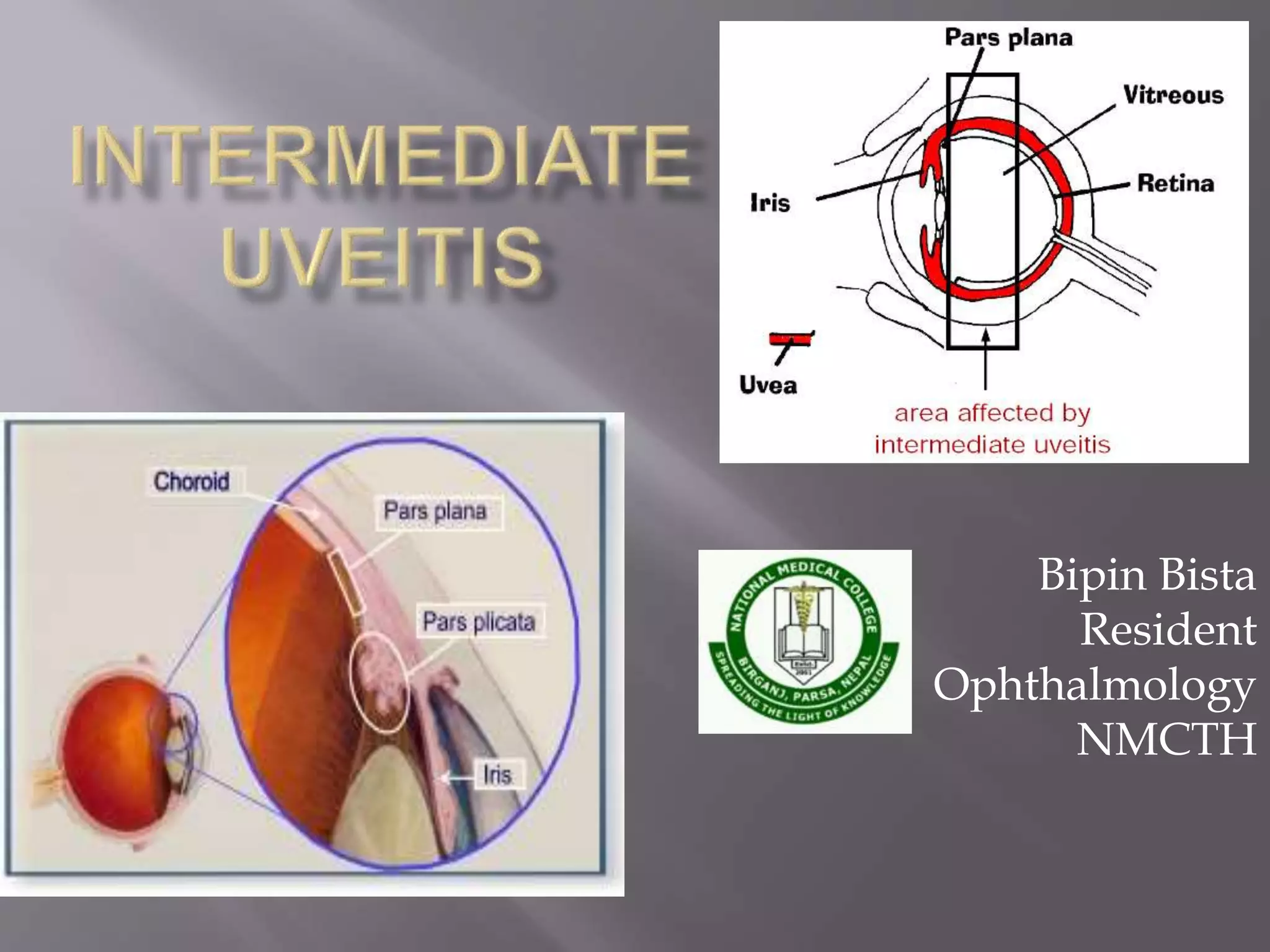
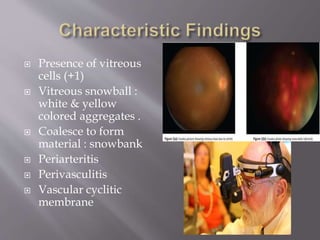
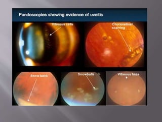
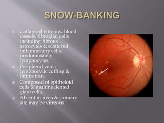
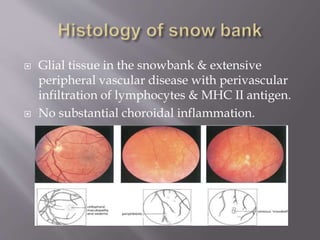
2. Diagnosis of intermediate uveitis is made by
inflammation primarily involves vitreous &
peripheral retina.
In 1960 Brockhurst & colleagues described it as
inflammation characterized by white
accumulation on pars plana.
International Uveitis Study Group : intraocular
inflammation predominantly involving
vitreous & peripheral retina.
3. National Institute of Health – 2.077 per 100000
persons.
No gender or race predilection.
Usually affects after the age of 40.
HLA Association.
4. Blurred vision
Floaters
Decreased visual acquity
If inflammation persists , severe visual loss.
Sudden loss of vision.
Anterior segment involvement – minimal.
AS involvement common in children.
5. Presence of vitreous
cells (+1)
Vitreous snowball :
white & yellow
colored aggregates .
Coalesce to form
material : snowbank
Periarteritis
Perivasculitis
Vascular cyclitic
membrane
6.
7. Snowbank may be discontinuous, usually
located inferiorly but can extend upto 3600
cystoid macular oedema – FA
Rhegmatogenous RD.
Chronic intermediate uveitis : long standing
macular oedema – development of retinal
pigment epithelial stippling in macula.
Disc oedema, optic atrophy & optic disc
neovascularization.
13. Collapsed vitreous, blood
vessels, fibroglial cells
including fibrous
astrocytes & scattered
inflammatory cells,
predominately
lymphocytes.
Peripheral vein :
lymphocytic cuffing &
infiltration.
Composed of epitheloid
cells & multinucleated
giant cells.
Absent in uvea & primary
site may be vitreous.
14. Glial tissue in the snowbank & extensive
peripheral vascular disease with perivascular
infiltration of lymphocytes & MHC II antigen.
No substantial choroidal inflammation.
21. Destroys the vascular component of peripheral
retinitis or vitreitis & eliminates the entrance
site for inflammatory mediators into the eye.
Combined with periocular steroid injections.
Evident within several weeks & lasts for 3-6
months.
Complication : transient increased vitreal
inflammation, reduced accomodation, cataract
& hyphaema.