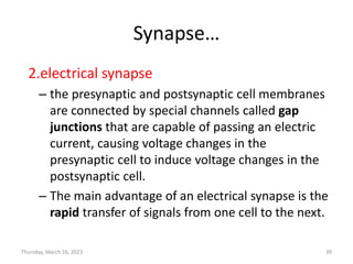
5. Introduction
• Human brain has some unique functions
• In human, nervous system makes it possible to have language,
abstract thinking, future planning etc.,
• Communication: a function of nervous system
– function of nervous system is to send signals from one cell
to other, or from one part of the body to other part.
– neurons communicate with other cells via synapses, for
rapid transmission of signals, mostly by chemical
transmission from one neuron to the next.
• Nervous system provides "point-to-point"
signals
5
Thursday, March 16, 2023
7. Neurons
• dendrites: receive signals from neighboring
neurons
• axon: transmit signals over a distance
• axon terminal: transmit signals to other
neuron dendrites or tissues
• myelin sheath: speeds up signal transmission
along the axon
Thursday, March 16, 2023 7
8. • There are three basic classes of neurons:
– Afferent neurons, Efferent neurons & Interneurons
• Afferent neurons
– are sensory neurons
– transmit sensory signals from receptors to central nervous
system.
8
Thursday, March 16, 2023
9. • Efferent neurons
– are motor neurons
– transmit motor signals from central nervous system to
effectors such as muscles and glands.
• Interneurons
– form complex networks within central nervous system.
– integrate the information received from afferent
neurons and to direct the function of the body through
efferent neurons.
9
Thursday, March 16, 2023
10. Neuroglia
• Neuroglia, also known as glial cells, act as “helper” cells of the
nervous system.
• Each neuron in the body is surrounded by 6 to 60 neuroglia
that protect, feed, and insulate neuron.
• Neuroglia are vital to maintain a functional nervous system.
• Though not taking part in conduction of neural signals.
• Three types of neuroglial cells have been identified:
– Microglia
– Oligodendrocytes
– Astrocytes
10
Thursday, March 16, 2023
11. Microglia
• Constitute 20% of total glial cells within the brain.
• These are the scavenger cells.
– Constantly moving and analyzing the CNS, for damaged
neurons and infectious agents.
• On activation, these cells migrate to area of tissue damage to
become macrophages, and remove cellular debris.
11
Thursday, March 16, 2023
12. Oligodendrogliocytes
• Oligodendrogliocytes possess very few cytoplasmic
processes.
• They are responsible for formation and maintenance of
myelin sheath around axons in central nervous system.
12
Thursday, March 16, 2023
13. Astrocytes
• Astrocytes have small cell bodies but have extensively
branching processes extending in all directions.
Two subtypes:
1. Fibrous astrocytes in white matter.
2. Protoplasmic astrocytes in gray matter
Functions:
• ‘End feet’ of astrocytes form tight junctions that surround
brain capillaries in Blood Brain Barrier (BBB).
• Also envelop synapses & surface of nerve cells.
13
Protoplasmic astrocyte
Fibrous astrocyte
Thursday, March 16, 2023
14. • Found throughout CNS and produce tropic substances to neurons.
• Help to maintain appropriate composition of interstitial fluid in CNS.
– Astrocytes take up K+ and neurotransmitters (glutamate and
GABA) released by neural activity into interstitial fluid.
14
Thursday, March 16, 2023
15. 15
Neuroglia cells Functions
Microglia • Act as phagocytes in nervous system and
remove degenerated tissue and foreign bodies
Oligodendroglia • Form myelination around axons in brain and
spinal cord
Astrocytes • Covers capillaries of brain to form BBB. Regulate
passage of molecules from blood to brain.
Other cells Functions
Ependymal cells • Line the surface of ventricles and central canal
of Spinal cord
Schwann cells • Surround axons of peripheral nerve fibres and
form myelin sheath
Thursday, March 16, 2023
17. Membrane potential
• Membrane potential (also transmembrane
potential or membrane voltage) is the
difference in electric potential between the
interior and the exterior of a biological cell.
• Differences in the concentrations of ions on
opposite sides of a cellular membrane lead to
the membrane potential.
Thursday, March 16, 2023 17
19. Resting membrane potential(RMP)
• The resting membrane potential of a cell is
defined as the electrical potential difference
across the plasma membrane when the cell is in
a non-excited state.
• Traditionally, the electrical potential difference
across a cell membrane is expressed by its
value inside the cell relative to the extracellular
cell.
• Neurons and muscle cells are excitable such that
these cell types can transition from a resting state
to an excited state.
Thursday, March 16, 2023 19
20. RMP
• The resting membrane potential is the result of
the movement of several different ion species
through various ion channels and transporters
(uniporters, cotransporters, and pumps) in the
plasma membrane.
• These movements result in different electrostatic
charges across the cell membrane.
• Neurons have a negative concentration gradient
most of the time, meaning there are more
positively charged ions outside than inside the
cell.
Thursday, March 16, 2023 20
21. RMP
• This regular state of a negative concentration
gradient is called resting membrane potential.
• During the resting membrane potential there
are:
– More sodium ions outside than inside the neuron
– More potassium ions inside than outside the
neuron
• A typical voltage across a cell membrane is
−70mV
Thursday, March 16, 2023 21
22. RMP
• The cell wants to maintain a negative resting
membrane potential, so it has a pump that
pumps potassium back into the cell and
pumps sodium out of the cell at the same
time.
Thursday, March 16, 2023 22
23. Action potential
• Action potentials are temporary shift (from
negative to positive) in the neuron’s
membrane potential caused by ions suddenly
flowing in and out of the neuron.
• Action potentials occur in several types of
cells, called excitable cells, which include
neurons, muscle cells
• Action potentials in neurons are also known as
"nerve impulses"
Thursday, March 16, 2023 23
24. Action potential…
• Action potentials are generated by special types of
voltage-gated ion channels embedded in a cell's
plasma membrane.
• During the resting state all of the voltage gated sodium
and potassium channels are closed.
• but they rapidly begin to open if the membrane
potential increases to a precisely defined threshold
voltage, depolarising the transmembrane potential
• When the channels open, they allow an inward flow of
sodium ions, which changes the electrochemical
gradient, which in turn produces a further rise in the
membrane potential towards zero.
Thursday, March 16, 2023 24
25. Action potential…
• The rapid influx of sodium ions causes the
polarity of the plasma membrane to reverse, and
the ion channels then rapidly inactivate.
• As the sodium channels close, sodium ions can no
longer enter the neuron, and they are then
actively transported back out of the plasma
membrane.
• Potassium channels are then activated, and there
is an outward current of potassium ions,
returning the electrochemical gradient to the
resting state.
Thursday, March 16, 2023 25
26. Action potential…
There are four main events that take place
during an action potential:
1. A triggering event(stimulus)
• This signal comes from other cells connecting to the
neuron, and it causes positively charged ions to flow
into the cell body.
• As positive ions flow into the negative cell, that
difference, and thus the cell’s polarity, decrease.
• If the cell body gets positive enough that it can trigger
the voltage-gated sodium channels found in the axon,
then the action potential will be sent.
Thursday, March 16, 2023 26
27. Action potential…
2.Depolarization
• makes the cell less polar (membrane potential gets less
negative as ions quickly begin to equalize the
concentration gradients) .
• This lets positively charged sodium ions flow into the
negatively charged axon, and depolarize the
surrounding axon.
• Though this stage is known as depolarization, the
neuron actually swings past equilibrium and becomes
positively charged as the action potential passes
through!
Thursday, March 16, 2023 27
28. Action potential…
3.Repolarization
• brings the cell back to resting potential.
• The inactivation gates of the sodium channels close,
stopping the inward rush of positive ions.
• At the same time, the potassium channels open.
• There is much more potassium inside the cell than out,
so when these channels open, more potassium exits.
• This means the cell loses positively charged ions, and
returns back toward its resting state.
Thursday, March 16, 2023 28
29. Action potential…
4.Hyperpolarization
• makes the cell more negative than its typical resting
membrane potential.
• As the action potential passes through, potassium
channels stay open a little bit longer, and continue to
let positive ions exit the neuron.
• This means that the cell temporarily hyperpolarizes, or
gets even more negative than its resting state.
• As the potassium channels close, the sodium-potassium
pump works to reestablish the resting state.
Thursday, March 16, 2023 29
32. Propagation AP
• The action potential generated at the axon
hillock propagates as a wave along the axon.
• The currents flowing inwards at a point on the
axon during an action potential spread out
along the axon, and depolarize the adjacent
sections of its membrane.
• If sufficiently strong, this depolarization
provokes a similar action potential at the
neighboring membrane patches.
Thursday, March 16, 2023 32
33. Synapse
• Synapse is a structure that permits a neuron to
pass an electrical or chemical signal to another
neuron or to the target effector cell.
• At a synapse, the plasma membrane of the signal-
passing neuron (the presynaptic neuron) comes
into close apposition with the membrane of the
target (postsynaptic)cell.
• In many synapses, the presynaptic part is located
on an axon and the postsynaptic part is located
on a dendrite or soma.
Thursday, March 16, 2023 33
35. Synapse…
• The vast majority of synapses in the nervous system are
classical axo-dendritic synapses (axon synapsing upon a
dendrite), however, a variety of other arrangements exist.
• These include
– axo-axonic,
– dendro-dendritic,
– axosecretory,
– axo-ciliary,
– somato-dendritic,
– dendro-somatic, and
– somato-somatic synapses.
• The axon can synapse onto a dendrite, onto a cell body, or
onto another axon or axon terminal, as well as into the
bloodstream or diffusely into the adjacent nervous tissue.
Thursday, March 16, 2023 35
36. Synapse…
There are two fundamentally different types of
synapses:
1. chemical synapse
– electrical activity in the presynaptic neuron is
converted (via the activation of voltage-gated calcium
channels) into the release of a chemical called a
neurotransmitter that binds to receptors located in
the plasma membrane of the postsynaptic cell.
– The neurotransmitter may initiate an electrical
response or a secondary messenger pathway that may
either excite or inhibit the postsynaptic neuron.
Thursday, March 16, 2023 36
38. Synapse…
Chemical synapses can be classified according to
the neurotransmitter released:
– glutamatergic (often excitatory)
– GABAergic (often inhibitory)
– cholinergic (e.g. vertebrate neuromuscular junction)
– adrenergic (releasing norepinephrine).
• Because of the complexity of receptor signal
transduction, chemical synapses can have
complex effects on the postsynaptic cell.
Thursday, March 16, 2023 38
39. Synapse…
2.electrical synapse
– the presynaptic and postsynaptic cell membranes
are connected by special channels called gap
junctions that are capable of passing an electric
current, causing voltage changes in the
presynaptic cell to induce voltage changes in the
postsynaptic cell.
– The main advantage of an electrical synapse is the
rapid transfer of signals from one cell to the next.
Thursday, March 16, 2023 39
40. Excitatory postsynaptic potential (EPSP)
• Arrival of an impulse at an axon terminal may result in a
transient depolarizing response ( For example membrane
potential from -70 to -65mV).
• The depolarizing response reaches a peak in 1-1.5 ms and
then disappears rapidly
• During this change, the excitability of postsynaptic neuron to
other stimuli is increased- Excitatory postsynaptic potential
(EPSP)
40
Thursday, March 16, 2023
41. Inhibitory postsynaptic potential (IPSP)
• Arrival of an impulse at an axon terminal may result
in a transient hyperpolarizing response ( For
example from -70 mv to -75 mV.
• The hyperpolarizing response reaches a peak in 1-1.5
ms and then disappears rapidly
• During this change, the excitability of postsynaptic
neuron to other stimuli is decreased- Inhibitory
postsynaptic potential (IPSP)
41
Thursday, March 16, 2023
42. Properties of EPSP
• No threshold level
• Decreases resting membrane
potential.
– Closer to firing level
• Can be graded in magnitude.
• Have no refractory period.
• Can summate.
42
Thursday, March 16, 2023
43. Synaptic Summation
• EPSPs can summate, producing
Action potential.
– Spatial summation:
• Numerous boutons
converge on a single
postsynaptic neuron .
– Temporal summation:
• Successive waves of
neurotransmitter release
(time).
43
Thursday, March 16, 2023
44. Generation of Action Potential
• Due to spatial or temporal summation, EPSP is strong enough
and generates an action potential in axon hillock region of
postsynaptic neuron.
• Axon hillock region has highest concentration of Na+ channels,
and hence has lowest threshold of excitation.
• Axon potential travels down the axon.
44
Thursday, March 16, 2023
That is, there is a difference in the energy required for electric charges to move from the internal to exterior cellular environments and vice versa, as long as there is no acquisition of kinetic energy or the production of radiation. The concentration gradients of the charges directly determine this energy requirement. For the exterior of the cell, typical values of membrane potential, normally given in units of milli volts and denoted as mV, range from –80 mV to –40 mV.
The point at which the forces of the electric fields completely counteract the force due to diffusion is called the equilibrium potential. At this point, the net flow of the specific ion (in this case sodium) is zero.
Action potentials (those electrical impulses that send signals around your body) are nothing more than a temporary shift (from negative to positive) in the neuron’s membrane potential caused by ions suddenly flowing in and out of the neuron. These gated channels are different from the leakage channels, and only open once an action potential has been triggered. Certain endocrine cells such as pancreatic beta cells, and certain cells of the anterior pituitary gland are also excitable cells
This then causes more channels to open, producing a greater electric current across the cell membrane and so on. The rapid influx of sodium ions causes the polarity of the plasma membrane to reverse, and the ion channels then rapidly inactivate.
An object is polar if there is some difference between more negative and more positive areas.
Positive ions still flow into the cell to depolarize it, but these ions pass through channels that open when a specific chemical, known as a neurotransmitter, binds to the channel and tells it to open. Neurotransmitters are released by cells near the dendrites, often as the end result of their own action potential! These incoming ions bring the membrane potential closer to 0, which is known as depolarization. An object is polar if there is some difference between more negative and more positive areas.
Voltage-gated sodium channels at the part of the axon closest to the cell body activate, thanks to the recently depolarized cell body.