Neuron
•Download as PPTX, PDF•
1 like•72 views

Neurons are electrically excitable cells that communicate with each other and the body. The human nervous system contains around 100 billion neurons. There are three main types of neurons - sensory neurons relay signals from sense organs to the central nervous system, motor neurons relay signals from the CNS to effector organs, and interneurons connect sensory and motor neurons. Each neuron has a cell body, dendrites that receive signals, and an axon that transmits signals. When a neuron is stimulated, it generates an action potential down its axon via changes in membrane potential. Neurotransmitters are released at synapses to transmit signals between neurons.

Recommended
More Related Content
What's hot
What's hot (20)
Similar to Neuron
Similar to Neuron (20)
Recently uploaded
Recently uploaded (20)
Neuron
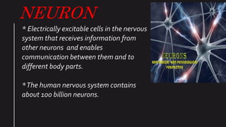
- 1. NEURON * Electrically excitable cells in the nervous system that receives information from other neurons and enables communication between them and to different body parts. *The human nervous system contains about 100 billion neurons.
- 2. TYPES OF NEURON *On the basis of functions 1. Sensory neuron – It relays impulse from sense organs to the CNS. 2. Motor neuron – It relays impulse from CNS to effector organs ( muscles ,glands, etc) 3. Interneuron – It is the link between sensory and motor neurons. • On the basis of structure 1. Multipolar – It has many dendrites extending from the cell body and one long axon. 2. Bipolar – It has one dendrite and one axon at opposite ends of the cell body 3. Unipolar – It has just have only one projection from the cell body which then further divide into two branches.
- 3. STRUCTURE OF NEURON • Each neuron comprise of three main parts – 1. Dendrites – Dendrites are thin widely branched projections from the cell body or soma. They are the major input points that receives information from other neurons. 2. Cyton /soma / cell body-The body of neuron from which the dendrites and axons project out . It contains aqueous material called cytoplasm and suspended structures inside it called cell organelles. 3. Axon- It is a single relatively thicker than dendrites fibre that relays information to muscle glands etc.
- 4. • Axon hillock – the axon is joint to the cell body at axon hillock where the nerve impulse originate. • Myelin sheath –A protective layer around the axon . • Those which are covered with myelin sheath are said to be myelinated. • Those which are not covered with myelin sheath are said to be non- myelinated. • Functions- It prevents ion leakage and protects the axon. • Glial cells –Their basic function is to provide nourishment and support to neurons .They are of 2 types • A) Oligodendrocytes- they produce myelin sheath in CNS. • B) Schwann cells- they produce myelin sheath in PNS. • Nodes of Ranvier – It is the gap in the axon, where myelin sheath is absent. • Function-They enables exchange of Intra cellular and extracellular fluids. • Telodendrion – the axon splits into many branches at the end of neuron which is called telodendrion • Synaptic knob – It is a bulb shaped swelling at the end of telodendrion that contains synaptic vesicles. • Synaptic vesicles –These are sac like structure present in synaptic knob that contains neurotransmitters which are chemical messengers. • Synapse –It consists of presynaptic membrane + synaptic cleft + post synaptic membrane, which acts as a point of contact neuron and its target
- 5. Types of synapse – There are 4 types of synapse – 1. Axodendritic synapse – Axon of one neuron synapses with the dendrite of subsequent neuron. 2. Axosoma synapse –Axon of one neuron synapse with the soma of subsequent neuron. 3. Dendrodendritic – Dendrites of one neuron synapses with the dendrite of subsequent neuron. 4. Axonaxionic – Axon of one neuron synapses with the axon of subsequent neuron.
- 6. EXCHANGE OF INFORMATION Exchange of information between neurons involves two types of transmission . 1. Axonal transmission –When the information travels along the length of neuron also called electrical transmission. 2. Synaptic transmission-When the information travels through the gap between the two neurons ( synaptic cleft ). It is also called chemical transmission. NEURAL MEMBRANE It is the site where most processes involved and functions are triggered. This lipid bilayer membrane have protein structure inside them which are of 3 types- A. Receptor protein – It is present to register the presence of neurotransmitter. B. Channel protein-These are responsible for the passage of ions and proteins. C. Pump proteins-To pass the ion/ protein from lower to higher concentration.
- 7. RESTING MEMBRANE POTENTIAL When a neuron is at rest – that is not receiving any information from other neurons that is the intracellular fluid is more negatively charged than the extracellular fluid.This difference in polarity between inside and outside the cell membrane is called RESTING MEMBRANE POTENTIAL. Using the intracellular fluid as a reference point the RMP of the neuron is -70 mV that means that inside of the cell is 70 mV more negatively charged than extracellular fluid. The outside of the cell contain high concentration of sodium ions( Na+), chloride ions (Cl-) and calcium ions (Ca+2) where as inside of the cell contains high concentration of potassium and negatively charged ions.
- 8. Two passive forces i.e. Diffusion and electrostatic pressure and one active force that is sodium potassium pump facilitate ion exchange across membrane 1. DIFFUSION –Tendency of molecule to move from higher concentration areas to low concentration areas till there is an even distribution of molecules. 2. Electrostatic pressure – It refers to the attraction of opposite polarity molecules and the repulsion of same polarity molecules. there are 3 cases a)The concentration of Cl- ions is higher outside the cell so they tend to diffuse in however the force of diffusion is counteracted by the electrostatic pressure inside the cell due to high negative charge inside the cell. Therefore Cl- remains OUTISDE. b) Similarly k+ ions will tend to diffuse outside the cell but will be counteracted electrostatic pressure created by the positively charged ions outside the cell. c) For Na+ ions, Both diffusion and electrostatic pressure would tend to move Na+ ions inside the cell but it remains outside due to activation of sodium potassium pump. 3. SODIUM POTASSIUM PUMP- It actively expels 3 Na+ ions outside for 2 k+ Ions inside the cell.
- 10. ACTION POTENTIAL The sequential events that occur when a resting neuron receives information is collectively referred to as Action Potential / Spike potential or firing of neuron. It occurs in 4 steps- 1. Depolarisation to threshold level 2. Reversal of Membrane polarity 3. Repolarisation to resting potential. 4. Refractory period.
- 11. DEPOLARISATION 1. Initially when the neuron is at rest, it exhibits RMP – (-70 mV) that is inside of the cell is more negative then outside. 2. On detecting a stimulus the sodium gates open temporarily as a result of which there is a rapid influx of sodium ions inside the cell, making it less negative. 3.When it reaches to (-55 mV), the cell membrane undergoes a more radical change in polarisation resulting in more number of sodium ions pouring inside the cell making It positive (+30 mV) 4. -55mV is the threshold level above which a neuron will pass a message 5. +30 mv is the peak potential. REVERSAL OF POLARITY Due to opening of many sodium channels the inside becomes temporarily positive bringing the membrane potential to +30mV.This is called the reversal of polarity.
- 12. REPOLARISATION TO RESTING POTENTIAL 1.The K+ channels open and k+ ions leave the cell Because they are repelled by higher levels of Na+ inside the cell. 2. Shortly after the K+ channels open , Na+ channels close and Na+ ions stop entering the cell. 3.The continuous exit of K+ ion outside the cell, causes the inside to become more negative then outside once again. 4.This phase is called Repolarisation. REFRACTORY PERIOD It consist of two phases- 1. Absolute refractory period - The brief period of time during which a neuron is completely resistant to further stimulation , no matter how intense the stimulus is, calledAbsolute R.P. 2. Relative refractory period –The period during which a neuron can generate an another action potential only in response to a more intense stimulus then normal. It corresponds to the part where the membrane potential actually becomes slightly more negative then normal (-90mV) , a state known as Hyperpolarisation
- 13. Restoring RMP – 1. Although the distribution of Na+ and K+ ions changes as a result of depolarisation and action potential the distribution quickly returns to normal through the sodium potassium pump that acts to maintain the ion concentration differences of RMP.(-70mV), until changed by a depolarising stimulus.
- 14. PROCESS OF NERVE IMPULSE CONDUCTION 1 . SALATORY CONDUCTION –The propagation of an action potential from node to node in myelinated axons is termed a saltatory conduction. . AtThe end of each telodendrion, the pre synaptic terminal Calcium channels open and Ca+2 ions enters inside the cell. . Calcium entry causes the synaptic vesicles to align themselves along the presynaptic membrane where they release the neurotransmitter molecules into the synaptic cleft . .Diffusion carries the neurotransmitter molecules across the synaptic cleft, and they encounter and attached to specific receptor site on the post synaptic membrane. .The neurotransmitter have either inhibitory effect or excitatory effect on the post synaptic membrane. A) Neurotransmitter that have excitatory influence produces DEPOLARISATION at the post synaptic membrane by increasing the number of sodium that opens, allowing the more Na+ ions to enters the cell.This depolarisation is called an Excitatory Post synaptic potential or EPSP. B) Neurotransmitter that have an inhibitory effect on the post synaptic membrane produces hyperpolarisation by causing K+ to leave the cell or Cl- to enter.This hyperpolarisation is called an Inhibitory post synaptic potential or IPSP
- 15. SUMMATION EFFECT The net effect of excitation and inhibition determines whether or not the neuron is activated and will fire. Unlike action potential, which are all or none events, EPSP and IPSP received by the neurons are GRADED POTENTIAL with different values at different times. The various EPSP is received by the post synaptic membrane can be added together to produce a combine effect of depolarisation, similarly IPSP produces a combine level of hyperpolarisation . This process of adding a positive or negative influences on the cell membrane is called summation. It is of two types- 1. Spatial summation- Occurs when multiple stimuli exerts their effects at the same time on different receptor site on the post synaptic membrane. 2. Temporal summation- Occurs when a stimuli (A twice)are close enough in time that their effects are additive.
- 16. PRESYNAPTIC EFFECTS POST SYNAPTIC RECEPTORS . Pres synaptic facilitation-The enhance release of neurotransmitter from the pre synaptic membrane caused by the action of another neuron. . Autoreceptors- A presynaptic receptors whose stimulation decreases the amount of neurotransmitter release by a pre synaptic neuron. .Ionotropic receptor – Receptor whose ion channel are rapidly open by the direct action of a neurotransmitter. . Metabotropic receptor- A receptor whose ion channel are indirectly open by a second messenger. .Second messenger- A chemical that causes changes inside the cell in response to a neurotransmitter that lead to ion channel opening. . Pre synaptic inhibition – It is the decrease in neurotransmitter from the pre synaptic membrane despite the occurrence of an action potential, caused by the action of another neuron. . AUTORECEPTORS – Released neurotransmitters acts as an autoreceptors to decrease the subsequent neurotransmitter release.
- 17. TERMINATION OF NEUROTRANSMITTER EFFECT It is done in two ways – 1. Enzymatic degradation- It is the process of breakdown and thus deactivation of neurotransmitter molecules by an enzyme ( acetylcholinesterase). 2. Neurotransmitter reuptake- It is the process of return of neurotransmitter to the pre synaptic neuron at the reuptake sites.
- 18. THANK YOU