
Recommended
More Related Content
What's hot
What's hot (20)
Similar to Nerve Cells Communicate via Synapses and Circuits
Similar to Nerve Cells Communicate via Synapses and Circuits (20)
More from Sabahat Ali
More from Sabahat Ali (20)
Recently uploaded
Recently uploaded (20)
Nerve Cells Communicate via Synapses and Circuits
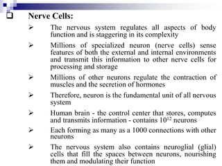
- 1. Nerve Cells: The nervous system regulates all aspects of body function and is staggering in its complexity Millions of specialized neuron (nerve cells) sense features of both the external and internal environments and transmit this information to other nerve cells for processing and storage Millions of other neurons regulate the contraction of muscles and the secretion of hormones Therefore, neuron is the fundamental unit of all nervous system Human brain - the control center that stores, computes and transmits information - contains 1012 neurons Each forming as many as a 1000 connections with other neurons The nervous system also contains neuroglial (glial) cells that fill the spaces between neurons, nourishing them and modulating their function
- 2. Despite the complexity of the nervous system as a whole, the structure and function of individual nerve cells is understood in great detail Perhaps in more detail than for any other type of cell The function of a neuron is to communicate information, which it does by two methods: Electric signals process and transmit information within a cell Chemical signals transmit information between cells, utilizing process similar to those employed by other types of cells to signal each other Information from the environment creates special problems because of the diverse types of signals that must be sensed - light, touch, pressure, sound, odorants, the stretching of muscles
- 3. Sensory neurons have specialized receptors that convert these stimuli into electric signals These electric signals are than converted into chemical signals that are passed on to other cells - called interneurons - that convert the information back into electric signals Ultimately, the information is transmitted to muscle- stimulating motor neuron or to other neurons that stimulate other types of cells such as glands Thus the out put of a nervous system is the result of its circuit properties, that is, the wiring, or interconnections, between neurons and the strength of these interconnections At times, the properties of a nervous system must change. For example: in the development of new memories
- 4. Such changes can be explained as alterations in the number and nature of the interconnections between individual neuron Neurons, Synapses and Nerve Circuits; Since neuron is the fundamental unit of all nervous systems, we will discuss the structural features that are unique to these cells and • the type of electrical signals that they use to process and transmit information Then synapses - the specialized sites where neurons send and receive information from other cells and Some of the circuits that allow groups of neurons to coordinate complex processes
- 5. Specialized Regions of Neurons Carry out Different Functions; • Most neuron contains four distinct regions with differing functions: - The cell body - The dendrites - The axon and - The axon terminals Fig 21.1 Lodish 3rd Ed • The cell body contains the nucleus and lysosomes and is the site where virtually all neuronal proteins and membranes are synthesized • Some proteins are synthesized in dendrites but axons and axon terminals do not contain ribosome. Therefore no protein synthesized there
- 6. Carries the nerve impulse From receptor cell to cell body Carries the nerve impulse from cell body to the spinal cord or brain
- 7. • Proteins and membranes that are required for renewal of the axon and nerve termini are synthesized in the cell body and assembled there in to membranous vesicles or multi-protein particles • These are transported along microtubules down the length of the axon • This process is called orthograde axoplasmic transport to the terminals • Where they are inserted in to plasma membrane or other organelles • Axonal microtubules are also the tracks along which damaged membrane and organelles move up the axon toward the cell body • Where they are degraded in lysosomes and this process is called retrograde transport
- 8. • Most neurons have a single axon • These are specialized for the conduction of a particular type of electrical impulse away from the body, called action potential • Action potential is series of sudden changes in the electric potential across the plasma membrane Fig 21.2 Lodish 3rd Ed • In resting or non-stimulated state the potential is ~ -60 mV (the inside is negative relative to the outside) • This is similar to the potential in most non-neuronal cells • At the peak of an action potential the membrane potential can be as much as +50 mV (inside positive), a net change of ~ 110 mV • This depolarization of the membrane is followed by a rapid re-polirization or return to the resting potential
- 10. • These characteristics distinguish an action potential from other types of changes in electrical potential across the plasma membrane and allow an action potential to move along an axon without diminution • These and other changes in membrane potential are generated and propagated by the opening and closing of specific ion channel proteins in the neuron plasma membrane • Many of which have been cloned and characterized in great detail Fig 21.3 Lodish 3rd Ed • Action potentials move rapidly, at speed up to 100 meters per second • In humans, axon may be more than a meter long, yet it takes only a few milliseconds for an action potential to move along their length
- 12. • An action potential originates at the axon hillox, the junction of axon and cell body and • is actively conducted down the axon into the axon terminals, small branches of the axon that form the synapses or connections with other cells • At synapses signals are passed: to other neurons to a muscle cell at a neuromuscular junction or to any of various other types of cells • A single axon in the central nervous system can synapse with many neurons and induce responses in all of them simultaneously • Most neurons have multiple dendrites, which extend outward from the cell body and are specialized to receive chemical signals from the axon termini of other neurons
- 13. • Dendrites convert these signals into small electric impulses and transmit them to cell body • Neuronal cell bodies can also form synapses and thus receive signals Fig 21.4 Lodish 3rd Ed • Particularly in the central nervous system, neurons have extremely long dendrites with complex branches • This allows them to form synapses with and receive signals from a large number of other neurons, perhaps up to a thousand • Electric disturbance generated in the dendrites or cell body spread to the axon hillox • If the electrical disturbance there is great enough, an action potential will originate and will be actively conducted down the axon
- 15. Synapses are Specialized Sites where Neurons Communicate with Other Cells; • Synapses are specialized sites where neurons communicate with each other • Synapses generally conduct signals in only one direction • An axon terminal from presynaptic cell sends signals that are picked up by the postsynaptic cell • There are two types of synapses which differ in both structure and function: i. Electrical ii. Chemical • Chemical synapses are much common than electric synapses
- 16. Fig 21.5a, b Lodish 3rd Ed • When action potential in the presynaptic cell reaches an axon terminal, some of the vesicles fuse with the plasma membrane, releasing their contents into the synaptic cleft • The neurotransmitter diffuses across the synaptic cleft and after a lag period of about 0.5 millisecond binds to receptors on the postsynaptic cells • The bound neurotransmitter changes the ion permeability of the postsynaptic plasma membrane • Which in turn, changes the membrane’s electrical potential at this point • If postsynaptic cell is neuron, this electric disturbance may be sufficient to induce an action potential • If postsynaptic cell is a muscle, the change in membrane potential following binding of the neurotransmitter may induce contraction
- 17. Epinephrine or acetylcholine Postsynaptic cell can be dendrite or cell body of another neuron, a muscle or gland cell or rarely even another axon. When postsynaptic is muscle cell, the synapse is called neuromuscular junction or motor end plate * * Filled with NT (E, Ach)
- 18. • If a gland cell - neurotransmitter may induce hormone secretion • Now the question is how then signal terminates? i. In some cases, enzymes attached to the fibrous network connecting the cells destroy the neurotransmitter after it has functioned ii. In other cases, the signal is terminated when the neurotransmitter diffuses away or is transported back into the presynaptic cell • In certain types of synapses, the postsynaptic neuron sends signals to the presynaptic one Fig 21.5c Lodish 3rd Ed • Such retrograde signals can be gases such as i. Nitric oxide (NO) ii. Carbon monoxide or iii. Peptide hormones
- 20. • This type of signaling modifies the ability of the presynaptic cell to signal the postsynaptic one • It is thought to be important in many types of learning • Sometimes an axon terminal of one neuron will synapse with the axon terminal of other neuron Fig 21.6 Lodish 3rd Ed • Such synapse may either inhibit or stimulate the ability of an axon terminal to secrete the contents of its synaptic vesicles and signal to a postsynaptic cell • Neurons communicating by an electrical synapse are connected by gap junctions • Through which electric impulse can pass directly from the presynaptic cell to the post synaptic one Fig 21.7 Lodish 3rd Ed
- 21. We will discuss later how the molecular properties of such synapses permit certain types of learning /Stimulatory neuron
- 23. • Electric synapses allow a presynaptic cell to induce an action potential in the postsynaptic cell with greater certainty then chemical synapse and with out lag period • Why….? Neurons are Organized into Circuits; • In complex multi-cellular animals, such as insects and mammals, signaling circuits consist of two or more neurons and • In some cases, highly specialized sensory receptor cells which respond to specific environmental stimuli such as: - light - heat - stretching - pressure and - concentrations of many chemical substances
- 24. • In the type of circuit called a reflex arc: • Interneurons connect multiple sensory and motor neurons, allowing one sensory neuron to affect multiple motor neurons and • One motor neuron to be affected by multiple sensory neurons • In this way interneurons integrate and enhance reflexes • For example, the knee-jerk reflex in humans involves a complex reflex arc in which one muscle is stimulated to contract while another is inhibited from contracting Fig 21.8 Lodish 3rd Ed • Such circuits allow an organism to respond to a sensory input by the coordinated action of sets of muscles that together achieve a single purpose
- 25. 1. Contraction 2. Inhibition of contraction Net result of 1 and 2 is extension of leg at knee joint
- 26. • The sensory and motor neurons of circuits such as the knee-jerk reflex are contained with in the peripheral nervous system • These circuits send information to and receive information from the central nervous system, which comprises the brain and spinal cord and is composed mainly of interneurons Fig 21.9 Lodish 3rd Ed • After having discussed the general features of neuron structure, interactions and circuits, we will try to understand mechanism by which a neuron generates and conducts electric impulses The Action Potential and Conduction of Electric Impulses: • We discussed earlier that an electric potential exists across the plasma membrane of all cells • The potential across the plasma membrane of cells can be measured as follows:
- 28. Fig 21.11 Lodish 3rd Ed • The potential across the surface membrane of most animal cells generally does not vary with time • In contrast, neurons and muscle cells- the principal types of electrically active cells – undergo controlled changes in their membrane potential • They conduct action potentials along their membrane by sequentially opening and closing ion channels that are specific for Na+ and K+ ions Fig 21.3 Lodish 3rd Ed • Thus, we need to explain how the opening and closing of ion channels and the resultant movement of small numbers of ions from one side of the membrane to the other causes changes in the membrane potential
- 31. Cellular Effects of Ca2+ Depends on its Cytosolic Level and often are Mediated by Calmodulin: Most intracellular Ca2+ ions sequestered in the mitochondria and endoplasmic reticulum (ER) or other cytoplasmic vesicles The concentration of free Ca2+ in the cytosol of any cell is extremely low i.e. ≤10-7 whereas its concentration in extra cellular fluid (~10-3 M) and in ER is high Thus there is a large gradient tending to drive Ca2+ into cytosol across both the plasma membrane and ER membrane When a signal transiently opens Ca2+ channels in either of these membranes, Ca2+ rushes into cytosol, dramatically increasing the local concentration and triggering Ca2+ responsive proteins in the cell
- 32. To use Ca2+ as an intracellular signal, cell must keep resting cytosolic Ca2+ levels low Fig 15.27 Alberts 3rd Ed Fig 24.18 Zubay Two common pathways by which Ca2+ can enter the cytosol in response to extra cellular signals Fig 15.28 Alberts 3rd Ed Release of intracellular Ca2+ stores also is mediated by ryanodine receptors in muscle cells and neurons In addition to IP3-sensitive Ca2+ channels, muscle cells and neurons possess other Ca2+ channels called ryanodine receptors (RYRs) This is because of their sensitivity to the plant alkaloids ryanodine Fig 20.44 Lodish 3rd Ed
- 37. Two mechanisms operate to terminate the initial Ca2+ response: IP3 is rapidly dephosphorylated and thereby inactivated by specific phosphatases Cytosolic Ca2+ is rapidly pumped out mainly out of the cell Ca2+ Oscillations often Prolong the Initial IP3 Induced Ca2+ Response Ca2+ sensitive fluorescent indicators such as aequorin or fura-2 are used to monitor cytosolic Ca2+ in individual cells In which the IP signaling pathway has been activated The initial Ca2+ signal is often seen to propagate as a wave through the cytosol from a localized region of the cell Fig 15.31 Alberts 3rd Ed
- 39. Biological significance of Ca2+ oscillations is uncertain Their frequency often depends on the concentration of the extra cellular signaling ligand and might, in principle, be translated into a frequency dependent cellular response In hormone secreting pituitary cells, for example, stimulation by an extra cellular signaling molecule induces repeated Ca2+ spikes Each of which is associated with a burst of hormone secretion It has been suggested that this arrangement might maximize secretory out put while avoiding the toxic effects of a sustained rise in cytosolic Ca2+
- 40. Calmodulin is a Ubiquitous Intracellular Ca2+ Receptor A typical animal cell contains more than 107 molecules of calmodulin, which can constitute as much as 1% of the total protein mass of the cell Highly conserved protein, single polypeptide chain containing 150 amino acids with four high affinity Ca2+ binding sites It undergoes a conformational change when it binds Ca2+ Fig 15.34 Alberts 3rd Ed The allosteric activation of calmodulin by Ca2+ is analogous to the allosteric activation of A kinase by cAMP except that
- 42. Ca2+/calmodulin has no enzyme activity itself but acts by binding to other proteins Targets regulated by Ca2+/calmodulin are: • various enzymes and • membrane transport proteins For example in many cells Ca2+/calmodulin binds to and activates the plasma membrane Ca2+-ATPase that pumps Ca2+ out of the cell Ca2+/Calmodulin-dependent Protein Kinases (CaM-Kinases) Mediate Most of the Actions of Ca2+ in Animal Cells Most of the effects of Ca2+ in cells are mediated by protein phosphorylation catalyzed by a family of Ca2+/calmodulin-dependent protein kinases (CaM- kinase
- 43. These kinases phosphorylate serine or threonine in proteins Like cAMP, the response of a target cell to an increase in free Ca2+ concentration in the cytosol depends on which CaM-kinase- regulated target proteins are present in the cell The first CaM-kinase to be discovered was myosin light chain kinase (MLCK) which activates smooth muscle contraction and phosphorylase kinase which activates glycogen breakdown The best studied example of such multifunctional CaM-kinase is CaM- kinase II, which is found in all animal cells but is specially enriched in the nervous system When neurons that use catecholamine (dopamine, nor-adrenaline or adrenaline) as their neurotransmitter are activated, for example, the influx of Ca2+ through voltage-gated Ca2+ channels in the plasma membrane stimulates the cells to secrete their neurotransmitter
- 44. The Ca2+ influx also activates CaM- kinase II to phosphorylate and their by activate tyrosine hydrolase, a rate limiting enzyme in catecholamine synthesis In this way both secretion and re-synthesis of the neurotransmitter are stimulated when the cell is activated Fig 24.10, Zubay CaM-kinase has a remarkable property: it can function as a molecular memory device, switching to an active state when exposed to Ca2+/calmodulin and then remaining active even after the Ca2+ is withdrawn This is because the kinase phosphorylate itself i.e. autophophorylation as well as other cell proteins when activated by Ca2+/ calmodulin
- 45. Tyrosine hydroxylase: The amount and activity are regulated by cAMP- dependent mechanism that are responsive to neurotransmitter (acetylcholine) CaM kinase II:
- 46. Fig 15.35 Alberts The cAMP and Ca2+ Pathways Interact: The cAMP and Ca2+ intracellular signaling pathways interacts at several levels in the hierarchy of control First, cytosolic Ca2+ and cAMP levels can influence each other. For example: • Some forms of the enzymes that break down and make cAMP are regulated by Ca2+/ calmodulin complexes - cAMP phosphodiestrase and adenyl cyclase • Similarly A-kinase can phosphorylate some Ca2+ channels and pumps and alter their activity for example:
- 48. • A-kinase phosphorylates the IP3 receptor in the ER which can either inhibit or promote IP3- induced Ca2+ release, depending on the cell type Second, the enzymes directly regulated by Ca2+ and cAMP can influence each other. For example: • Some CaM-kinases are phosphorylated by A- kinase Third, these enzymes can have interacting effects on shared downstream target molecules. Thus • A-kinase and CaM-kinases frequently phosphorylate different sites on the same proteins, which are thereby regulated by both cAMP and Ca2+. For example: - CREB (cAMP responsive element binding protein) gene regulatory protein
- 49. Fig 20.41, Lodish 3rd Ed Fig 15.36 Alberts 3rd Ed Fig 15.16 Alberts 3rd Ed Table 15.1 Lodish 6th Ed G Protein-Linked Cell-Surface Receptors that Regulate Ion Channels Trimeric G proteins do not act exclusively by regulating the activity of enzymes and altering the concentration of cyclic nucleotides or Ca2+ in cytosol In some cases, they directly activate or inactivate ion channels in the plasma membrane of the target cell
- 50. Epinephrine SRnot only triggers muscle contraction but also * *
- 54. Mice Defective in the Hippocampal α-Ca2+- Calmodulin Activated Protein Kinase are Impaired in Long-Term Potentiation and in Spatial Learning – the Beginnings of a Molecular Psychology: As observed, opening of the NMDA receptors is associated with an increase in cytosolic Ca2+ and an activation of several Ca2+-dependent enzymes Among these is the α-isoform of Ca2+-calmodulin- dependent protein kinase II An enzyme found in abundance in neurons in the hippocampus For this reason, workers suspected that this enzyme was involved in induction of long-term potentiation and perhaps in certain types of learning To test this hypothesis, mice were genetically engineered to have a deletion in the gene coding this enzyme In most respect these mice grow and behave normally. For example:
- 55. Their abilities to eat and mate are normal and they have the coordinated motor skills to swim normally in water However, cultured hippocampus neurons from these mice are defective in the induction of long-term potentiation More strikingly, the mice are impaired in a particular type learning process, supporting the notion that α-isoform of Ca2+-calmodulin-dependent protein kinase II is essential for induction of long-term potentiation and that Long-term potentiation, in turn, is the electro- physiological basis of a particular type of learning In a critical psychological test, mice are placed in a round pool of opaque water To escape the water, the mice must swim to a submerged platform
- 56. The mutant mice can find the platform normally if it is made visible by a flag, indicating that they can learn to associate the flag with the submerged platform In contrast, when platform is hidden, the mice must learn to find it from integrating multiple spatial relationships between objects in the room surrounding the pool (e.g., pictures on the wall) and the position of the platform The mice defective with Ca2+-calmodulin-dependent protein kinase take longer to learn to find the platform than do their normal littermates This demonstrates that the α-isoform of Ca 2+- calmodulin-dependent protein kinase II plays an important role in spatial learning and That it is not essential for some types of non-spatial learning As more hippocampus-specific proteins are identified by molecular cloning techniques, scientists may identify other enzymes or receptors essential for other types of learning processes in mice
- 57. Memory and Neurotransmitters: In its most general sense, learning is a process by which human and other animals modify their behavior as a result of experience or as a result of acquisition of information about the environment Memory is a process by which this information is stored and retrieved Psychologists have defined two types of memory depending on how long it persists: Short term i.e. minutes to hours Long term i.e. days to years It is generally accepted that memory results from changes in the structure or function of particular synapses But until recently memory and learning could not be studies with the tools of cell biology or genetics
- 58. Most researchers believe that long term memory involves the formation or elimination of specific synapses in the brain and The synthesis of new mRNAs and proteins Because short term memory is too rapid to be attributed to such gross alterations Some have suggested that changes in the release and function of neurotransmitters at particular synapses are the basis of short term memory Indeed, recent work has identified several types of proteins that function to modify synaptic activity These proteins integrate two different signaling pathways: they respond to two (or more) different but coincident signals by generating an output that is different from that produced by either signal acting separately
- 59. We shall see how two such proteins are involved in elemental forms of learning? We begin with a molecular analysis of elemental forms of learning in the fruit fly Drosophila and the sea slug Aplysia and Then turn to long term potentiation, a form of learning exhibited by many synapses in the mammalian brain Mutation in Drosophila Affect Learning and Memory: Remarkable as it sounds, fruit flies can be trained to avoid certain noxious stimuli During the training period, a population of flies is exposed to two different stimuli Either two odoriferous chemicals or two colors of light One of the two is associated with an electric shock The flies are then removed and placed in a new apparatus and
- 60. The two stimuli are repeated but with out the electric shock The flies are tested for their avoidance of the stimulus associated with the shock About half of the flies learn to avoid the stimulus associated with shock and This memory persist for at lease 24 hr Pain staking observation of mutagenized flies has led to the identification of six different genes in which mutation cause defects in this learning process Two mutations that disrupt memory affect cAMP levels are: One dunce, is due to a mutation in one of two isoforms of the enzyme cAMP phosphodiesterase