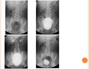
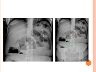
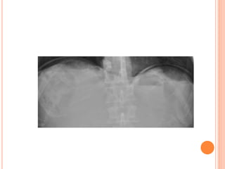
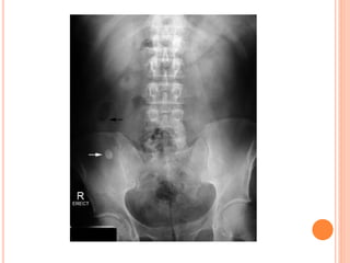
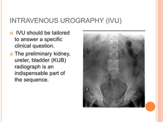
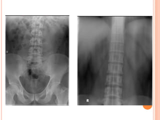
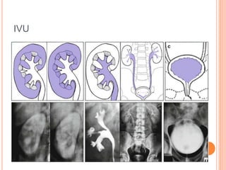
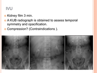
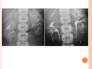
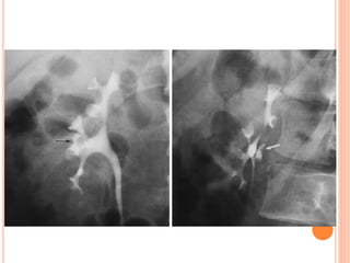
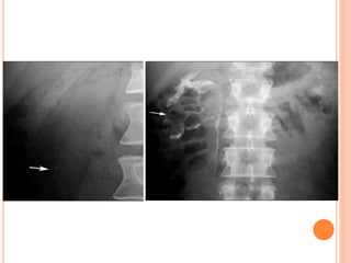
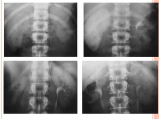
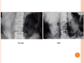
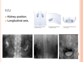
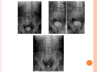
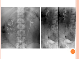
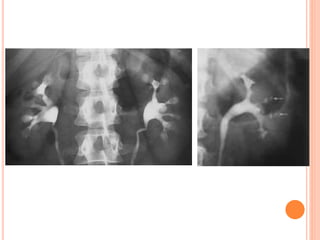
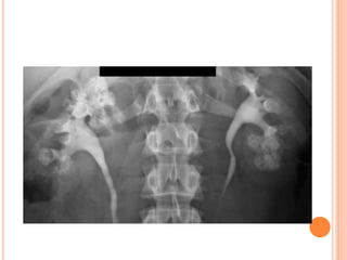
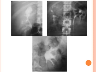
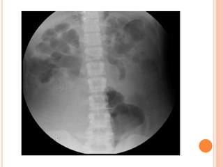
The document provides an overview of conventional imaging techniques of the urinary tract. It discusses conventional radiography, intravenous urography (IVU), voiding cystourethrography (VCUG), and contrast media. IVU involves injecting contrast dye intravenously to image the kidneys, ureters, and bladder. It allows evaluation of renal size and function. VCUG assesses the bladder, urethra, and detects vesicoureteral reflux by filling the bladder with contrast under fluoroscopy during voiding. The document outlines procedures, interpretations, and risks of these techniques, emphasizing their role in evaluating the urinary system.
A presentation about Intravenous Urography (Also known as Intravenous Pyeography).
The presentation contains 41 slides, and is divided into 4 parts :
1 - Introduction.
2 - The procedure.
3 - Examples for abnormal findings.
4 - Studies comparing IVU accuracy with KUB & USG with CT Scan.
This presentation was prepared and presented by me in the tutorials of the Radiology Department of Sebha Medical Center.
An IVU (Intravenous Urography) is an x-ray of your urinary tract (consisting of kidneys, ureters and bladder) following an injection of a clear dye called contrast into a vein in your arm.
The pictures produced are called intravenous urograms (IVU) or intravenous pyelograms (IVP).
A series of x-rays are taken of the abdomen at various time intervals. This usually takes up to an hour, but occasionally it may be necessary to take additional delayed images, which may continue for several hours.
Routine IVP[edit]
This procedure is most common for patients who have unexplained microscopic or macroscopic hematuria. It is used to ascertain the presence of a tumour or similar anatomy-altering disorders. The sequence of images is roughly as follows:
plain or Control KUB image;
immediate X-ray of just the renal area;
5 minute X-ray of just the renal area.
15 minute X-ray of just the renal area.
At this point, compression may or may not be applied (this is contraindicated in cases of obstruction).
In pyelography, compression involves pressing on the lower abdominal area, which results in distension of the upper urinary tract.[1]
If compression is applied: a 10 minutes post-injection X-ray of the renal area is taken, followed by a KUB on release of the compression.
If compression is not given: a standard KUB is taken to show the ureters emptying. This may sometimes be done with the patient lying in a prone position.
A post-micturition X-ray is taken afterwards. This is usually a coned bladder view.
Image Assessment[edit]
Ivu is a radiological investigation for visualization and assessment of the urinary tract.This presentation covers brief anatomy of urinary tract, indication and contraindication,contrast media dose and administration, routine and modified ivu procedure,its complication,ctivu and some abnormalities in the urinary tract.
Tom Selleck Health: A Comprehensive Look at the Iconic Actor’s Wellness Journeygreendigital
Tom Selleck, an enduring figure in Hollywood. has captivated audiences for decades with his rugged charm, iconic moustache. and memorable roles in television and film. From his breakout role as Thomas Magnum in Magnum P.I. to his current portrayal of Frank Reagan in Blue Bloods. Selleck's career has spanned over 50 years. But beyond his professional achievements. fans have often been curious about Tom Selleck Health. especially as he has aged in the public eye.
Follow us on: Pinterest
Introduction
Many have been interested in Tom Selleck health. not only because of his enduring presence on screen but also because of the challenges. and lifestyle choices he has faced and made over the years. This article delves into the various aspects of Tom Selleck health. exploring his fitness regimen, diet, mental health. and the challenges he has encountered as he ages. We'll look at how he maintains his well-being. the health issues he has faced, and his approach to ageing .
Early Life and Career
Childhood and Athletic Beginnings
Tom Selleck was born on January 29, 1945, in Detroit, Michigan, and grew up in Sherman Oaks, California. From an early age, he was involved in sports, particularly basketball. which played a significant role in his physical development. His athletic pursuits continued into college. where he attended the University of Southern California (USC) on a basketball scholarship. This early involvement in sports laid a strong foundation for his physical health and disciplined lifestyle.
Transition to Acting
Selleck's transition from an athlete to an actor came with its physical demands. His first significant role in "Magnum P.I." required him to perform various stunts and maintain a fit appearance. This role, which he played from 1980 to 1988. necessitated a rigorous fitness routine to meet the show's demands. setting the stage for his long-term commitment to health and wellness.
Fitness Regimen
Workout Routine
Tom Selleck health and fitness regimen has evolved. adapting to his changing roles and age. During his "Magnum, P.I." days. Selleck's workouts were intense and focused on building and maintaining muscle mass. His routine included weightlifting, cardiovascular exercises. and specific training for the stunts he performed on the show.
Selleck adjusted his fitness routine as he aged to suit his body's needs. Today, his workouts focus on maintaining flexibility, strength, and cardiovascular health. He incorporates low-impact exercises such as swimming, walking, and light weightlifting. This balanced approach helps him stay fit without putting undue strain on his joints and muscles.
Importance of Flexibility and Mobility
In recent years, Selleck has emphasized the importance of flexibility and mobility in his fitness regimen. Understanding the natural decline in muscle mass and joint flexibility with age. he includes stretching and yoga in his routine. These practices help prevent injuries, improve posture, and maintain mobilit
NVBDCP.pptx Nation vector borne disease control programSapna Thakur
NVBDCP was launched in 2003-2004 . Vector-Borne Disease: Disease that results from an infection transmitted to humans and other animals by blood-feeding arthropods, such as mosquitoes, ticks, and fleas. Examples of vector-borne diseases include Dengue fever, West Nile Virus, Lyme disease, and malaria.
Basavarajeeyam is a Sreshta Sangraha grantha (Compiled book ), written by Neelkanta kotturu Basavaraja Virachita. It contains 25 Prakaranas, First 24 Chapters related to Rogas& 25th to Rasadravyas.
Local Advanced Lung Cancer: Artificial Intelligence, Synergetics, Complex Sys...Oleg Kshivets
Overall life span (LS) was 1671.7±1721.6 days and cumulative 5YS reached 62.4%, 10 years – 50.4%, 20 years – 44.6%. 94 LCP lived more than 5 years without cancer (LS=2958.6±1723.6 days), 22 – more than 10 years (LS=5571±1841.8 days). 67 LCP died because of LC (LS=471.9±344 days). AT significantly improved 5YS (68% vs. 53.7%) (P=0.028 by log-rank test). Cox modeling displayed that 5YS of LCP significantly depended on: N0-N12, T3-4, blood cell circuit, cell ratio factors (ratio between cancer cells-CC and blood cells subpopulations), LC cell dynamics, recalcification time, heparin tolerance, prothrombin index, protein, AT, procedure type (P=0.000-0.031). Neural networks, genetic algorithm selection and bootstrap simulation revealed relationships between 5YS and N0-12 (rank=1), thrombocytes/CC (rank=2), segmented neutrophils/CC (3), eosinophils/CC (4), erythrocytes/CC (5), healthy cells/CC (6), lymphocytes/CC (7), stick neutrophils/CC (8), leucocytes/CC (9), monocytes/CC (10). Correct prediction of 5YS was 100% by neural networks computing (error=0.000; area under ROC curve=1.0).
Ozempic: Preoperative Management of Patients on GLP-1 Receptor Agonists Saeid Safari
Preoperative Management of Patients on GLP-1 Receptor Agonists like Ozempic and Semiglutide
ASA GUIDELINE
NYSORA Guideline
2 Case Reports of Gastric Ultrasound
These simplified slides by Dr. Sidra Arshad present an overview of the non-respiratory functions of the respiratory tract.
Learning objectives:
1. Enlist the non-respiratory functions of the respiratory tract
2. Briefly explain how these functions are carried out
3. Discuss the significance of dead space
4. Differentiate between minute ventilation and alveolar ventilation
5. Describe the cough and sneeze reflexes
Study Resources:
1. Chapter 39, Guyton and Hall Textbook of Medical Physiology, 14th edition
2. Chapter 34, Ganong’s Review of Medical Physiology, 26th edition
3. Chapter 17, Human Physiology by Lauralee Sherwood, 9th edition
4. Non-respiratory functions of the lungs https://academic.oup.com/bjaed/article/13/3/98/278874
Title: Sense of Smell
Presenter: Dr. Faiza, Assistant Professor of Physiology
Qualifications:
MBBS (Best Graduate, AIMC Lahore)
FCPS Physiology
ICMT, CHPE, DHPE (STMU)
MPH (GC University, Faisalabad)
MBA (Virtual University of Pakistan)
Learning Objectives:
Describe the primary categories of smells and the concept of odor blindness.
Explain the structure and location of the olfactory membrane and mucosa, including the types and roles of cells involved in olfaction.
Describe the pathway and mechanisms of olfactory signal transmission from the olfactory receptors to the brain.
Illustrate the biochemical cascade triggered by odorant binding to olfactory receptors, including the role of G-proteins and second messengers in generating an action potential.
Identify different types of olfactory disorders such as anosmia, hyposmia, hyperosmia, and dysosmia, including their potential causes.
Key Topics:
Olfactory Genes:
3% of the human genome accounts for olfactory genes.
400 genes for odorant receptors.
Olfactory Membrane:
Located in the superior part of the nasal cavity.
Medially: Folds downward along the superior septum.
Laterally: Folds over the superior turbinate and upper surface of the middle turbinate.
Total surface area: 5-10 square centimeters.
Olfactory Mucosa:
Olfactory Cells: Bipolar nerve cells derived from the CNS (100 million), with 4-25 olfactory cilia per cell.
Sustentacular Cells: Produce mucus and maintain ionic and molecular environment.
Basal Cells: Replace worn-out olfactory cells with an average lifespan of 1-2 months.
Bowman’s Gland: Secretes mucus.
Stimulation of Olfactory Cells:
Odorant dissolves in mucus and attaches to receptors on olfactory cilia.
Involves a cascade effect through G-proteins and second messengers, leading to depolarization and action potential generation in the olfactory nerve.
Quality of a Good Odorant:
Small (3-20 Carbon atoms), volatile, water-soluble, and lipid-soluble.
Facilitated by odorant-binding proteins in mucus.
Membrane Potential and Action Potential:
Resting membrane potential: -55mV.
Action potential frequency in the olfactory nerve increases with odorant strength.
Adaptation Towards the Sense of Smell:
Rapid adaptation within the first second, with further slow adaptation.
Psychological adaptation greater than receptor adaptation, involving feedback inhibition from the central nervous system.
Primary Sensations of Smell:
Camphoraceous, Musky, Floral, Pepperminty, Ethereal, Pungent, Putrid.
Odor Detection Threshold:
Examples: Hydrogen sulfide (0.0005 ppm), Methyl-mercaptan (0.002 ppm).
Some toxic substances are odorless at lethal concentrations.
Characteristics of Smell:
Odor blindness for single substances due to lack of appropriate receptor protein.
Behavioral and emotional influences of smell.
Transmission of Olfactory Signals:
From olfactory cells to glomeruli in the olfactory bulb, involving lateral inhibition.
Primitive, less old, and new olfactory systems with different path
Basavarajeeyam is an important text for ayurvedic physician belonging to andhra pradehs. It is a popular compendium in various parts of our country as well as in andhra pradesh. The content of the text was presented in sanskrit and telugu language (Bilingual). One of the most famous book in ayurvedic pharmaceutics and therapeutics. This book contains 25 chapters called as prakaranas. Many rasaoushadis were explained, pioneer of dhatu druti, nadi pareeksha, mutra pareeksha etc. Belongs to the period of 15-16 century. New diseases like upadamsha, phiranga rogas are explained.
2. CONVENTIONAL RADIOGRAPHY
Images produced through the use of ionizing
radiation are called conventional radiographs.
It is relatively inexpensive to produce, can be
obtained almost anywhere by using portable or
mobile machines, and are still the most widely
obtained imaging studies.
They require a source to produce the x-rays (the
“x-ray machine”), a method to record the image.
The major disadvantages of conventional
radiography are the limited range of densities it
can demonstrate and that it uses ionizing
radiation.
5. BIOLOGICAL EFFECTS OF RADIATION
Radiation causes biological effects on a cellular
level either (1) by directly damaging molecules or
(2) by indirectly creating free radicals to disrupt
cellular metabolism.
Deterministic effects (nonrandom): This is
damage that occurs when a threshold level is
met. Both the probability and the severity of the
effect are proportional to increasing dose, where
the dose is usually given in one exposure or several
exposures over a very short period of time.
6. BIOLOGICAL EFFECTS OF RADIATION
Stochastic effects (random): Damage that may
occur at any level of exposure, without a
threshold dose.
These effects occur by chance, and while their
probability increases with an increasing dose,
their severity is independent of the dose.
These effects are due to damage of cellular
components, usually DNA, by free radicals, leading
to abnormal cell function if repair is incomplete or
incorrect.
13. INTRAVENOUS UROGRAPHY (IVU)
IVU should be tailored
to answer a specific
clinical question.
The preliminary kidney,
ureter, bladder (KUB)
radiograph is an
indispensable part of
the sequence.
14. PLAIN RADIOGRAPHY(UTP)
- from the suprarenal
region to a level below
the symphysis pubis.
- The patient should void
immediately prior to
examination.
- may require additional
images
19. KUB ANALYSIS
Musculoskeletal: evaluate all bone elements.
Psoas muscle margin: straight, convex or absent.
Intestinal gas: overlap, displaced.
Kidneys
Calcifications: overlying the UT or outside.
Gas shadow: abnormal air at UT.
24. IVU
Kidney film 3 min.
A KUB radiograph is obtained to assess temporal
symmetry and opacification.
Compression? (Contraindications ).
25.
26.
27.
28. IVU
Bladder film early
(suspected bladder
lesion).
KUB after release of
compression( 15 min).
delayed images for
bladder distention, and
oblique, prone, or post-
void images.
29. INTERPRETATION OF IVU
Renal size.
position of the kidney.
Renal parenchyma at
nephrographic phase.
Renal contour
(interpapillary line).
41. IVU
Evaluation of the PCS.
- Obstruction (round
forniceal margin).
- CM inside the papillae.
- Parenchymal cavities
filled with CM.
- Filling defect.
- Phantom calyx.
53. Medial deviation of the ureter should be considered
when the ureter overlies the ipsilateral lumbar
pedicle.
lateral deviation should be considered when the
ureter lies more than 1.5 cm beyond the tip of the
transverse process.
56. IODINATED CONTRAST MEDIA
Water soluble, they
into negative
and positive ions which
attract the negative and
positive poles of the
water molecules.
Do not dissociate and
are rendered water
soluble by their polar OH
groups.
Ionic contrast media Nonionic contrast media
60. TYPES AND FREQUENCY OF ACUTE
REACTIONS
Acute idiosyncratic systemic reactions (also described as
allergy-like or anaphylactoid) are defined as
unpredictable reactions which occur within 1 h.
Chemotoxic reactions, are dose-related and dependent
on the physico-chemical properties of the contrast
medium.
61. ACUTE IDIOSYNCRATIC REACTIONS
Mild or minor reactions include nausea, mild vomiting,
urticaria and itching.
Moderate reactions include more severe vomiting,
marked urticaria, bronchospasm, facial or laryngeal
oedema, and vasovagal reactions.
Severe reactions include hypotensive shock, respiratory
arrest, cardiac arrest and convulsions.
62. RISK FACTORS FOR ACUTE IDIOSYNCRATIC
REACTIONS
Type of Contrast Agent.
Previous Contrast Medium Reaction.
Asthma.
Allergy.
Drugs( B Blockers, Ca channel antagonist
and Inter leukin).
63. PREVENTION OF ACUTE IDIOSYNCRATIC
REACTIONS
In any patient at increased risk of contrast medium
reaction, especially if there has been a previous reaction
to an iodinated contrast agent, use other modalities.
If iodinated contrast medium is still deemed essential the
risk of an acute reaction can be reduced by an
appropriate choice of contrast medium and
premedication.
64. CHOICE OF CONTRAST MEDIUM
Nonionic low osmolality agents which are
associated with a four to five times lower risk
of reactions.
Nonionic CM is preferred specially if there is
history of previous reaction, allergy or
asthma.
Previous history to nonionic CM?.
65. PREMEDICATION
Most frequently steroids with or without additional H1
antihistamines have been recommended.
In severe reactors to ionic CM, steroids should be given
12&2 h before contrast medium.
The minimal effective time interval between steroids and
CM is considered unlikely to be less than 6 h.
67. GENERAL CONSIDERATIONS
Use nonionic contrast media.
Keep the patient in the Radiology Department for
30 min after contrast medium injection.
Have the drugs and equipment for resuscitation
readily available.
68. MANAGEMENT OF ACUTE CM ADVERSE REACTIONS
The adverse event that occurs within 60 min of an
injection of contrast medium.
The mild reactions include flushing, nausea, arm
pain, pruritus, vomiting, headache, and mild urticaria.
They are usually of short duration, self-limiting and
generally require no specific treatment.
69. TREATMENT OF SPECIFIC REACTIONS
Nausea and Vomiting ( self limited, ? Anti-emetic).
Cutaneous Reactions ( treat if extensive).
Bronchospasm ( O2, bronchodil., ? Adrenaline).
Laryngeal Edema ( i.m adrenaline, O2).
Hypotension ( rise leg, rapid IV fluid).
Vagal Reaction ( as hypotension+ I.V atropine).
Generalized Anaphylactoid Reactions ( airway, O2,
rapid IV fluid, i.m adrenaline, ECG monitor).
70. LATE ADVERSE REACTIONS
Reactions occurring between 1 h
and 1 week after contrast medium
injection.
They are mainly mild or moderate
skin reactions and usually resolve
within 3–7 days.
71. CI NEPHROPATHY
Thee reduction in renal function induced by contrast
media which occurs within 3 days following
administration of contrast media in the absence of
an alternative etiology.
Most episodes of CIN are self-limited and resolve
within 1–2 weeks.
A persistent nephrogram on plain radiography or
CT of the abdomen at 24–48 h.
72. PREDISPOSING FACTORS
Pre-existing renal impairment [> 1.5 mg/dl], particularly when
secondary to diabetic nephropathy.
Large doses of contrast media and multiple injections within
72 h.
The route of administration (IA > IV).
Dehydration and CHF. Hypertension, hyperuricemia and
prteinuria. Multiple Myeloma.
Nephrotoxic drugs.
Type of CM.
73. PREVENTION OF CIN
Extracellular volume expansion, the choice of
normal (0.9%) saline when intravenous hydration is
used,
The choice of low or iso-osmolar nonionic contrast
medium,
Lowest contrast medium dose consistent with a
diagnostic conclusion or a therapeutic goal.
74. EXTRAVASATION OF CM
Elevation of the Affected Limb.
Application of hot & cold fomentations.
Topical antibiotic.
Topical Hyaluronidase.
Aspiration of subcutaneous fluid.
76. VCUG is commonly performed in children
with prenatally diagnosed hydronephrosis,
urinary tract infections, and voiding
abnormalities.
The procedure should include assessment
of the spine and pelvis; masses or opaque
calculi; bladder capacity, contour, and
emptying capability; presence and grade of
reflux; and urethral appearance.
77. PRELIMINARY IMAGING
Clinical data and results of prior
imaging studies should be reviewed
before starting the examination.
Preliminary abdominal imaging usually
precedes catheterization.
80. BLADDER FILLING
Early Filling
Several seconds after the contrast
material begins to flow, the minimally
filled bladder is imaged in the AP view.
A ureterocele or bladder tumor that is
well seen during early filling may
become obscured as more contrast
material enters the bladder
81.
82. INTERMEDIATE FILLING
Vesicoureteral reflux can be
seen on oblique radiographs
obtained just before voiding
and can be graded after
voiding with the International
Reflux System.
83.
84.
85. PREVOIDING IMAGING
If reflux is observed during late bladder
filling, the ipsilateral renal fossa may be
imaged in the anteroposterior projection
prior to voiding.
86.
87.
88. IMAGING DURING VOIDING
Bladder capacity={Age(ys)+2}x30.
A smaller than expected voiding volume
may also indicate a neurologic abnormality
(spastic bladder) or active bladder
infection.
89.
90. POSTVOIDING IMAGING
At the conclusion of voiding, each
renal fossa should be imaged. Still
images may demonstrate reflux that is
not appreciated at fluoroscopy as well
as other anomalies or abnormalities.