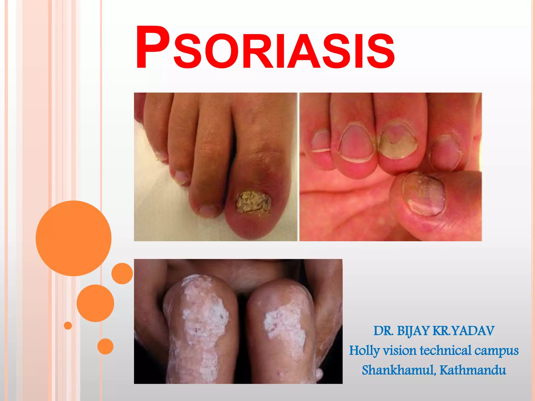
2. PSORIASIS :
Greek word; psora-itching,
iasis-condition
Challenge to the physician
Definition :
It is a common, genetically determined, inflammatory and
chronic disease of the skin with unpredictable course of
remission and relapses, the most characteristic lesions
consisting of sharply demarcated, dull red, scaly papule and
plaque particularly on the extensor prominences and the
scalp.
4. EPIDEMIOLOGY
Incidence :1-3% of population.
Age of onset: Peak incidence at 22.5 years and
55 years of life.
Early onset predicts: more severe form &
Probability of positive family hx.
Race : Low incidence in Asian pop.
Sex: Equal
Season: Worse in winter.
5. CLINICAL FEATURES :
1. Sites of psoriasis :
Mainly on extenser surface of limbs i.e Knee, elbow, sacral area.
Nails involvement (30% cases)
Flexure surface
2. Clinical types of psoriasis :
a. Plaque type :- most common & symmetrical over extensor surface
of limbs, Itching is variable
b. Guttate type :- Sudden onset eruptions of small lesions.
c. Pustular type :- Severe form of diseases, numerous small
pustules occurs in previous lesions
d. Erythrodermic psoriasis :- also severe form of diseases, lesions
involve all the body surface area.
e. Psoriasis ungum :- lesions involve nails.
f. Arthopathic psoriasis :- in this skin lesion are associated with
joints & bone involvement.
8. SKIN LESIONS
Usually well defined, salmon-pink papules and
plaques, sharply marginated with marked silvery
white scale which are lameller, loose and easily
removable (mica-like)
9. BED SIDE TESTS
Grattage test: Scales in a psoriatic plaques can be
accentuated by grating with glass slide.
Auspitz sign :
Step A: Positive grattage test
Step B: Burkley’s membrane
Step C: Puntate visible bleeding points.
17. JOINT
10% of patients with psoriasis have joint
involvement.
Four patterns
Polyarticular variety
Monoarticular variety
Rheumatoid arthritis like
Axial variety
18. DIAGNOSIS
Typical distribution
Well defined, dry, erythematous areas with silvery white
scales.
Grattage test, Koebner’s phenomenon, Auspitz’s sign.
Mild itching
H/O prev. attack, seasonal variation, Fam. Hx
Typical histopathology-
19. MANAGEMENT
1. Topical therapy :-
a. Emollients – reduces scales scales & decreases itching
b. Dithranol – decreases the proliferation of keratinocytes
Ingram regimen : Dithranol + Zinc oxide application over
lesion
Uv radiation & tar bath
Application of talcum powder & bandage
c. Coal tar – Decrease DNA synthesis ( it will decrease the cell
devision)
d. Topical steroids – betamethasone / Hydrocortisone
e. Salicyclic acid – causes softening of lesions & scales.
20. 2. PUVA / Psoralen / Ultravoilet therapy :
In moderate to severe form of disease
Psoralen is given orally & lesion exposed to
ultravoilet light or sunlight
3. Systemic therapy :
Useful in failure of topical therapy & in cases of
severe disease as pustular or erythrodermic
Drugs used are :
a. Methotrexate (5-25 mg / week)
b. Oral retinoids
c. Hydroxyurea
d. Corticosteroids – prednisolone .
21. MANAGEMENT IN HEALTH POST
After diagnosis of the lesions mainly by clinical ways, initially the lesions
should be treated with topical therapy.
Most commonly used is psoriatic cream
composition is – coal tar 6% + Salicyclic acid 3% + Ammoniated mercury 1%
The cases should be followed regularly & according to the progression of
lesion additional agents as topical steroid can be added.
If the lesions become extensive or generalized or turn into pustular form then
the case should be reffered to higher center as further management will be
required.
In case of following condition special attention should be given :
o pregnancy
o Infection
o Local irritation
o Sudden steroid withdrawl
o Hypocalcemia