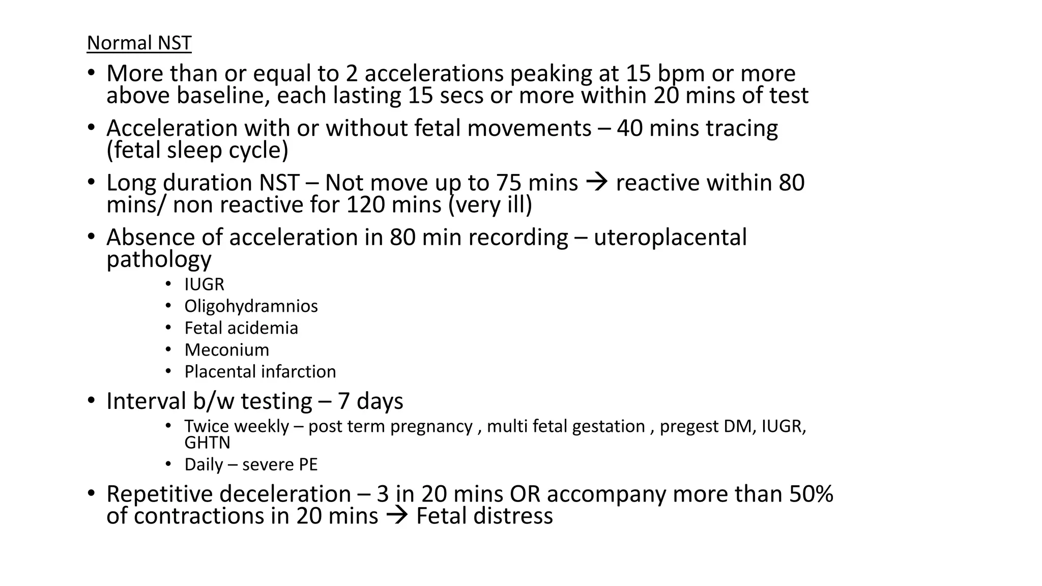
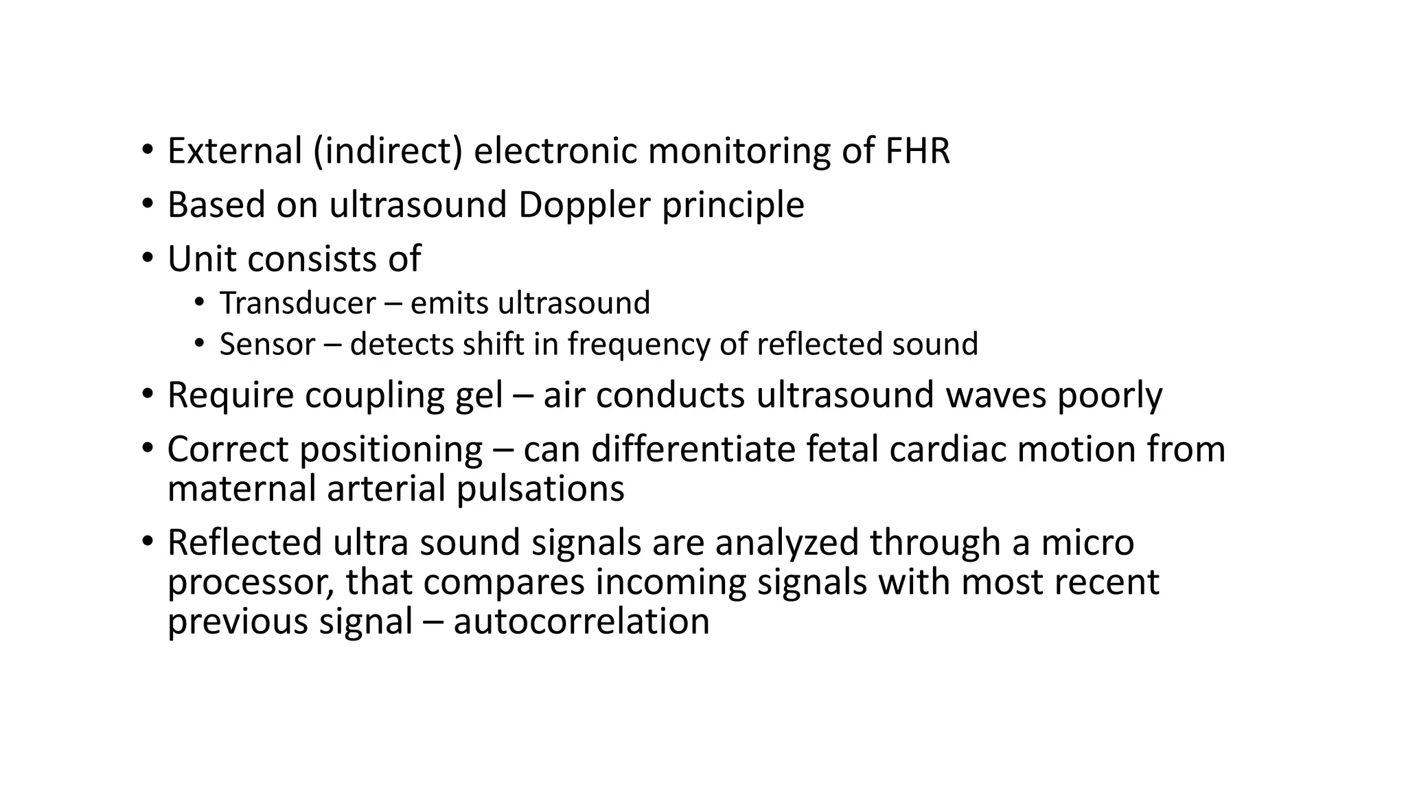
Non Stress Test
N S T
• External (indirect) electronic monitoring of FHR
• Based on ultrasound Doppler principle
• Unit consists of
• Transducer – emits ultrasound
• Sensor – detects shift in frequency of reflected sound
• Require coupling gel – air conducts ultrasound waves poorly
• Correct positioning – can differentiate fetal cardiac motion from
maternal arterial pulsations
• Reflected ultra sound signals are analyzed through a micro
processor, that compares incoming signals with most recent
previous signal – autocorrelation
FHR Patterns
• Mean FHR with increments of 5 bpm in 10mins excluding
- Periodic/ episodic changes
- Marked FHR variability
- Baseline segment differ by more than 25 bpm
• Minimum 2 min in 10 min segment
• Normal FHR – 110 to160 bpm
• Tachycardia > 160 bpm
• Bradycardia < 110 bpm
Baseline
• Baseline fluctuations with irregular amplitude and
frequency
• Amplitude of peak to trough in bpm
• Absent – amplitude range is undetectable
• Minimal – detectable but less than equal to 5 bpm
• Moderate (Normal) – 6 to 25 bpm
• Marked – more than 25 bpm
Baseline Variability
• Abrupt increase in FHR (Onset to peak in less than 30
sec)
• At or beyond 32 wk – peak of 15 bpm or more lasting
for 15 sec or more but less than 2 mins from onset to
return
• Before 32 weeks – peak of 10 bpm or more lasting for
10 sec or more but less than 2 mins from onset to
return
• Prolonged acceleration – lasts 2 mins or more but less
than 10 mins
• 10 mins or longer – baseline change
Acceleration
• Symmetrical gradual decrease (>= 30 sec) and return of FHR
associated with uterine contraction
• Lowest point of deceleration coincide with peak of contraction
Early Deceleration
• Lowest point of deceleration occur after the peak of
contraction
• Onset, lowest point and recovery of deceleration occur
after the beginning, peak & ending of the contraction
respectively
Late Deceleration
• Abrupt fall in FHR
• From Onset to the lowest point of FHR within 30 sec
• Decrease in FHR is 15 bpm or greater , lasting 15 sec or
greater & less than 2 mins in duration
• Onset, depth and duration vary with successive uterine
contractions
Variable
Deceleration
• Decrease in FHR below baseline i.e. 15 bpm or more and
more than 2 mins duration
• Deceleration last 10 mins or longer – baseline change
Prolonged
Deceleration
• Apparent, smooth, sine wave pattern in FHR baseline
with a cyclic frequency of 3- 5 per mins which persists
for 20 mins or more
Sinusoidal pattern
NICHD – propose standardized definitions for interpretation of FHR pattern during labor.
- modified in 2008  adopted by ACOG
30 bpm per vertical cm (Range – 30 to 240bpm) and 3 cm per min chart recorder paper speed
Baseline Fetal Heart Activity
Rate
• With an increase in fetal maturation – FHR decreases, continues post natally
• Decline an average of 24 bpm b/w 16 wk to term
• Due maturation of parasympathetic (vagal) heart control
• Avg FHR – result of tonic balance b/w symp and parasymp influences on
pacemaker cells
– under the control of arterial chemoreceptors[ prolonged hypoxia with
rising blood lactate level & severe metabolic acidemia  fall FHR
Bradycardia
• T3 – normal FHR 120 to 160 bpm
• 100 to 119 bpm in the absence of other changes – not represent
fetal compromise
• 80 to 120 bpm with good variability – reassuring
• < 80 bpm – non reassuring
• Causes
• Congenital heart block
• Serious fetal compromise – placental abruption/hypoxia/sepsis
• Maternal hypoglycemia /hypothermia
• Drugs to mother-pethidine,antihypertensive,mgso4
• Severe pyelonephritis
Tachycardia
Causes
 Maternal fever
 Fetal compromise-hypoxia,infection
 Cardiac arrhythmia
 Drugs to mother-
sympathomimetic[isoxsuprine,ritodrine],parasymp
inhibiting[atropine]
 Maternal hypotension
 Anemia-maternal/fetal
Beat to Beat variability
• Important index of cardio vascular function
• Regulated by ANS
• Short term variability – instantaneous change from 1 beat to the next
– measure of time interval b/w cardiac systoles
– most reliably determined by scalp electrode
• Long term variability – oscillatory changes during 1min & result in the
waviness of the baseline
– normal frequency 3 to 5 cycles per min
• Increased variability – fetal breathing and body movements
– advancing gestation
• Up to 30 wks - similar baseline characteristics in fetal rest and activity
• After 30 wks – fetal inactivity  diminished variability
– fetal activity  enhance
• Baseline FHR becomes less variable as rate rises
• Decreased variability – causes
• Analgesics
• CNS depressant drugs (transient variation) – Narcotics, barbiturates,
phenothiazine's, tranquilizers, general anaesthetics
• Corticosteroids
• MgSO4 – without adverse neonatal effects
• Severe maternal acidemia – DKA  Depression of fetal brainstem or the heart itself
creates loss of variability
• Diminished BBV with fetal compromise – due to acidemia (not hypoxia)
• Absent variability – within the normal baseline rate range and without
deceleration  previous fetal insult resulted in neurological damage
Sinusoidal heart rate
Seen in
• Fetal intracranial hemorrhage
• Severe fetal asphyxia
• Severe fetal anemia – anti D alloimmunization, feto maternal
hemorrhage, TTTS, fetal parvo viral inf, vasaprevia with bleeding
• Narcotics – sine frequency of 6 cycles per min
• Chorioamnionitis
• Fetal distress
• Umbilical cord occlusion
Features of sinusoidal pattern
• Stable baseline FHR of 120 to 160 bpm with regular oscillations
• Amplitude of 5 to 15 bpm
• Long term variability frequency of 2 to 5 cycles per min
• Fixed or flat short term variability
• Oscillations of sinusoidal waveform above or below a baseline
• Absent acceleration
Intrapartum sine wave like baseline variation with periods of
acceleration – pseudo sinusoidal
• Mild – epidural
• Intermediate – transient episodes of fetal hypoxia caused by
umbilical cord compression
Periodic FHR changes
Accelerations
• Represent intact neurohormonal cardio vascular control mechanisms linked to fetal
behavioral states
• Always reassuring – no acidemia
• Seen in
• Fetal movement
• Stimulation by uterine contraction
• Umbilical cord occlusion
• Fetal stimulation during pelvic examination
• Scalp blood sampling
• Acoustic stimulation
Early Deceleration
• Head compression  Dural stimulation  vagal nerve activation  early
deceleration
• Degree of deceleration is proportional to contraction strength
• Rarely falls below 100 to 110 bpm or 20 to 30 bpm below baseline
• Common during active labor
• Not associated with fetal hypoxia or acidemia
Late Deceleration
• Utero placental insufficiency
• Magnitude is not more than 30 to 40 bpm below baseline (typically 10 to 20 bpm)
• Seen in
• Maternal Hypotension – epidural
• Uterine hyper activity – oxytocin stimulation
• Chronic placental dysfunction – maternal disease (HTN, DM, collagen vascular disorders)
• Placental abruption – acute late deceleration
Variable Deceleration
• Cord compression
• 2 types
a) Type A – seen in complete occlusion
b) Type B – due to differing degrees of partial occlusion
– Acceleration before and after
– Vein occlusion  reduce fetal blood return  trigger baroreceptor mediated
acceleration
– With increase in intrauterine pressure  complete cord occlusion  obstruction of
umbilical artery flow  fetal systemic HTN  baroreceptor mediated deceleration
– vagal response due to chemo/baro receptor activity
• Recurrent variable deceleration with minimal to moderate BBV are indeterminate whereas
those with absent variability are abnormal
Other FHR patterns
(associated with umbilical cord compression)
• Saltatory baseline HR – rapidly recurring couplets of acceleration & deceleration
– seen in post term pregnancies
• Lambda – acceleration followed by a variable deceleration with no acceleration at the end
of deceleration
– seen in early labor
– Result from mild cord compression or stretch
• Overshoot - variable deceleration followed by acceleration
Prolonged deceleration
Causes
• Cervical examination
• Uterine Hyperactivity
• Cord Entanglement
• Maternal Supine Hypotension
• Epidural / Spinal / Paracervical analgesia
• Maternal hypo perfusion / Hypoxia from any cause
• Placental abruption
• Umbilical cord knots / prolapse
• Maternal Seizure
• Application of fetal scalp electrode
• Impending birth or maternal valsalva maneuver
• Placenta is effective in resuscitating the fetus if the original insult does not recur
immediately
• Fetus may die during prolonged decelerations
Normal NST
• More than or equal to 2 accelerations peaking at 15 bpm or more
above baseline, each lasting 15 secs or more within 20 mins of test
• Acceleration with or without fetal movements – 40 mins tracing
(fetal sleep cycle)
• Long duration NST – Not move up to 75 mins  reactive within 80
mins/ non reactive for 120 mins (very ill)
• Absence of acceleration in 80 min recording – uteroplacental
pathology
• IUGR
• Oligohydramnios
• Fetal acidemia
• Meconium
• Placental infarction
• Interval b/w testing – 7 days
• Twice weekly – post term pregnancy , multi fetal gestation , pregest DM, IUGR,
GHTN
• Daily – severe PE
• Repetitive deceleration – 3 in 20 mins OR accompany more than 50%
of contractions in 20 mins  Fetal distress
• False normal NST – fetal death within 7 days (post term pregnancy)
• Mean interval b/w testing and death - 4 days (1 to 7 days)
• Causes of fetal death
• Intrauterine infection
• Abnormal cord position
• Malformation
• Placental abruption
FHR patterns during second stage of labor
• Decelerations are common due to both cord and fetal head compression
• If prolonged  still birth and neonatal death
• If total no. of decelerations < 70 bpm increased, persistent baseline brady /tachy –
decreased APGAR
3 tier FHR interpretation system
Category 1 – normal
• Baseline rate – 110 to 160 bpm
• Baseline FHR variability – moderate
• Late or variable decelerations- absent
• Early decelerations – present / absent
• Accelerations – present / absent
Category 2 – Indeterminate
• Baseline rate
• Bradycardia not accompanied by absent baseline variability
• Tachycardia
• Baseline FHR variability
• Minimal baseline variability
• Absent variability not accompanied by recurrent decelerations
• Marked baseline variability
• Accelerations
• Absence of induced accelerations after fetal stimulation
• Periodic / Episodic decelerations
• Recurrent variable decelerations with minimal or moderate baseline variability
• Prolonged deceleration
• Recurrent late decelerations with moderate baseline variability
• Variable decelerations with other characteristics – slow return to baseline, overshoot,
shoulders
Category 3 – Abnormal
Include either
• Absent baseline FHR variability & any of the following
• Recurrent late decelerations
• Recurrent variable decelerations
• Bradycardia
• Sinusoidal pattern
Resuscitative measures for Category II or III Tracings
FHR abnormality Interventions
Recurrent late decelerations
Prolonged decelerations or Brady
Minimal/Absent FHR variability
Lateral decubitus positioning, administer
maternal oxygen, IV fluid bolus, reduce
uterine contraction frequency
Tachysystole with category II or III tracing Discontinue oxytocin or PG, give tocolytics –
MgSO4, Terbutaline
Recurrent variable decelerations
Prolonged decelerations or brady
Reposition mother, amnioinfusion, with cord
prolapse – manually elevate the presenting
part while preparing for immediate delivery
Thank You

Non stress test gynaecology presentation

  • 1.
  • 2.
    • External (indirect)electronic monitoring of FHR • Based on ultrasound Doppler principle • Unit consists of • Transducer – emits ultrasound • Sensor – detects shift in frequency of reflected sound • Require coupling gel – air conducts ultrasound waves poorly • Correct positioning – can differentiate fetal cardiac motion from maternal arterial pulsations • Reflected ultra sound signals are analyzed through a micro processor, that compares incoming signals with most recent previous signal – autocorrelation
  • 3.
    FHR Patterns • MeanFHR with increments of 5 bpm in 10mins excluding - Periodic/ episodic changes - Marked FHR variability - Baseline segment differ by more than 25 bpm • Minimum 2 min in 10 min segment • Normal FHR – 110 to160 bpm • Tachycardia > 160 bpm • Bradycardia < 110 bpm Baseline • Baseline fluctuations with irregular amplitude and frequency • Amplitude of peak to trough in bpm • Absent – amplitude range is undetectable • Minimal – detectable but less than equal to 5 bpm • Moderate (Normal) – 6 to 25 bpm • Marked – more than 25 bpm Baseline Variability
  • 5.
    • Abrupt increasein FHR (Onset to peak in less than 30 sec) • At or beyond 32 wk – peak of 15 bpm or more lasting for 15 sec or more but less than 2 mins from onset to return • Before 32 weeks – peak of 10 bpm or more lasting for 10 sec or more but less than 2 mins from onset to return • Prolonged acceleration – lasts 2 mins or more but less than 10 mins • 10 mins or longer – baseline change Acceleration • Symmetrical gradual decrease (>= 30 sec) and return of FHR associated with uterine contraction • Lowest point of deceleration coincide with peak of contraction Early Deceleration
  • 6.
    • Lowest pointof deceleration occur after the peak of contraction • Onset, lowest point and recovery of deceleration occur after the beginning, peak & ending of the contraction respectively Late Deceleration • Abrupt fall in FHR • From Onset to the lowest point of FHR within 30 sec • Decrease in FHR is 15 bpm or greater , lasting 15 sec or greater & less than 2 mins in duration • Onset, depth and duration vary with successive uterine contractions Variable Deceleration
  • 7.
    • Decrease inFHR below baseline i.e. 15 bpm or more and more than 2 mins duration • Deceleration last 10 mins or longer – baseline change Prolonged Deceleration • Apparent, smooth, sine wave pattern in FHR baseline with a cyclic frequency of 3- 5 per mins which persists for 20 mins or more Sinusoidal pattern NICHD – propose standardized definitions for interpretation of FHR pattern during labor. - modified in 2008  adopted by ACOG 30 bpm per vertical cm (Range – 30 to 240bpm) and 3 cm per min chart recorder paper speed
  • 10.
    Baseline Fetal HeartActivity Rate • With an increase in fetal maturation – FHR decreases, continues post natally • Decline an average of 24 bpm b/w 16 wk to term • Due maturation of parasympathetic (vagal) heart control • Avg FHR – result of tonic balance b/w symp and parasymp influences on pacemaker cells – under the control of arterial chemoreceptors[ prolonged hypoxia with rising blood lactate level & severe metabolic acidemia  fall FHR
  • 11.
    Bradycardia • T3 –normal FHR 120 to 160 bpm • 100 to 119 bpm in the absence of other changes – not represent fetal compromise • 80 to 120 bpm with good variability – reassuring • < 80 bpm – non reassuring • Causes • Congenital heart block • Serious fetal compromise – placental abruption/hypoxia/sepsis • Maternal hypoglycemia /hypothermia • Drugs to mother-pethidine,antihypertensive,mgso4 • Severe pyelonephritis
  • 12.
    Tachycardia Causes  Maternal fever Fetal compromise-hypoxia,infection  Cardiac arrhythmia  Drugs to mother- sympathomimetic[isoxsuprine,ritodrine],parasymp inhibiting[atropine]  Maternal hypotension  Anemia-maternal/fetal
  • 13.
    Beat to Beatvariability • Important index of cardio vascular function • Regulated by ANS • Short term variability – instantaneous change from 1 beat to the next – measure of time interval b/w cardiac systoles – most reliably determined by scalp electrode • Long term variability – oscillatory changes during 1min & result in the waviness of the baseline – normal frequency 3 to 5 cycles per min
  • 15.
    • Increased variability– fetal breathing and body movements – advancing gestation • Up to 30 wks - similar baseline characteristics in fetal rest and activity • After 30 wks – fetal inactivity  diminished variability – fetal activity  enhance • Baseline FHR becomes less variable as rate rises • Decreased variability – causes • Analgesics • CNS depressant drugs (transient variation) – Narcotics, barbiturates, phenothiazine's, tranquilizers, general anaesthetics • Corticosteroids • MgSO4 – without adverse neonatal effects • Severe maternal acidemia – DKA  Depression of fetal brainstem or the heart itself creates loss of variability
  • 16.
    • Diminished BBVwith fetal compromise – due to acidemia (not hypoxia) • Absent variability – within the normal baseline rate range and without deceleration  previous fetal insult resulted in neurological damage Sinusoidal heart rate Seen in • Fetal intracranial hemorrhage • Severe fetal asphyxia • Severe fetal anemia – anti D alloimmunization, feto maternal hemorrhage, TTTS, fetal parvo viral inf, vasaprevia with bleeding • Narcotics – sine frequency of 6 cycles per min • Chorioamnionitis • Fetal distress • Umbilical cord occlusion
  • 17.
    Features of sinusoidalpattern • Stable baseline FHR of 120 to 160 bpm with regular oscillations • Amplitude of 5 to 15 bpm • Long term variability frequency of 2 to 5 cycles per min • Fixed or flat short term variability • Oscillations of sinusoidal waveform above or below a baseline • Absent acceleration Intrapartum sine wave like baseline variation with periods of acceleration – pseudo sinusoidal • Mild – epidural • Intermediate – transient episodes of fetal hypoxia caused by umbilical cord compression
  • 18.
    Periodic FHR changes Accelerations •Represent intact neurohormonal cardio vascular control mechanisms linked to fetal behavioral states • Always reassuring – no acidemia • Seen in • Fetal movement • Stimulation by uterine contraction • Umbilical cord occlusion • Fetal stimulation during pelvic examination • Scalp blood sampling • Acoustic stimulation
  • 19.
    Early Deceleration • Headcompression  Dural stimulation  vagal nerve activation  early deceleration • Degree of deceleration is proportional to contraction strength • Rarely falls below 100 to 110 bpm or 20 to 30 bpm below baseline • Common during active labor • Not associated with fetal hypoxia or acidemia Late Deceleration • Utero placental insufficiency • Magnitude is not more than 30 to 40 bpm below baseline (typically 10 to 20 bpm) • Seen in • Maternal Hypotension – epidural • Uterine hyper activity – oxytocin stimulation • Chronic placental dysfunction – maternal disease (HTN, DM, collagen vascular disorders) • Placental abruption – acute late deceleration
  • 20.
    Variable Deceleration • Cordcompression • 2 types a) Type A – seen in complete occlusion b) Type B – due to differing degrees of partial occlusion – Acceleration before and after – Vein occlusion  reduce fetal blood return  trigger baroreceptor mediated acceleration – With increase in intrauterine pressure  complete cord occlusion  obstruction of umbilical artery flow  fetal systemic HTN  baroreceptor mediated deceleration – vagal response due to chemo/baro receptor activity • Recurrent variable deceleration with minimal to moderate BBV are indeterminate whereas those with absent variability are abnormal
  • 22.
    Other FHR patterns (associatedwith umbilical cord compression) • Saltatory baseline HR – rapidly recurring couplets of acceleration & deceleration – seen in post term pregnancies • Lambda – acceleration followed by a variable deceleration with no acceleration at the end of deceleration – seen in early labor – Result from mild cord compression or stretch • Overshoot - variable deceleration followed by acceleration
  • 23.
    Prolonged deceleration Causes • Cervicalexamination • Uterine Hyperactivity • Cord Entanglement • Maternal Supine Hypotension • Epidural / Spinal / Paracervical analgesia • Maternal hypo perfusion / Hypoxia from any cause • Placental abruption • Umbilical cord knots / prolapse • Maternal Seizure • Application of fetal scalp electrode • Impending birth or maternal valsalva maneuver • Placenta is effective in resuscitating the fetus if the original insult does not recur immediately • Fetus may die during prolonged decelerations
  • 24.
    Normal NST • Morethan or equal to 2 accelerations peaking at 15 bpm or more above baseline, each lasting 15 secs or more within 20 mins of test • Acceleration with or without fetal movements – 40 mins tracing (fetal sleep cycle) • Long duration NST – Not move up to 75 mins  reactive within 80 mins/ non reactive for 120 mins (very ill) • Absence of acceleration in 80 min recording – uteroplacental pathology • IUGR • Oligohydramnios • Fetal acidemia • Meconium • Placental infarction • Interval b/w testing – 7 days • Twice weekly – post term pregnancy , multi fetal gestation , pregest DM, IUGR, GHTN • Daily – severe PE • Repetitive deceleration – 3 in 20 mins OR accompany more than 50% of contractions in 20 mins  Fetal distress
  • 25.
    • False normalNST – fetal death within 7 days (post term pregnancy) • Mean interval b/w testing and death - 4 days (1 to 7 days) • Causes of fetal death • Intrauterine infection • Abnormal cord position • Malformation • Placental abruption FHR patterns during second stage of labor • Decelerations are common due to both cord and fetal head compression • If prolonged  still birth and neonatal death • If total no. of decelerations < 70 bpm increased, persistent baseline brady /tachy – decreased APGAR
  • 26.
    3 tier FHRinterpretation system Category 1 – normal • Baseline rate – 110 to 160 bpm • Baseline FHR variability – moderate • Late or variable decelerations- absent • Early decelerations – present / absent • Accelerations – present / absent Category 2 – Indeterminate • Baseline rate • Bradycardia not accompanied by absent baseline variability • Tachycardia • Baseline FHR variability • Minimal baseline variability • Absent variability not accompanied by recurrent decelerations • Marked baseline variability
  • 27.
    • Accelerations • Absenceof induced accelerations after fetal stimulation • Periodic / Episodic decelerations • Recurrent variable decelerations with minimal or moderate baseline variability • Prolonged deceleration • Recurrent late decelerations with moderate baseline variability • Variable decelerations with other characteristics – slow return to baseline, overshoot, shoulders Category 3 – Abnormal Include either • Absent baseline FHR variability & any of the following • Recurrent late decelerations • Recurrent variable decelerations • Bradycardia • Sinusoidal pattern
  • 28.
    Resuscitative measures forCategory II or III Tracings FHR abnormality Interventions Recurrent late decelerations Prolonged decelerations or Brady Minimal/Absent FHR variability Lateral decubitus positioning, administer maternal oxygen, IV fluid bolus, reduce uterine contraction frequency Tachysystole with category II or III tracing Discontinue oxytocin or PG, give tocolytics – MgSO4, Terbutaline Recurrent variable decelerations Prolonged decelerations or brady Reposition mother, amnioinfusion, with cord prolapse – manually elevate the presenting part while preparing for immediate delivery
  • 32.