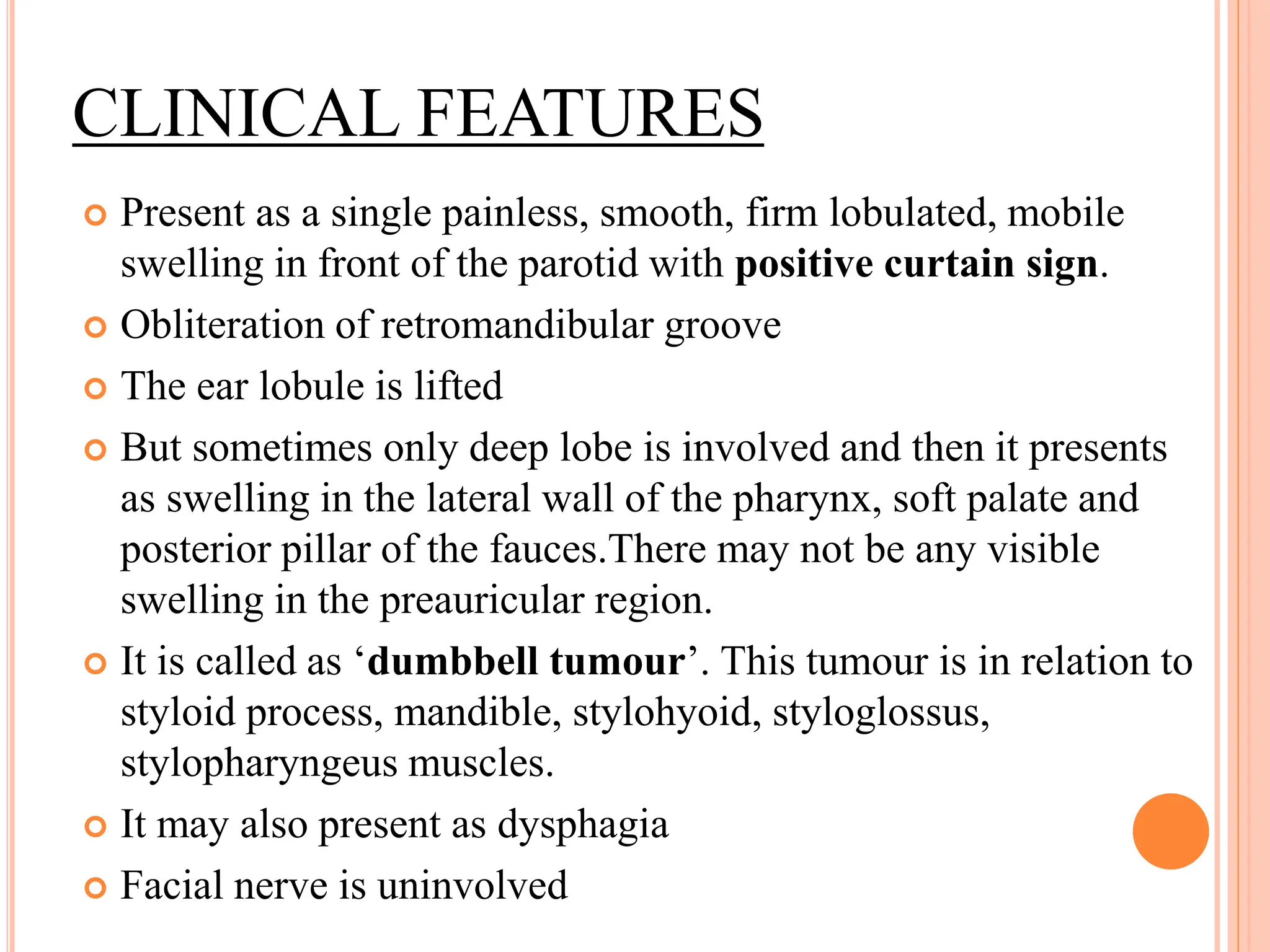
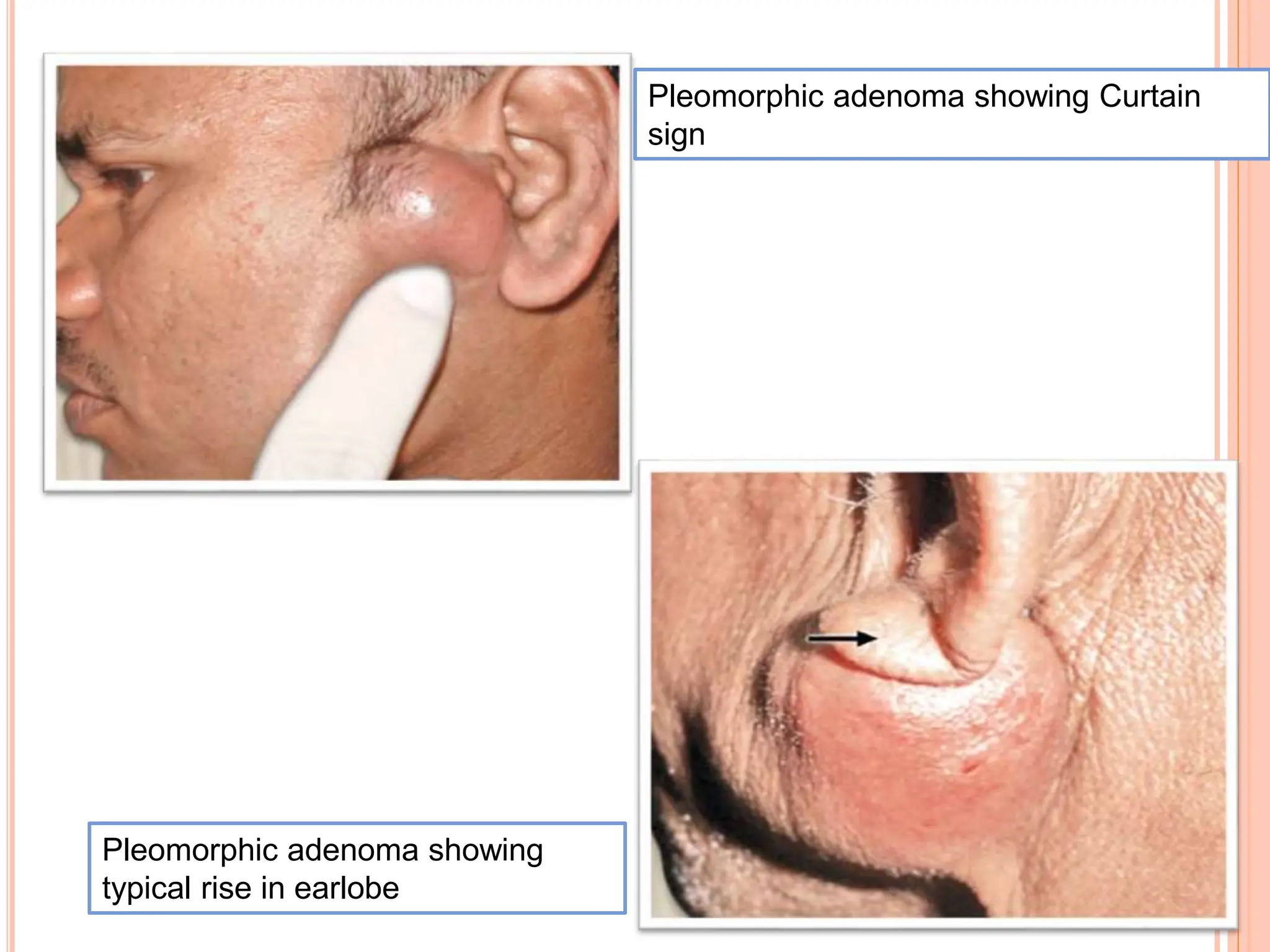
PLEOMORPHIC
ADENOMA
RAJALEKSHMI P A
ROLL NO: 74
PLEOMORPHIC ADENOMA
Commonest salivary gland tumour in
adults
80% of all salivary gland tumors
80% in parotids, 10% in submandibular,
0.5% in sublingual salivary gland
 Benign tumors that consists of a mixture of
ductal(epithelial), myoepithelial and
mesenchymal cells.
 Grossly it contains cartilages, cystic spaces, solid
tissues.
 Microscopically it is biphasic in nature with
epithelial and stromal components- mixed
tumors
GROSS MORPHOLOGY
 Round, well demarcated masses
 Rarely exceeds 6 cm in greatest dimension
 Even though it is capsulated, tumour may come
out as pseudopods and may extend beyond the
main limit of the tumour tissue.
 Cut surface- grey white with myxoid and blue
translucent areas of chondroid stroma.
HISTOLOGY
 Dominant histologic feature- heterogeneity
 Epithelial elements – ductal cells or
myoepithelial cells- arranged as ducts, acini,
irregular tubules, strands or sheets of cells.
 These elements are typically dispersed within a
background of loose myxoid and hyaline tissue
containing islands of cartilage of foci of bone.
 Most cases, there is no mitotic activity or
epithelial dysplasia.
EPIDEMIOLOGY
 80% common.
 Common in females (3:1).
 Occurs in any age group. But common in 4th and
5th decade.
 Usually unilateral
CLINICAL FEATURES
 Present as a single painless, smooth, firm lobulated, mobile
swelling in front of the parotid with positive curtain sign.
 Obliteration of retromandibular groove
 The ear lobule is lifted
 But sometimes only deep lobe is involved and then it presents
as swelling in the lateral wall of the pharynx, soft palate and
posterior pillar of the fauces.There may not be any visible
swelling in the preauricular region.
 It is called as ‘dumbbell tumour’. This tumour is in relation to
styloid process, mandible, stylohyoid, styloglossus,
stylopharyngeus muscles.
 It may also present as dysphagia
 Facial nerve is uninvolved
Pleomorphic adenoma showing
typical rise in earlobe
Pleomorphic adenoma showing Curtain
sign
COMPLICATIONS
 Recurrence—5–50%.
 Malignancy.
 3–5% in early tumours.
 10% in long duration (15 or more years)
tumours.
CARCINOMA IN EX PLEOMORPHIC
ADENOMA
Long-standing pleomorphic adenoma may turn
into carcinoma. Its features are:
• ™
Pain and nodularity
• ™
Involvement of skin, ulceration
• ™
Involvement of masseter
• ™
Involvement of facial nerve—lower facial
nerve palsy—(Difficulty in closing eyelid,
difficulty in blowing and clenching teeth)
• ™
Involvement of neck lymph node
• ™
Restriction of jaw movements
INVESTIGATIONS
 FNAC is very important and diagnostic.
 CT scan to know the status of deep lobe, local
extension and spread.
 MRI is better method
Open biopsy is contraindicated in parotid
tumours due to:
 ™
. Chance of injury to facial nerve
 ™
. Seedling and high chance of recurrence
 ™
. Chance of parotid fistula formation
TREATMENT
 Surgery—first line treatment.
 If only superficial lobe is involved, then
superficial parotidectomy is done wherein
parotid superficial to facial nerve is removed.
 If both lobes are involved, then total
conservative parotidectomy is done by retaining
facial nerve.
 Enucleation is avoided as it causes high
recurrence due to extension of tumour outside as
pseudopods across the capsule.
REFERENCES
 Bailey and Love- Short Practice Of Surgery 28th
edition
 SRB Manual Of Surgery 7th edition
 Robbins Pathologic Basis Of Disease
PLEOMORPHIC ADENOMA presentaion sss.pptx

PLEOMORPHIC ADENOMA presentaion sss.pptx

  • 1.
  • 2.
    PLEOMORPHIC ADENOMA Commonest salivarygland tumour in adults 80% of all salivary gland tumors 80% in parotids, 10% in submandibular, 0.5% in sublingual salivary gland
  • 3.
     Benign tumorsthat consists of a mixture of ductal(epithelial), myoepithelial and mesenchymal cells.  Grossly it contains cartilages, cystic spaces, solid tissues.  Microscopically it is biphasic in nature with epithelial and stromal components- mixed tumors
  • 4.
    GROSS MORPHOLOGY  Round,well demarcated masses  Rarely exceeds 6 cm in greatest dimension  Even though it is capsulated, tumour may come out as pseudopods and may extend beyond the main limit of the tumour tissue.  Cut surface- grey white with myxoid and blue translucent areas of chondroid stroma.
  • 6.
    HISTOLOGY  Dominant histologicfeature- heterogeneity  Epithelial elements – ductal cells or myoepithelial cells- arranged as ducts, acini, irregular tubules, strands or sheets of cells.  These elements are typically dispersed within a background of loose myxoid and hyaline tissue containing islands of cartilage of foci of bone.  Most cases, there is no mitotic activity or epithelial dysplasia.
  • 8.
    EPIDEMIOLOGY  80% common. Common in females (3:1).  Occurs in any age group. But common in 4th and 5th decade.  Usually unilateral
  • 9.
    CLINICAL FEATURES  Presentas a single painless, smooth, firm lobulated, mobile swelling in front of the parotid with positive curtain sign.  Obliteration of retromandibular groove  The ear lobule is lifted  But sometimes only deep lobe is involved and then it presents as swelling in the lateral wall of the pharynx, soft palate and posterior pillar of the fauces.There may not be any visible swelling in the preauricular region.  It is called as ‘dumbbell tumour’. This tumour is in relation to styloid process, mandible, stylohyoid, styloglossus, stylopharyngeus muscles.  It may also present as dysphagia  Facial nerve is uninvolved
  • 10.
    Pleomorphic adenoma showing typicalrise in earlobe Pleomorphic adenoma showing Curtain sign
  • 11.
    COMPLICATIONS  Recurrence—5–50%.  Malignancy. 3–5% in early tumours.  10% in long duration (15 or more years) tumours.
  • 12.
    CARCINOMA IN EXPLEOMORPHIC ADENOMA Long-standing pleomorphic adenoma may turn into carcinoma. Its features are: • ™ Pain and nodularity • ™ Involvement of skin, ulceration • ™ Involvement of masseter • ™ Involvement of facial nerve—lower facial nerve palsy—(Difficulty in closing eyelid, difficulty in blowing and clenching teeth) • ™ Involvement of neck lymph node • ™ Restriction of jaw movements
  • 13.
    INVESTIGATIONS  FNAC isvery important and diagnostic.  CT scan to know the status of deep lobe, local extension and spread.  MRI is better method Open biopsy is contraindicated in parotid tumours due to:  ™ . Chance of injury to facial nerve  ™ . Seedling and high chance of recurrence  ™ . Chance of parotid fistula formation
  • 14.
    TREATMENT  Surgery—first linetreatment.  If only superficial lobe is involved, then superficial parotidectomy is done wherein parotid superficial to facial nerve is removed.  If both lobes are involved, then total conservative parotidectomy is done by retaining facial nerve.  Enucleation is avoided as it causes high recurrence due to extension of tumour outside as pseudopods across the capsule.
  • 15.
    REFERENCES  Bailey andLove- Short Practice Of Surgery 28th edition  SRB Manual Of Surgery 7th edition  Robbins Pathologic Basis Of Disease