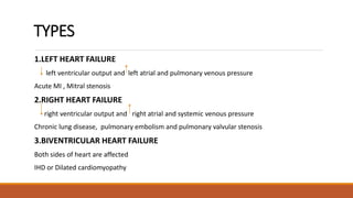
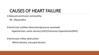
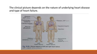
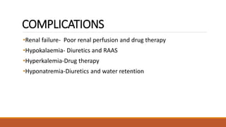
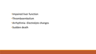
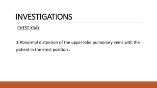
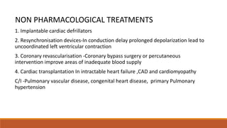
HEART FAILURE
REF:DAVIDSON’S PRINCIPLES AND PRACTICE OF MEDICINE
ABIYA THOMAS
ADITHYA S BABU
CONTENTS
DEFINITION
TYPES
CAUSES
PATHOGENESIS
CLINICAL ASSESSMENT
COMPLICATIONS
INVESTIGATIONS
MANAGEMENT
HEART FAILURE
Definition
Heart failure describes the clinical syndrome that develops
when the heart cannot maintain adequate output, or can do
so only at the expense of elevated ventricular filling
pressure.
•mild to moderate heart failure = symptoms occur only when the
metabolic demand increases during exercise and some other form of
stress
•severe heart failure= symptoms may be present at rest
TYPES
1.LEFT HEART FAILURE
left ventricular output and left atrial and pulmonary venous pressure
Acute MI , Mitral stenosis
2.RIGHT HEART FAILURE
right ventricular output and right atrial and systemic venous pressure
Chronic lung disease, pulmonary embolism and pulmonary valvular stenosis
3.BIVENTRICULAR HEART FAILURE
Both sides of heart are affected
IHD or Dilated cardiomyopathy
CAUSES OF HEART FAILURE
1.Reduced ventricular contractility-
MI , Myocarditis
2.Ventricular outflow obstruction(pressure overload)-
. Hypertension, aortic stenosis (LHF);Pulmonary hypertension(RHF)
3.Ventricular inflow obstruction-
Mitral stenosis, tricuspid stenosis
4.volume overload-
Left ventricular volume overload (mitral or aortic Regurgitation);Ventricular
septal defect;Right ventricular volume overload (atrial septal defect);increased
metabolic demand (high output)
5.Arrhythmia-
Atrial fibrillation;Tachycardia;Complete heart block
6.Diastolic dysfunction-
Constrictive pericarditis,Restrictive cardiomyopathy,Left ventricular hypertrophy
and fibrosis; Cardiac tamponade.
PATHOGENESIS
Cardiac output is determined by
preload (the volume and pressure of blood in the ventricles at the end of
diastole),
afterload (the volume and pressure of blood in the ventricles during systole)
myocardial contractility
PATHOGENESIS
•Ventricular dysfunction-impaired systolic contraction or diastolic
dysfunction- seen in ventricular hypertrophy
•High output failure-due to large arteriovenous shunt in patients w/o
heart disease;or due to high cardiac output
•Valvular disease-impaired filling of ventricles due to mitral or
tricuspid stenosis;aortic and tricuspid stenosis,hypertrophic
cardiomyopathy
CLINICAL ASSESSMENT
•Heart failure may develop suddenly as in MI, or gradually as in valvular heart
disease.
•Compensated heart failure- those with impaired cardiac function, in whom
adaptive changes have prevented the development of overt heart failure.
•atrial fibrillation or infection may precipitate heart failure in these.
•acute heart failure sometimes come along with a decompensating episode, on a
background of chronic heart failure-acute-on-chronic heart failure.
Clinical assessment of acute heart failure
•Presents with a sudden onset of dyspnea at rest that progresses to acute
respiratory distress, orthopnoea & prostration.
• H/o acute MI
•patient appears agitated, pale, clammy;
•peripheries cool to the touch
•pulse –rapid
•BP usually high.
•JVP usually elevated
•gallop rhythm, a 3rd heart sound heard quite early
•chest examination-crepitations at lung bases or throughout the lungs.
•Expiratory wheeze
•potential precipitants- URTI or inappropriate cessation of diuretic medication.
Clinical assessment of chronic heart
failure
•Relapsing and remitting course
•Periods of stability and episodes of decompensation
•Low cardiac output- fatigue , listlessness, poor effort tolerance, weakness
•Periphery – cold ; BP low
•Oliguria, uremia
The clinical picture depends on the nature of underlying heart disease
and type of heart failure.
COMPLICATIONS
•Renal failure- Poor renal perfusion and drug therapy
•Hypokalaemia- Diuretics and RAAS
•Hyperkalemia-Drug therapy
•Hyponatremia-Diuretics and water retention
•Impaired liver function
•Thromboembolism
•Arrhythmia -Electrolyte changes
•Sudden death
INVESTIGATIONS
CHEST XRAY
1.Abnormal distension of the upper lobe pulmonary veins with the
patient in the erect position .
2.KERLEY B LINES
-Vascularity of the lung fields becomes more prominent and the right and Ieft
pulmonary arteries dilate.
-Interstitial oedema causes thickened interlobular septa and dilated lymphatics.
-These are evident as horizontal lines in the costophrenic angles
ECHOCARDIOGRAPHY
Determine aetiology
Detect valvular heart disease
Identify patients with benefits from long term treatment
UREA, CREATINE, ELECTROLYTE, HB,TFT
MANAGEMENT PLAN
Management of acute heart failure
•Medical emergency
•If the above measures are ineffective then inotropic agents such as
dobutamine (2.5-10 ug/kg/min) may be required to augment cardiac
output, particularly in hypotensive patients.
•Insertion of an intra-aortic balloon pump may be beneficial in patients
with acute cardiogenic pulmonary oedema and shock.
Management of chronic heart failure
Aims of treatment
-Increase contractlity
-Optimising preload or decreasing afterload
-Control cardiac rate and rhythm
1. Education
2. Drug treatment
a.Diuretics
•Diuretics promote urinary sodium and water excretion leading to a reduction in
blood plasma volume
•Inturn reduces preload and improves pulmonary and systemic venous
congestion.
•Also reduce afterload and ventricular volume, leading to a fall in ventricular wall
tension and increased cardiac efficiency.
•In case of severe chronic heart failure with renal impairment, oedema may
persist even after loop diuretic therapy so furosemide combined with thiazide
diuretic such as bendroflumethiazide is used.
•K sparing diuretics such as spironolactone and eplerenone is also used in severe
HF cases
b.ACE INHIBITORS
•Prevent conversion of angiotensin I to angiotensin II
•reduces peripheral vasoconstriction
•activation of the sympathetic nervous system and salt and water retention due to
aldosterone release
•as well as preventing the activation of the renin- angiotensin system caused by
diuretic therapy.
•Adverse effects of ACE inhibitors include symptomatic hypotension and
impairment of renal function, especially in patients with bilateral renal
artery stenosis or those with pre-existing renal disease.
•Short-acting ACE inhibitors can cause marked falls in BP, particularly in
the elderly or when started in the presence of hypotension,
hypovolaemia and ,hyponatraemia.
c.ARB
•Act by blocking action of angiotensin II on heart , peripheral vasculature and kidney
•Adverse effects
•Renal dysfunction and Hyperkalemia
•similar to ACE INHIBITORS
d.Neprilysin inhibitors
-a small-molecule inhibitor of neprilysin, which is responsible for breakdown of
endogenous diuretics ANP and BNP
-Used in combination with ARB (Valsartan) additional symptomatic and mortality
benefit over ACE INHIBITORS
-only drug is sacubitril
•e.Vasodilators
•When ACE INHIBITORS and ARB contraindicated
•Vasodilators nitrates reduce preload
•Arterial dilator hydralazine reduce afterload
•Limited use
•f. Beta blockers
•Beta-blockade helas io_counteract
•the deleterious effects of enhanced sympathetic stimulation and
•reduces the risk of arrhythmias and sudden death.
G.IVABRADINE
-Act on inward current in SA node reduce HR
-Used when beta blockers and contraindicated or not effective
-Ineffective in atrial fibrillation
H.DIGOXIN
-control HR
-no effect on long term survival
I.AMIODARONE
-Potent Anti Arrhythmic drug
-Last option
NON PHARMACOLOGICAL TREATMENTS
1. Implantable cardiac defrillators
2. Resynchronisation devices-In conduction delay prolonged depolarization lead to
uncoordinated left ventricular contraction
3. Coronary revascularisation -Coronary bypass surgery or percutaneous
intervention improve areas of inadequate blood supply
4. Cardiac transplantation In intractable heart failure ,CAD and cardiomyopathy
C/I -Pulmonary vascular disease, congenital heart disease, primary Pulmonary
hypertension
THANK YOU

heart failure final.. presentation medicine pptx

  • 1.
    HEART FAILURE REF:DAVIDSON’S PRINCIPLESAND PRACTICE OF MEDICINE ABIYA THOMAS ADITHYA S BABU
  • 2.
  • 3.
    HEART FAILURE Definition Heart failuredescribes the clinical syndrome that develops when the heart cannot maintain adequate output, or can do so only at the expense of elevated ventricular filling pressure.
  • 4.
    •mild to moderateheart failure = symptoms occur only when the metabolic demand increases during exercise and some other form of stress •severe heart failure= symptoms may be present at rest
  • 5.
    TYPES 1.LEFT HEART FAILURE leftventricular output and left atrial and pulmonary venous pressure Acute MI , Mitral stenosis 2.RIGHT HEART FAILURE right ventricular output and right atrial and systemic venous pressure Chronic lung disease, pulmonary embolism and pulmonary valvular stenosis 3.BIVENTRICULAR HEART FAILURE Both sides of heart are affected IHD or Dilated cardiomyopathy
  • 6.
    CAUSES OF HEARTFAILURE 1.Reduced ventricular contractility- MI , Myocarditis 2.Ventricular outflow obstruction(pressure overload)- . Hypertension, aortic stenosis (LHF);Pulmonary hypertension(RHF) 3.Ventricular inflow obstruction- Mitral stenosis, tricuspid stenosis
  • 7.
    4.volume overload- Left ventricularvolume overload (mitral or aortic Regurgitation);Ventricular septal defect;Right ventricular volume overload (atrial septal defect);increased metabolic demand (high output) 5.Arrhythmia- Atrial fibrillation;Tachycardia;Complete heart block 6.Diastolic dysfunction- Constrictive pericarditis,Restrictive cardiomyopathy,Left ventricular hypertrophy and fibrosis; Cardiac tamponade.
  • 8.
    PATHOGENESIS Cardiac output isdetermined by preload (the volume and pressure of blood in the ventricles at the end of diastole), afterload (the volume and pressure of blood in the ventricles during systole) myocardial contractility
  • 10.
    PATHOGENESIS •Ventricular dysfunction-impaired systoliccontraction or diastolic dysfunction- seen in ventricular hypertrophy •High output failure-due to large arteriovenous shunt in patients w/o heart disease;or due to high cardiac output •Valvular disease-impaired filling of ventricles due to mitral or tricuspid stenosis;aortic and tricuspid stenosis,hypertrophic cardiomyopathy
  • 11.
    CLINICAL ASSESSMENT •Heart failuremay develop suddenly as in MI, or gradually as in valvular heart disease. •Compensated heart failure- those with impaired cardiac function, in whom adaptive changes have prevented the development of overt heart failure. •atrial fibrillation or infection may precipitate heart failure in these. •acute heart failure sometimes come along with a decompensating episode, on a background of chronic heart failure-acute-on-chronic heart failure.
  • 12.
    Clinical assessment ofacute heart failure •Presents with a sudden onset of dyspnea at rest that progresses to acute respiratory distress, orthopnoea & prostration. • H/o acute MI •patient appears agitated, pale, clammy; •peripheries cool to the touch •pulse –rapid
  • 13.
    •BP usually high. •JVPusually elevated •gallop rhythm, a 3rd heart sound heard quite early •chest examination-crepitations at lung bases or throughout the lungs. •Expiratory wheeze •potential precipitants- URTI or inappropriate cessation of diuretic medication.
  • 14.
    Clinical assessment ofchronic heart failure •Relapsing and remitting course •Periods of stability and episodes of decompensation •Low cardiac output- fatigue , listlessness, poor effort tolerance, weakness •Periphery – cold ; BP low •Oliguria, uremia
  • 15.
    The clinical picturedepends on the nature of underlying heart disease and type of heart failure.
  • 16.
    COMPLICATIONS •Renal failure- Poorrenal perfusion and drug therapy •Hypokalaemia- Diuretics and RAAS •Hyperkalemia-Drug therapy •Hyponatremia-Diuretics and water retention
  • 17.
  • 18.
    INVESTIGATIONS CHEST XRAY 1.Abnormal distensionof the upper lobe pulmonary veins with the patient in the erect position .
  • 19.
    2.KERLEY B LINES -Vascularityof the lung fields becomes more prominent and the right and Ieft pulmonary arteries dilate. -Interstitial oedema causes thickened interlobular septa and dilated lymphatics. -These are evident as horizontal lines in the costophrenic angles
  • 21.
    ECHOCARDIOGRAPHY Determine aetiology Detect valvularheart disease Identify patients with benefits from long term treatment UREA, CREATINE, ELECTROLYTE, HB,TFT
  • 22.
    MANAGEMENT PLAN Management ofacute heart failure •Medical emergency
  • 23.
    •If the abovemeasures are ineffective then inotropic agents such as dobutamine (2.5-10 ug/kg/min) may be required to augment cardiac output, particularly in hypotensive patients. •Insertion of an intra-aortic balloon pump may be beneficial in patients with acute cardiogenic pulmonary oedema and shock.
  • 24.
    Management of chronicheart failure Aims of treatment -Increase contractlity -Optimising preload or decreasing afterload -Control cardiac rate and rhythm
  • 25.
  • 26.
  • 27.
    a.Diuretics •Diuretics promote urinarysodium and water excretion leading to a reduction in blood plasma volume •Inturn reduces preload and improves pulmonary and systemic venous congestion. •Also reduce afterload and ventricular volume, leading to a fall in ventricular wall tension and increased cardiac efficiency.
  • 28.
    •In case ofsevere chronic heart failure with renal impairment, oedema may persist even after loop diuretic therapy so furosemide combined with thiazide diuretic such as bendroflumethiazide is used. •K sparing diuretics such as spironolactone and eplerenone is also used in severe HF cases
  • 29.
    b.ACE INHIBITORS •Prevent conversionof angiotensin I to angiotensin II •reduces peripheral vasoconstriction •activation of the sympathetic nervous system and salt and water retention due to aldosterone release •as well as preventing the activation of the renin- angiotensin system caused by diuretic therapy.
  • 30.
    •Adverse effects ofACE inhibitors include symptomatic hypotension and impairment of renal function, especially in patients with bilateral renal artery stenosis or those with pre-existing renal disease. •Short-acting ACE inhibitors can cause marked falls in BP, particularly in the elderly or when started in the presence of hypotension, hypovolaemia and ,hyponatraemia.
  • 31.
    c.ARB •Act by blockingaction of angiotensin II on heart , peripheral vasculature and kidney •Adverse effects •Renal dysfunction and Hyperkalemia •similar to ACE INHIBITORS
  • 32.
    d.Neprilysin inhibitors -a small-moleculeinhibitor of neprilysin, which is responsible for breakdown of endogenous diuretics ANP and BNP -Used in combination with ARB (Valsartan) additional symptomatic and mortality benefit over ACE INHIBITORS -only drug is sacubitril
  • 33.
    •e.Vasodilators •When ACE INHIBITORSand ARB contraindicated •Vasodilators nitrates reduce preload •Arterial dilator hydralazine reduce afterload •Limited use
  • 34.
    •f. Beta blockers •Beta-blockadehelas io_counteract •the deleterious effects of enhanced sympathetic stimulation and •reduces the risk of arrhythmias and sudden death.
  • 35.
    G.IVABRADINE -Act on inwardcurrent in SA node reduce HR -Used when beta blockers and contraindicated or not effective -Ineffective in atrial fibrillation H.DIGOXIN -control HR -no effect on long term survival I.AMIODARONE -Potent Anti Arrhythmic drug -Last option
  • 36.
    NON PHARMACOLOGICAL TREATMENTS 1.Implantable cardiac defrillators 2. Resynchronisation devices-In conduction delay prolonged depolarization lead to uncoordinated left ventricular contraction 3. Coronary revascularisation -Coronary bypass surgery or percutaneous intervention improve areas of inadequate blood supply 4. Cardiac transplantation In intractable heart failure ,CAD and cardiomyopathy C/I -Pulmonary vascular disease, congenital heart disease, primary Pulmonary hypertension
  • 37.