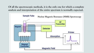
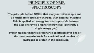
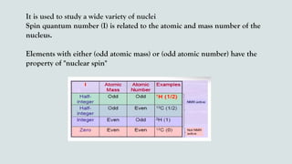
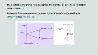
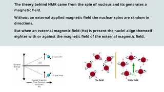
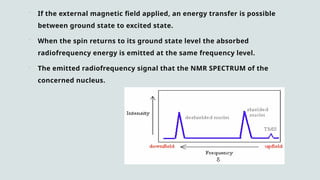
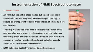
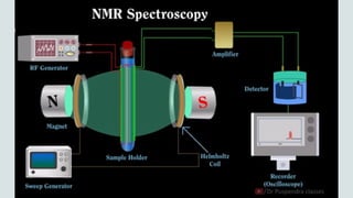
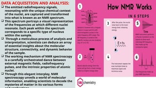
GURU GHASIDAS VISHWAVIDYALAYA
NMR SPECTROSCOPY
M.SC BIOTECHNOLOGY (SEMESTER-1)
SUBMITTED TO:
Dr. Santosh Kumar
Associate Professor
Department of Biotechnology
Guru Ghasidas Vishwavidyalaya , Bilaspur (C.G.)
PRESENTED BY:
Dharun Sao
M.Sc. Biotechnology
Department of Biotechnology
Guru Ghasidas Vishwavidyalaya , Bilaspur (C.G.)
CONTENTS
 HISTORY
 INTRODUCTION OF NMR (Spectroscopy)
 PRINCIPLE OF NMR (Spectroscopy)
 WORKING OF NMR (Spectroscopy)
 INSTRUMENTATION OF NMR (Spectroscopy)
 APPLICATION OF NMR (Spectroscopy)
 NMR SPECTROSCOPY TECHNIQUES
 REFERENCES
HISTORY
 The first NMR spectrum was
first published in the same
issue of the Physical Review
in January 1946.
 Bloch and Purcell were
jointly awarded the 1952
Nobel Prize in Physics for
their research of Nuclear
Magnetic Resonance
Spectroscopy.
INTRODUCTION
Nuclear magnetic resonance spectroscopy, most commonly
known as NMR spectroscopy or magnetic resonance
spectroscopy (MRS), is a spectroscopic technique to observe
local magnetic fields around atomic nuclei.
It is a spectroscopy technique that is based on the absorption
of electromagnetic radiation in the radiofrequency region 4 to
900 MHz by nuclei of the atoms.
Over the past fifty years, NMR has become the preeminent
technique for determining the structure of organic compounds.
Of all the spectroscopic methods, it is the only one for which a complete
analysis and interpretation of the entire spectrum is normally expected.
PRINCIPLE OF NMR
SPECTROSCOPY
The principle behind NMR is that many nuclei have spin and
all nuclei are electrically charged. If an external magnetic
field is applied, an energy transfer is possible between
the base energy to a higher energy level (generally a
single energy gap).
Proton Nuclear magnetic resonance spectroscopy is one of
the most powerful tools for elucidation of number of
hydrogen or proton in the compound.
It is used to study a wide variety of nuclei
Spin quantum number (I) is related to the atomic and mass number of the
nucleus.
Elements with either (odd atomic mass) or (odd atomic number) have the
property of "nuclear spin"
 If an external magnetic field is applied the number of possible orientation
calculate by (2I+1).
 Hydrogen has spin quantum number I=½ and possible orientation is
(2*½+1=2) two +½ and –½.
 The theory behind NMR came from the spin of nucleus and its generates a
magnetic field.
 Without an external applied magnetic field the nuclear spins are random in
directions.
 But when an external magnetic field (Ho) is present the nuclei align themself
eighter with or against the magnetic field of the external magnetic field.
 If the external magnetic field applied, an energy transfer is possible
between ground state to excited state.
 When the spin returns to its ground state level the absorbed
radiofrequency energy is emitted at the same frequency level.
 The emitted radiofrequency signal that the NMR SPECTRUM of the
concerned nucleus.
Instrumentation of NMR Spectrophotometer
1. SAMPLE TUBE :
 An NMR tube is a thin glass walled tube used to contain
samples in nuclear magnetic resonance spectroscopy. It
should be transparent to radio frequencies, chemically inert
and durable.
 Typically NMR tubes are 5 mm diameters but 10 mm and 3
mm samples are known. It is important that the tubes are
uniformly thick and well-balanced to ensure that NMR tube
spins at a regular rate (i.e., they do not wobble), usually
about 20 Hz in the NMR spectrometer.
 NMR tubes are typically made of borosilicate glass.
2. MAGNET SYSTEM:
 Permanent magnets: magnetic field strengths up to 2 Tesla, typically used in benchtop NMRs.
 Electromagnets: higher fields, need high & stable electrical power, problematic field stability and
uniformity.
 Superconducting magnets: very homogenous (uniform) magnetic fields up to 25 Tesla,
Superconducting magnets are also called “cryomagnets” because of their need for cryogenic
liquids (liquid Helium and liquid Nitrogen) to keep the magnet coil in a superconducting state.
Loss of cryogens will lead to a “magnet quench”, a loss of the magnetic field, requiring very
expensive repairs.
3. SWEEP GENERATOR:
 A sweep generator is an electronic device that generates a waveform with a constant amplitude
and a linearly varying frequency. The main role of the sweep generator in the NMR spectroscopy
is that it allows the equal magnitude magnetic field to pass through the sample.
4. SAMPLE PROBE:
 It accepts the sample, sends RF energy into the sample and detects the signal emanating from
the sample.
 3. RADIO FREQUENCY SYSTEM:
 RF coils are used to generate the RF pulses that excite the nuclei in the sample and to
detect the resulting NMR signals. There are usually two types of coils: the transmitter
coil, which sends out the RF pulses, and the receiver coil, which detects the
resulting NMR signals.
 4. AMPLIFIER:
 The purpose of the amplifier is to amplify the obtained NMR signal, it improves the
visibility of the NMR signals by adjusting the frequencies by using the upper pass and
lower pass filters. Usually, the SRS 560 amplifier is used in NMR spectroscopy.
 5. CONSOLE AND COMPUTER:
 The NMR console serves as the control center, where parameters like magnetic field
strength, pulse sequences, and data acquisition settings are programmed. Data is
collected and processed using software on a connected computer.
Working of NMR Spectrophotometer
 Place the sample in a magnetic field.
 Excite the nuclei sample into nuclear magnetic resonance with the help of radio waves to produce NMR
signals.
 These NMR signals are detected with sensitive radio receivers.
 The resonance frequency of an atom in a molecule is changed by the intramolecular magnetic field
surrounding it.
 In specific condition when the magnetic field strength exceeds radio frequency , proton will align in the
direction of the magnetic field. This is known as resonance.
 When this resonance in protons takes place then the receiver coils will receive the signal and it will pass
the signal to the amplifier.
 Amplifier will amplify the signal and the detector will detect the signal and pass it to the recorder, where
we will get the NMR signal.
 This gives details of a molecule’s individual functional groups and its electronic structure.
DATA ACQUISITION AND ANALYSIS:
 The emitted radiofrequency signals,
resonating with the unique chemical context
of the nuclei, are captured and transformed
into what is known as an NMR spectrum.
 This spectrum portrays a visual representation
of the frequencies at which different nuclei
resonate. Each peak within the spectrum
corresponds to a specific type of nucleus
within the sample.
 Through a meticulous process of analysis and
interpretation, scientists can deduce an array
of essential insights about the molecular
structure, connectivity, and dynamic behavior
of the sample.
 The working mechanism of NMR spectroscopy
is a carefully orchestrated dance between
external magnetic fields, radiofrequency
pulses, and the intrinsic properties of atomic
nuclei.
 Through this elegant interplay, NMR
spectroscopy unveils a world of molecular
information, enabling scientists to decode the
mysteries of matter in its various forms
Chemical Shift in NMR Spectroscopy
 A spinning charge generates a magnetic field that results in a magnetic
moment proportional to the spin. In the presence of an external magnetic
field, two spin states exist; one spin up and one spin down, where one aligns
with the magnetic field and the other opposes it .
 Chemical shift is characterized as the difference between the resonant
frequency of the spinning protons and the signal of the reference molecule.
Nuclear magnetic resonance chemical change is one of the most important
properties usable for molecular structure determination .
 There are also different nuclei that can be detected by NMR spectroscopy,
1H (proton), 13C (carbon 13), 15N (nitrogen 15), 19F (fluorine 19), among
many more. 1H and 13C are the most widely used.
 The definition of 1H as it is very descriptive of the spectroscopy of the
NMR. Both the nuts have a good charge and are constantly revolving like a
cloud. Through mechanics, we learn that a charge in motion produces a
magnetic field.
 In NMR, when we reach the radio frequency (Rf) radiation nucleus, it
causes the nucleus and its magnetic field to turn (or it causes the nuclear
magnet to pulse, thus the term NMR).
Application of NMR
 Spectroscopy is the study of the interaction of electromagnetic radiation with matter. NMR
spectroscopy is the use of the NMR phenomenon to study the physical, chemical, and biological
properties of matter.
 It is an analytical chemistry technique used in quality control.
 It is used in research for determining the content and purity of a sample as well as its molecular
structure. For example, NMR can quantitatively analyze mixtures containing known compounds.
 NMR spectroscopy is routinely used by chemists to study chemical structure using simple one-
dimensional techniques. Two-dimensional techniques are used to determine the structure of more
complicated molecules.
 These techniques are replacing x-ray crystallography for the determination of protein structure.
 Time domain NMR spectroscopy techniques are used to probe molecular dynamics in solution.
 Solid state NMR spectroscopy is used to determine the molecular structure of solids.
 Other scientists have developed NMR methods-of measuring diffusion coefficients.
Some other Applications
 NMR spectroscopy is a Spectroscopy technique used by chemists and biochemists to
investigate the properties of organic molecules, although it is applicable to any kind
of sample that contains nuclei possessing spin.
 For example, the NMR can quantitatively analyze mixtures containing known
compounds. NMR can either be used to match against spectral libraries or to infer
the basic structure directly for unknown compounds.
 Once the basic structure is known, NMR can be used to determine molecular
conformation in solutions as well as in studying physical properties at the molecular
level such as conformational exchange, phase changes, solubility, and diffusion.
Techniques of NMR
NMR spectroscopy employs various techniques to analyze the structure dynamics and
introductions of molecules.
i) 1D NMR Spectroscopy: most basic form where only one frequency axis is measured,
which corresponds to the resonance frequencies of nuclei in a sample.
ii) 2D NMR Spectroscopy: Two different frequency axes are used to plot the data allowing
for correlation between different nuclei in a molecule to be observed. The most popular
technique among 2D NMR spectroscopy are:
 COSY (Correlation Spectroscopy): correlates coupled nuclear spins and provides
information about which protons in a molecule are located close to each other.
 NOESY (Nuclear Overhauser Effect Spectroscopy): detects spatial proximities between
nuclear spins, and provides structure of molecules and distance between atoms.
Techniques of NMR
iii) Solid-State NMR Spectroscopy: used to study materials in their solid form, and
provides information about molecular dynamics in solid state samples.
iv) Zero-Field NMR spectroscopy: operates in the absence of a strong external
magnetic field.
v) In-cell NMR spectroscopy: investigates the biomolecular structure and interaction
within living organisms. It also provides in sigights into cellular processes and disease
mechanism.
References
1) https://www.sciencedirect.com/topics/materials-science/nuclear-magnetic-resonance-spec
troscopy
2).
https://en.wikipedia.org/wiki/File:Nuclear_Magnetic_Resonance_(NMR)_basic_principles.web
m
3). https://en.wikipedia.org/wiki/Nuclear_magnetic_resonance
4). https://byjus.com/chemistry/nmr-spectroscopy/
THANK YOU

NMR Spectroscopy: Principles, Techniques, and Applicationsy.pptx

  • 1.
    GURU GHASIDAS VISHWAVIDYALAYA NMRSPECTROSCOPY M.SC BIOTECHNOLOGY (SEMESTER-1) SUBMITTED TO: Dr. Santosh Kumar Associate Professor Department of Biotechnology Guru Ghasidas Vishwavidyalaya , Bilaspur (C.G.) PRESENTED BY: Dharun Sao M.Sc. Biotechnology Department of Biotechnology Guru Ghasidas Vishwavidyalaya , Bilaspur (C.G.)
  • 2.
    CONTENTS  HISTORY  INTRODUCTIONOF NMR (Spectroscopy)  PRINCIPLE OF NMR (Spectroscopy)  WORKING OF NMR (Spectroscopy)  INSTRUMENTATION OF NMR (Spectroscopy)  APPLICATION OF NMR (Spectroscopy)  NMR SPECTROSCOPY TECHNIQUES  REFERENCES
  • 3.
    HISTORY  The firstNMR spectrum was first published in the same issue of the Physical Review in January 1946.  Bloch and Purcell were jointly awarded the 1952 Nobel Prize in Physics for their research of Nuclear Magnetic Resonance Spectroscopy.
  • 4.
    INTRODUCTION Nuclear magnetic resonancespectroscopy, most commonly known as NMR spectroscopy or magnetic resonance spectroscopy (MRS), is a spectroscopic technique to observe local magnetic fields around atomic nuclei. It is a spectroscopy technique that is based on the absorption of electromagnetic radiation in the radiofrequency region 4 to 900 MHz by nuclei of the atoms. Over the past fifty years, NMR has become the preeminent technique for determining the structure of organic compounds.
  • 5.
    Of all thespectroscopic methods, it is the only one for which a complete analysis and interpretation of the entire spectrum is normally expected.
  • 6.
    PRINCIPLE OF NMR SPECTROSCOPY Theprinciple behind NMR is that many nuclei have spin and all nuclei are electrically charged. If an external magnetic field is applied, an energy transfer is possible between the base energy to a higher energy level (generally a single energy gap). Proton Nuclear magnetic resonance spectroscopy is one of the most powerful tools for elucidation of number of hydrogen or proton in the compound.
  • 7.
    It is usedto study a wide variety of nuclei Spin quantum number (I) is related to the atomic and mass number of the nucleus. Elements with either (odd atomic mass) or (odd atomic number) have the property of "nuclear spin"
  • 8.
     If anexternal magnetic field is applied the number of possible orientation calculate by (2I+1).  Hydrogen has spin quantum number I=½ and possible orientation is (2*½+1=2) two +½ and –½.
  • 9.
     The theorybehind NMR came from the spin of nucleus and its generates a magnetic field.  Without an external applied magnetic field the nuclear spins are random in directions.  But when an external magnetic field (Ho) is present the nuclei align themself eighter with or against the magnetic field of the external magnetic field.
  • 10.
     If theexternal magnetic field applied, an energy transfer is possible between ground state to excited state.  When the spin returns to its ground state level the absorbed radiofrequency energy is emitted at the same frequency level.  The emitted radiofrequency signal that the NMR SPECTRUM of the concerned nucleus.
  • 11.
    Instrumentation of NMRSpectrophotometer 1. SAMPLE TUBE :  An NMR tube is a thin glass walled tube used to contain samples in nuclear magnetic resonance spectroscopy. It should be transparent to radio frequencies, chemically inert and durable.  Typically NMR tubes are 5 mm diameters but 10 mm and 3 mm samples are known. It is important that the tubes are uniformly thick and well-balanced to ensure that NMR tube spins at a regular rate (i.e., they do not wobble), usually about 20 Hz in the NMR spectrometer.  NMR tubes are typically made of borosilicate glass.
  • 12.
    2. MAGNET SYSTEM: Permanent magnets: magnetic field strengths up to 2 Tesla, typically used in benchtop NMRs.  Electromagnets: higher fields, need high & stable electrical power, problematic field stability and uniformity.  Superconducting magnets: very homogenous (uniform) magnetic fields up to 25 Tesla, Superconducting magnets are also called “cryomagnets” because of their need for cryogenic liquids (liquid Helium and liquid Nitrogen) to keep the magnet coil in a superconducting state. Loss of cryogens will lead to a “magnet quench”, a loss of the magnetic field, requiring very expensive repairs. 3. SWEEP GENERATOR:  A sweep generator is an electronic device that generates a waveform with a constant amplitude and a linearly varying frequency. The main role of the sweep generator in the NMR spectroscopy is that it allows the equal magnitude magnetic field to pass through the sample. 4. SAMPLE PROBE:  It accepts the sample, sends RF energy into the sample and detects the signal emanating from the sample.
  • 13.
     3. RADIOFREQUENCY SYSTEM:  RF coils are used to generate the RF pulses that excite the nuclei in the sample and to detect the resulting NMR signals. There are usually two types of coils: the transmitter coil, which sends out the RF pulses, and the receiver coil, which detects the resulting NMR signals.  4. AMPLIFIER:  The purpose of the amplifier is to amplify the obtained NMR signal, it improves the visibility of the NMR signals by adjusting the frequencies by using the upper pass and lower pass filters. Usually, the SRS 560 amplifier is used in NMR spectroscopy.  5. CONSOLE AND COMPUTER:  The NMR console serves as the control center, where parameters like magnetic field strength, pulse sequences, and data acquisition settings are programmed. Data is collected and processed using software on a connected computer.
  • 16.
    Working of NMRSpectrophotometer  Place the sample in a magnetic field.  Excite the nuclei sample into nuclear magnetic resonance with the help of radio waves to produce NMR signals.  These NMR signals are detected with sensitive radio receivers.  The resonance frequency of an atom in a molecule is changed by the intramolecular magnetic field surrounding it.  In specific condition when the magnetic field strength exceeds radio frequency , proton will align in the direction of the magnetic field. This is known as resonance.  When this resonance in protons takes place then the receiver coils will receive the signal and it will pass the signal to the amplifier.  Amplifier will amplify the signal and the detector will detect the signal and pass it to the recorder, where we will get the NMR signal.  This gives details of a molecule’s individual functional groups and its electronic structure.
  • 18.
    DATA ACQUISITION ANDANALYSIS:  The emitted radiofrequency signals, resonating with the unique chemical context of the nuclei, are captured and transformed into what is known as an NMR spectrum.  This spectrum portrays a visual representation of the frequencies at which different nuclei resonate. Each peak within the spectrum corresponds to a specific type of nucleus within the sample.  Through a meticulous process of analysis and interpretation, scientists can deduce an array of essential insights about the molecular structure, connectivity, and dynamic behavior of the sample.  The working mechanism of NMR spectroscopy is a carefully orchestrated dance between external magnetic fields, radiofrequency pulses, and the intrinsic properties of atomic nuclei.  Through this elegant interplay, NMR spectroscopy unveils a world of molecular information, enabling scientists to decode the mysteries of matter in its various forms
  • 19.
    Chemical Shift inNMR Spectroscopy  A spinning charge generates a magnetic field that results in a magnetic moment proportional to the spin. In the presence of an external magnetic field, two spin states exist; one spin up and one spin down, where one aligns with the magnetic field and the other opposes it .  Chemical shift is characterized as the difference between the resonant frequency of the spinning protons and the signal of the reference molecule. Nuclear magnetic resonance chemical change is one of the most important properties usable for molecular structure determination .  There are also different nuclei that can be detected by NMR spectroscopy, 1H (proton), 13C (carbon 13), 15N (nitrogen 15), 19F (fluorine 19), among many more. 1H and 13C are the most widely used.
  • 20.
     The definitionof 1H as it is very descriptive of the spectroscopy of the NMR. Both the nuts have a good charge and are constantly revolving like a cloud. Through mechanics, we learn that a charge in motion produces a magnetic field.  In NMR, when we reach the radio frequency (Rf) radiation nucleus, it causes the nucleus and its magnetic field to turn (or it causes the nuclear magnet to pulse, thus the term NMR).
  • 21.
    Application of NMR Spectroscopy is the study of the interaction of electromagnetic radiation with matter. NMR spectroscopy is the use of the NMR phenomenon to study the physical, chemical, and biological properties of matter.  It is an analytical chemistry technique used in quality control.  It is used in research for determining the content and purity of a sample as well as its molecular structure. For example, NMR can quantitatively analyze mixtures containing known compounds.  NMR spectroscopy is routinely used by chemists to study chemical structure using simple one- dimensional techniques. Two-dimensional techniques are used to determine the structure of more complicated molecules.  These techniques are replacing x-ray crystallography for the determination of protein structure.  Time domain NMR spectroscopy techniques are used to probe molecular dynamics in solution.  Solid state NMR spectroscopy is used to determine the molecular structure of solids.  Other scientists have developed NMR methods-of measuring diffusion coefficients.
  • 22.
    Some other Applications NMR spectroscopy is a Spectroscopy technique used by chemists and biochemists to investigate the properties of organic molecules, although it is applicable to any kind of sample that contains nuclei possessing spin.  For example, the NMR can quantitatively analyze mixtures containing known compounds. NMR can either be used to match against spectral libraries or to infer the basic structure directly for unknown compounds.  Once the basic structure is known, NMR can be used to determine molecular conformation in solutions as well as in studying physical properties at the molecular level such as conformational exchange, phase changes, solubility, and diffusion.
  • 23.
    Techniques of NMR NMRspectroscopy employs various techniques to analyze the structure dynamics and introductions of molecules. i) 1D NMR Spectroscopy: most basic form where only one frequency axis is measured, which corresponds to the resonance frequencies of nuclei in a sample. ii) 2D NMR Spectroscopy: Two different frequency axes are used to plot the data allowing for correlation between different nuclei in a molecule to be observed. The most popular technique among 2D NMR spectroscopy are:  COSY (Correlation Spectroscopy): correlates coupled nuclear spins and provides information about which protons in a molecule are located close to each other.  NOESY (Nuclear Overhauser Effect Spectroscopy): detects spatial proximities between nuclear spins, and provides structure of molecules and distance between atoms.
  • 24.
    Techniques of NMR iii)Solid-State NMR Spectroscopy: used to study materials in their solid form, and provides information about molecular dynamics in solid state samples. iv) Zero-Field NMR spectroscopy: operates in the absence of a strong external magnetic field. v) In-cell NMR spectroscopy: investigates the biomolecular structure and interaction within living organisms. It also provides in sigights into cellular processes and disease mechanism.
  • 25.
  • 26.