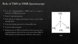
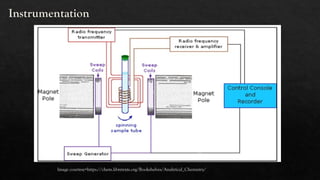
Nuclear Magnetic Resonance
(NMR) Spectroscopy
Powerful Tool For Biology
Efforts by – Gunali Chaudhari (16108)
Contents
Introduction
Principle
TMS reference
Instrumentation
NMR spectrum with example
References
Application
Introduction
 NMR spectroscopy can be defined as an indispensable tool which
applies a magnetic field to an atomic nucleus.
 Felix Bloch and Edward Purcell discovered NMR spectroscopy.
 An NMR instrument allows the molecular structure of a material to
be analysed by observing and measuring the interaction of nuclear
spins when placed in a powerful magnetic field.
 Radio waves= wavelength= 109
nm to 1012
nm
 NMR ACTIVE MOLECULE=
 Spin described the nature of the magnetic field surrounding the
nucleus of an atom and it is denoted by I.
 It can be 0, +1/2, -1/2.
 If 0 or no. of proton + no. of neutron is even no, then the molecule
is NMR inactive. For e.g. 12
C
 If no 0 or no.of proton + no.of neutron is odd no, then the
molecule is NMR active. For e.g. 13
C
No. of possible orientations= 2I+1
Here I = 1/2
Principle
 The principle behind NMR includes the spinning of nuclei and generating a magnetic field.
 Without an external applied magnetic field, the nuclear spins are random in directions.
 But when an external magnetic field is present the nuclei align themselves either with or against the field of the
external magnet.
 Aligned with field= low strength magnetic field is required for excitation.
 Aligned against field = high strength magnetic field is required for excitation.
Image courtesy= https://www.chemistrysteps.com/nmr-chemical-shift
 Absorption of photon of energy and transition of nuclear
spin from ground to excited state leads to NMR signal.
 Nuclear transitions differ in frequency from one nucleus
to other.
 As strength of magnetic field increases, excitation of
group of nuclei in two different orientations will create
difference in energy levels and that excitation difference
leads to signal which will recorded.
 Nature of chemical group also affects these transitions.
For e.g. methyl protons resonate at different frequency
than amide protons. This is called chemical shift.
 Shielding = more electron in the proton’s vicinity =
opposing magnetic field increases.
 Deshielding = less electron in the proton’s vicinity =
opposing magnetic field decreases.
Chemical shift
Image courtesy= https://www.chemistrysteps.com/nmr-chemical-shift/
Role of TMS in NMR Spectroscopy
 It is the Tetramethylsilane (TMS) and it is used as
reference in NMR spectroscopy.
 It has 12 equivalent proton.
 Each proton in single environment, hence it gives single
intensity peak.
 It has low boiling point and it is chemically inert.
 It can be easily recovered.
 Methyl groups in TMS are shielded by their electrons from
external magnetic field and this is the reason that NMR of
TMS obtained at high magnetic field than other proton.
TMS
Instrumentation
Image courtesy=https://chem.libretexts.org/Bookshelves/Analytical_Chemistry/
 Sample holder - Glass tube with 8.5 cm long, 0.3 cm in diameter.
 Permanent magnet - It provides homogeneous magnetic field at 60 – 100
MHZ.
 Magnetic coils - These coils induce magnetic field when current flows
through them.
 Sweep generator - To produce the equal amount of magnetic field pass
through the sample.
 Radio-frequency transmitter - A radio transmitter coil that produces a short
powerful pulse of radio waves.
 Radiofrequency - A radio receiver coil that detects radio frequencies emitted
as nuclei relax to a lower energy level.
 Readout system - A computer that analyses and record the data.
NMR spectrum
 The NMR spectrum is a plot of intensity of NMR signals VS magnetic field (frequency) in reference to TMS.
 The emitted radio frequency is directly proportional to the strength of the applied field.
 Signals produced radiofrequency radiations are converted to the normal spectrum by mathematical process of fulliar
transformation.
 1945= First NMR ethanol spectrum
 1957= First protein NMR spectrum
 Ethanol NMR spectrum=
 Protons of same group do not interact with themselves
because observative splitting.
 Multiplicity of the peak of group of equivalent proton
is determining by neighbouring proton.
 Hence, methyl group = 3 peaks (because of
neighbouring protons of methylene group).
 Methylene group= 8 peaks ( because 2 peaks from
neighbouring single proton of OH and 4 peaks from
neighbouring 3 protons of CH3, hence 4*2=8).
 Hydroxyl group= 3 peaks (because of neighbouring
protons of methylene group).
We can also directly use this to construct or interpret
NMR spectrum
Pascal’s triangle of NMR
Task
Draw the NMR spectrum of the methanol
using pascal’s triangle of NMR.
Application
 Nuclear magnetic resonance spectroscopy is widely used to determine the structure of organic
molecules in solution and study molecular physics and crystals as well as non-crystalline materials.
 NMR is also routinely used in advanced medical imaging techniques, such as in magnetic resonance
imaging (MRI).
 The ability to measure the chemical shift was a boon to chemists; it meant they could perform non-
destructive chemical analyses of samples to determine molecular identity and structure much faster
and more simply than before.
 NMR methods have been introduced to quantitative analysis in order to determine the impurity
profile of a drug to characteristic the composition of drug products and to investigate metabolites of
drugs in body fluids.
 NMR spectroscopy is used to determine structure of proteins, amino acid profile, carotenoids,
organic acids, lipid fractions, the mobility of the water in foods. NMR spectroscopy is also used to
identify and quantify the metabolites in foods.
References
 Principles and Techniques of Biochemistry and Molecular Biology, 7th
edition by Wilson, k and Walker, J.
 Biophysical chemistry principles and techniques by Upadhyay and Nath.
 Tampieri, Alberto, Szabó, Márk, Medina, Francesc and Gulyás, Henrik. "A brief introduction to the basics of
NMR spectroscopy and selected examples of its applications to materials characterization" Physical Sciences
Reviews, vol. 6, no. 1, 2021, pp. 20190086.
 Mukesh Kumar Singh, Annika Singh, Nuclear magnetic resonance spectroscopy overview.
 Marion D. An introduction to biological NMR spectroscopy. Mol Cell Proteomics. 2013 Nov;12(11):3006-25.
doi: 10.1074/mcp.O113.030239. Epub 2013 Jul 6. PMID: 23831612; PMCID: PMC3820920.
 O'Connell MR, Gamsjaeger R, Mackay JP. The structural analysis of protein-protein interactions by NMR
spectroscopy. Proteomics. 2009 Dec;9(23):5224-32. doi: 10.1002/pmic.200900303. PMID: 19834907.
Special thanks to =
Dr. Sanket Tembe
Dr. Revati wanikar

NMR Spectroscopy- bioanalytical techniques

  • 1.
    Nuclear Magnetic Resonance (NMR)Spectroscopy Powerful Tool For Biology Efforts by – Gunali Chaudhari (16108)
  • 2.
  • 3.
    Introduction  NMR spectroscopycan be defined as an indispensable tool which applies a magnetic field to an atomic nucleus.  Felix Bloch and Edward Purcell discovered NMR spectroscopy.  An NMR instrument allows the molecular structure of a material to be analysed by observing and measuring the interaction of nuclear spins when placed in a powerful magnetic field.  Radio waves= wavelength= 109 nm to 1012 nm  NMR ACTIVE MOLECULE=  Spin described the nature of the magnetic field surrounding the nucleus of an atom and it is denoted by I.  It can be 0, +1/2, -1/2.  If 0 or no. of proton + no. of neutron is even no, then the molecule is NMR inactive. For e.g. 12 C  If no 0 or no.of proton + no.of neutron is odd no, then the molecule is NMR active. For e.g. 13 C No. of possible orientations= 2I+1 Here I = 1/2
  • 4.
    Principle  The principlebehind NMR includes the spinning of nuclei and generating a magnetic field.  Without an external applied magnetic field, the nuclear spins are random in directions.  But when an external magnetic field is present the nuclei align themselves either with or against the field of the external magnet.  Aligned with field= low strength magnetic field is required for excitation.  Aligned against field = high strength magnetic field is required for excitation. Image courtesy= https://www.chemistrysteps.com/nmr-chemical-shift
  • 5.
     Absorption ofphoton of energy and transition of nuclear spin from ground to excited state leads to NMR signal.  Nuclear transitions differ in frequency from one nucleus to other.  As strength of magnetic field increases, excitation of group of nuclei in two different orientations will create difference in energy levels and that excitation difference leads to signal which will recorded.  Nature of chemical group also affects these transitions. For e.g. methyl protons resonate at different frequency than amide protons. This is called chemical shift.  Shielding = more electron in the proton’s vicinity = opposing magnetic field increases.  Deshielding = less electron in the proton’s vicinity = opposing magnetic field decreases.
  • 6.
    Chemical shift Image courtesy=https://www.chemistrysteps.com/nmr-chemical-shift/
  • 7.
    Role of TMSin NMR Spectroscopy  It is the Tetramethylsilane (TMS) and it is used as reference in NMR spectroscopy.  It has 12 equivalent proton.  Each proton in single environment, hence it gives single intensity peak.  It has low boiling point and it is chemically inert.  It can be easily recovered.  Methyl groups in TMS are shielded by their electrons from external magnetic field and this is the reason that NMR of TMS obtained at high magnetic field than other proton. TMS
  • 8.
  • 9.
     Sample holder- Glass tube with 8.5 cm long, 0.3 cm in diameter.  Permanent magnet - It provides homogeneous magnetic field at 60 – 100 MHZ.  Magnetic coils - These coils induce magnetic field when current flows through them.  Sweep generator - To produce the equal amount of magnetic field pass through the sample.  Radio-frequency transmitter - A radio transmitter coil that produces a short powerful pulse of radio waves.  Radiofrequency - A radio receiver coil that detects radio frequencies emitted as nuclei relax to a lower energy level.  Readout system - A computer that analyses and record the data.
  • 10.
    NMR spectrum  TheNMR spectrum is a plot of intensity of NMR signals VS magnetic field (frequency) in reference to TMS.  The emitted radio frequency is directly proportional to the strength of the applied field.  Signals produced radiofrequency radiations are converted to the normal spectrum by mathematical process of fulliar transformation.  1945= First NMR ethanol spectrum  1957= First protein NMR spectrum  Ethanol NMR spectrum=
  • 11.
     Protons ofsame group do not interact with themselves because observative splitting.  Multiplicity of the peak of group of equivalent proton is determining by neighbouring proton.  Hence, methyl group = 3 peaks (because of neighbouring protons of methylene group).  Methylene group= 8 peaks ( because 2 peaks from neighbouring single proton of OH and 4 peaks from neighbouring 3 protons of CH3, hence 4*2=8).  Hydroxyl group= 3 peaks (because of neighbouring protons of methylene group).
  • 12.
    We can alsodirectly use this to construct or interpret NMR spectrum Pascal’s triangle of NMR
  • 13.
    Task Draw the NMRspectrum of the methanol using pascal’s triangle of NMR.
  • 14.
    Application  Nuclear magneticresonance spectroscopy is widely used to determine the structure of organic molecules in solution and study molecular physics and crystals as well as non-crystalline materials.  NMR is also routinely used in advanced medical imaging techniques, such as in magnetic resonance imaging (MRI).  The ability to measure the chemical shift was a boon to chemists; it meant they could perform non- destructive chemical analyses of samples to determine molecular identity and structure much faster and more simply than before.  NMR methods have been introduced to quantitative analysis in order to determine the impurity profile of a drug to characteristic the composition of drug products and to investigate metabolites of drugs in body fluids.  NMR spectroscopy is used to determine structure of proteins, amino acid profile, carotenoids, organic acids, lipid fractions, the mobility of the water in foods. NMR spectroscopy is also used to identify and quantify the metabolites in foods.
  • 15.
    References  Principles andTechniques of Biochemistry and Molecular Biology, 7th edition by Wilson, k and Walker, J.  Biophysical chemistry principles and techniques by Upadhyay and Nath.  Tampieri, Alberto, Szabó, Márk, Medina, Francesc and Gulyás, Henrik. "A brief introduction to the basics of NMR spectroscopy and selected examples of its applications to materials characterization" Physical Sciences Reviews, vol. 6, no. 1, 2021, pp. 20190086.  Mukesh Kumar Singh, Annika Singh, Nuclear magnetic resonance spectroscopy overview.  Marion D. An introduction to biological NMR spectroscopy. Mol Cell Proteomics. 2013 Nov;12(11):3006-25. doi: 10.1074/mcp.O113.030239. Epub 2013 Jul 6. PMID: 23831612; PMCID: PMC3820920.  O'Connell MR, Gamsjaeger R, Mackay JP. The structural analysis of protein-protein interactions by NMR spectroscopy. Proteomics. 2009 Dec;9(23):5224-32. doi: 10.1002/pmic.200900303. PMID: 19834907. Special thanks to = Dr. Sanket Tembe Dr. Revati wanikar