Download to read offline

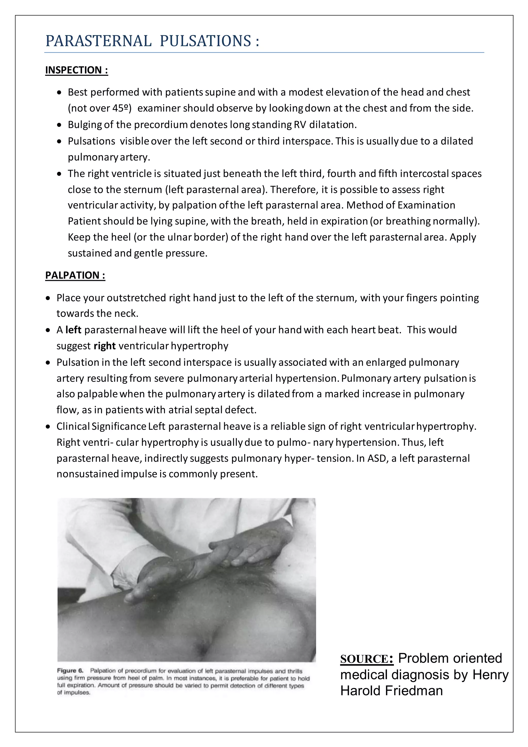

Parasternal pulsations can be observed by having the patient lie supine with a modest head elevation. The examiner should look down at the chest and from the side to see bulging of the precordium, which indicates long-standing right ventricular dilation, or pulsations over the left second or third interspace, usually due to a dilated pulmonary artery. Palpation of the left parasternal area can assess right ventricular activity, with a left parasternal heave lifting the heel of the hand with each heartbeat, suggesting right ventricular hypertrophy. Pulsations in the left second interspace are often associated with enlarged pulmonary arteries from severe pulmonary hypertension.


