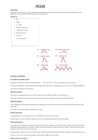
SlideShare a Scribd company logo
PULSE
DEFINITION:
EXPANSION AND ELONGATION OF THE ARTERIAL WALL IMPARTED BY THE COLUMN OF BLOOD AND IS PASSIVELY PRODUCED BY THE
PRESSURE CHANGES DURING VENTRICULAR SYSTOLE AND DIASTOLE.
Points to note:
07 Lippincott Williams & Wilkins
CLINICAL NOTES:
Irregularly irregular pulse:
Amplitude of each beat varies with the filling interval. Seen in AF, PVCs, PACs, and multifocal atrial tachycardia.
PVCs are recognized by a regular rhythm with a dropped beat followed by a compensatory pause, then a beat of increased amplitude.
PACs throw the rhythm out of synchrony.
Bisferiens pulse :
The pattern is a tapping percussion wave with a rapid early rise, a decline, and then a second tidal wave.
This pulse is classically found in hypertrophic cardiomyopathy and in combined aortic stenosis and regurgitation.
Bigeminal pulse :
This is palpable as a strong pulse alternating with a weak one, the second beat caused by decreased ventricular filling with an early
contraction.
It is found in ventricular bigeminy and digoxin overdose.
Pulsus alternans :
The amplitude varies with each pulse and is accentuated after a premature contraction.
Usually due to severe left ventricular dysfunction, also seen in hypertrophic obstructive cardiomyopathy.
Pulsus paradoxus :
The easiest way to obtain this measurement is to start above systole, deflating the cuff until the first sounds are heard, with the pulse
disappearing during inspiration. Continue to deflate the cuff until the heart sounds are rapid and regular. A difference between these
measures of more than 10 mm Hg is abnormal.
Increased pulsus paradoxus may be observed in severe asthma, pericardial tamponade, pulmonary embolism, and hypovolemic shock.
It does not occur with constrictive pericarditis, severe CHF, or right ventricular failure.
Pulsus parvus et tardus:
• Rate
• Rhythm
• Volume
• Condition of arterial wall
• Comparisons b/n radials
• Radio femoral delay
• Character
• Peripheral pulsesto Bedside
The classic finding in hemodynamically significant aortic stenosis, the carotid pulse is low in volume and has a slowly rising upstroke
with a prolonged plateau. A “shudder” may also be felt.
Asymmetric pulses: Consider subclavian artery atherosclerosis, arterial thrombosis (especially with atrial fibrillation), thoracic
outlet compression, or aortic dissection.
Thready pulse : A low-volume thready pulse is found in hypovolemic or septic shock, severe aortic stenosis, and severe left
ventricular dysfunction. Intense vasoconstriction may produce a diminished pulse with normal stroke volume.
Bounding pulses: Hyperkinetic pulse with a rapid large-amplitude upstroke and rapid collapse is associated with increased stroke
volume or decreased arterial compliance. The classic “collapsing pulse” is found in aortic regurgitation. It also occurs in thyrotoxicosis.

More Related Content

What's hot

Acute coronary syndrome
Acute coronary syndrome Acute coronary syndrome
Acute coronary syndrome
Dee Evardone
 
Aortic Stenosis
Aortic StenosisAortic Stenosis
Aortic Stenosis
Abdullatif Al-Rashed
 
GEMC: Case Presentation- Pericarditis: Resident Training
GEMC: Case Presentation- Pericarditis: Resident TrainingGEMC: Case Presentation- Pericarditis: Resident Training
GEMC: Case Presentation- Pericarditis: Resident Training
Open.Michigan
 
Pericardial diseases
Pericardial diseasesPericardial diseases
Pericardial diseases
DrArpan Chouhan
 
Complications of acute mi
Complications of acute miComplications of acute mi
Complications of acute mi
Amir Mahmoud
 
Myocardial infarction
Myocardial infarctionMyocardial infarction
Myocardial infarction
Dr. Sobia Khalid
 
Myocardial infarction
Myocardial infarctionMyocardial infarction
Myocardial infarctionadolescent4u
 
Cardiomegaly
CardiomegalyCardiomegaly
Cardiomegaly
SheelamannilJohn
 
pericarditis.ppt
pericarditis.pptpericarditis.ppt
pericarditis.ppt
icdlab
 
Acute Coronary Syndrome (NSTEMI)
Acute Coronary Syndrome (NSTEMI) Acute Coronary Syndrome (NSTEMI)
Acute Coronary Syndrome (NSTEMI)
Muhammad Asim Rana
 
Right and left ventricular hypertrophy
Right and left ventricular hypertrophyRight and left ventricular hypertrophy
Right and left ventricular hypertrophy
Rawalpindi Medical College
 
Heart failure
Heart failureHeart failure
Heart failure
BSMMU
 
Wpw case presentation by dr adeel
Wpw case presentation by dr adeelWpw case presentation by dr adeel
Wpw case presentation by dr adeel
West Medicine Ward
 
Aortic dissection nikku ppt
Aortic dissection nikku pptAortic dissection nikku ppt
Aortic dissection nikku ppt
Nikhil Vaishnav
 
Cardiac tamponade BY PANKAJ
Cardiac tamponade BY PANKAJCardiac tamponade BY PANKAJ
Cardiac tamponade BY PANKAJ
pankaj rana
 
CABG
CABGCABG
ECG
ECGECG
Myocardial infarction (MI) ecg localisation
Myocardial infarction (MI) ecg localisationMyocardial infarction (MI) ecg localisation
Myocardial infarction (MI) ecg localisation
Malleswara rao Dangeti
 
Conduction Disorders
Conduction DisordersConduction Disorders
Conduction Disorders
Eneutron
 

What's hot (20)

Acute coronary syndrome
Acute coronary syndrome Acute coronary syndrome
Acute coronary syndrome
 
Aneurysms & dissection 7
Aneurysms & dissection 7Aneurysms & dissection 7
Aneurysms & dissection 7
 
Aortic Stenosis
Aortic StenosisAortic Stenosis
Aortic Stenosis
 
GEMC: Case Presentation- Pericarditis: Resident Training
GEMC: Case Presentation- Pericarditis: Resident TrainingGEMC: Case Presentation- Pericarditis: Resident Training
GEMC: Case Presentation- Pericarditis: Resident Training
 
Pericardial diseases
Pericardial diseasesPericardial diseases
Pericardial diseases
 
Complications of acute mi
Complications of acute miComplications of acute mi
Complications of acute mi
 
Myocardial infarction
Myocardial infarctionMyocardial infarction
Myocardial infarction
 
Myocardial infarction
Myocardial infarctionMyocardial infarction
Myocardial infarction
 
Cardiomegaly
CardiomegalyCardiomegaly
Cardiomegaly
 
pericarditis.ppt
pericarditis.pptpericarditis.ppt
pericarditis.ppt
 
Acute Coronary Syndrome (NSTEMI)
Acute Coronary Syndrome (NSTEMI) Acute Coronary Syndrome (NSTEMI)
Acute Coronary Syndrome (NSTEMI)
 
Right and left ventricular hypertrophy
Right and left ventricular hypertrophyRight and left ventricular hypertrophy
Right and left ventricular hypertrophy
 
Heart failure
Heart failureHeart failure
Heart failure
 
Wpw case presentation by dr adeel
Wpw case presentation by dr adeelWpw case presentation by dr adeel
Wpw case presentation by dr adeel
 
Aortic dissection nikku ppt
Aortic dissection nikku pptAortic dissection nikku ppt
Aortic dissection nikku ppt
 
Cardiac tamponade BY PANKAJ
Cardiac tamponade BY PANKAJCardiac tamponade BY PANKAJ
Cardiac tamponade BY PANKAJ
 
CABG
CABGCABG
CABG
 
ECG
ECGECG
ECG
 
Myocardial infarction (MI) ecg localisation
Myocardial infarction (MI) ecg localisationMyocardial infarction (MI) ecg localisation
Myocardial infarction (MI) ecg localisation
 
Conduction Disorders
Conduction DisordersConduction Disorders
Conduction Disorders
 

Similar to Pulse

Examination of pulse
Examination of pulseExamination of pulse
Examination of pulseBasem Enany
 
Hypertrophic cardiomyopathy
Hypertrophic cardiomyopathyHypertrophic cardiomyopathy
Hypertrophic cardiomyopathy
Vijay Balaji
 
MED Cvs
MED CvsMED Cvs
MED CvsO J
 
Cvs examination nov 2020
Cvs examination nov 2020Cvs examination nov 2020
Cvs examination nov 2020
rajasthan govt
 
CHRONIC RHEUMATIC FEVER.pptx
CHRONIC RHEUMATIC FEVER.pptxCHRONIC RHEUMATIC FEVER.pptx
CHRONIC RHEUMATIC FEVER.pptx
miroofafrika
 
Disturbance of heart rhythm
Disturbance of heart rhythmDisturbance of heart rhythm
Disturbance of heart rhythm
BPT4thyearJamiaMilli
 
Cardiac Physical Exam and Innocent Murmurs Presentation.pdf
Cardiac Physical Exam and Innocent Murmurs Presentation.pdfCardiac Physical Exam and Innocent Murmurs Presentation.pdf
Cardiac Physical Exam and Innocent Murmurs Presentation.pdf
sonijayprakash28
 
Pericardial compressive syndromes
Pericardial compressive syndromesPericardial compressive syndromes
Pericardial compressive syndromes
SLNursesAssociation
 
Types of Arterial pulses
Types of  Arterial  pulsesTypes of  Arterial  pulses
Types of Arterial pulses
Dr. Amit Anand
 
Pericarditis
PericarditisPericarditis
Pericarditis
Sakina Musa
 
Hemodynamics of pericardial constriction
Hemodynamics of pericardial constrictionHemodynamics of pericardial constriction
Hemodynamics of pericardial constriction
Suheil Dhanse
 
History & physical examination of cvs
History & physical examination of cvsHistory & physical examination of cvs
History & physical examination of cvsMandeep Duarah
 
Aortic stenosis
Aortic stenosis Aortic stenosis
Aortic stenosis
Pratap Tiwari
 
Continuous Murmurs
Continuous MurmursContinuous Murmurs
Continuous Murmurs
Sujay Iyer
 
Pericardial diseases
Pericardial diseasesPericardial diseases
Pericardial diseases
Dilmo Yeldo
 
Cvs examination may 2021
Cvs examination   may 2021Cvs examination   may 2021
Cvs examination may 2021
rajasthan govt
 
Clinical Value of Examination of Radial and Carotid Pulses at Bedside
Clinical Value of Examination of Radial and Carotid Pulses at BedsideClinical Value of Examination of Radial and Carotid Pulses at Bedside
Clinical Value of Examination of Radial and Carotid Pulses at Bedside
Ayesha Bukhari
 

Similar to Pulse (20)

Examination of pulse
Examination of pulseExamination of pulse
Examination of pulse
 
Hypertrophic cardiomyopathy
Hypertrophic cardiomyopathyHypertrophic cardiomyopathy
Hypertrophic cardiomyopathy
 
MED Cvs
MED CvsMED Cvs
MED Cvs
 
pulses
pulsespulses
pulses
 
Cvs examination nov 2020
Cvs examination nov 2020Cvs examination nov 2020
Cvs examination nov 2020
 
CHRONIC RHEUMATIC FEVER.pptx
CHRONIC RHEUMATIC FEVER.pptxCHRONIC RHEUMATIC FEVER.pptx
CHRONIC RHEUMATIC FEVER.pptx
 
Disturbance of heart rhythm
Disturbance of heart rhythmDisturbance of heart rhythm
Disturbance of heart rhythm
 
Aortic Stenosis
Aortic StenosisAortic Stenosis
Aortic Stenosis
 
Cardiac Physical Exam and Innocent Murmurs Presentation.pdf
Cardiac Physical Exam and Innocent Murmurs Presentation.pdfCardiac Physical Exam and Innocent Murmurs Presentation.pdf
Cardiac Physical Exam and Innocent Murmurs Presentation.pdf
 
Pericardial compressive syndromes
Pericardial compressive syndromesPericardial compressive syndromes
Pericardial compressive syndromes
 
Aortic Regurgitation
Aortic RegurgitationAortic Regurgitation
Aortic Regurgitation
 
Types of Arterial pulses
Types of  Arterial  pulsesTypes of  Arterial  pulses
Types of Arterial pulses
 
Pericarditis
PericarditisPericarditis
Pericarditis
 
Hemodynamics of pericardial constriction
Hemodynamics of pericardial constrictionHemodynamics of pericardial constriction
Hemodynamics of pericardial constriction
 
History & physical examination of cvs
History & physical examination of cvsHistory & physical examination of cvs
History & physical examination of cvs
 
Aortic stenosis
Aortic stenosis Aortic stenosis
Aortic stenosis
 
Continuous Murmurs
Continuous MurmursContinuous Murmurs
Continuous Murmurs
 
Pericardial diseases
Pericardial diseasesPericardial diseases
Pericardial diseases
 
Cvs examination may 2021
Cvs examination   may 2021Cvs examination   may 2021
Cvs examination may 2021
 
Clinical Value of Examination of Radial and Carotid Pulses at Bedside
Clinical Value of Examination of Radial and Carotid Pulses at BedsideClinical Value of Examination of Radial and Carotid Pulses at Bedside
Clinical Value of Examination of Radial and Carotid Pulses at Bedside
 

More from Kurian Joseph

Vj tongue
Vj tongueVj tongue
Vj tongue
Kurian Joseph
 
Urine casts &crystals
Urine casts &crystalsUrine casts &crystals
Urine casts &crystals
Kurian Joseph
 
Urinary bladder nerve supply
Urinary bladder nerve supply Urinary bladder nerve supply
Urinary bladder nerve supply
Kurian Joseph
 
Types of portal hypertension
Types of portal hypertensionTypes of portal hypertension
Types of portal hypertension
Kurian Joseph
 
Trigeminal
TrigeminalTrigeminal
Trigeminal
Kurian Joseph
 
Faces in medicine
Faces in medicine Faces in medicine
Faces in medicine
Kurian Joseph
 
Superficial reflexes
Superficial reflexesSuperficial reflexes
Superficial reflexesKurian Joseph
 
Signs of aortic regurgitation
Signs of aortic regurgitationSigns of aortic regurgitation
Signs of aortic regurgitationKurian Joseph
 
S3 and s4 heart sounds
S3 and s4 heart soundsS3 and s4 heart sounds
S3 and s4 heart soundsKurian Joseph
 
Percussion in respiratory system
Percussion in respiratory systemPercussion in respiratory system
Percussion in respiratory systemKurian Joseph
 
Para sternal puls (bavya)
Para sternal puls (bavya)Para sternal puls (bavya)
Para sternal puls (bavya)Kurian Joseph
 
Palpation of spleen final
Palpation of spleen  finalPalpation of spleen  final
Palpation of spleen finalKurian Joseph
 
Neurocutaneous markers
Neurocutaneous markersNeurocutaneous markers
Neurocutaneous markersKurian Joseph
 

More from Kurian Joseph (20)

Vj tongue
Vj tongueVj tongue
Vj tongue
 
Urine casts &crystals
Urine casts &crystalsUrine casts &crystals
Urine casts &crystals
 
Urinary bladder nerve supply
Urinary bladder nerve supply Urinary bladder nerve supply
Urinary bladder nerve supply
 
Types of portal hypertension
Types of portal hypertensionTypes of portal hypertension
Types of portal hypertension
 
Trigeminal
TrigeminalTrigeminal
Trigeminal
 
Faces in medicine
Faces in medicine Faces in medicine
Faces in medicine
 
Superficial reflexes
Superficial reflexesSuperficial reflexes
Superficial reflexes
 
Signs of aortic regurgitation
Signs of aortic regurgitationSigns of aortic regurgitation
Signs of aortic regurgitation
 
Sensory dermatomes
Sensory dermatomesSensory dermatomes
Sensory dermatomes
 
Second heart sound
Second heart soundSecond heart sound
Second heart sound
 
S3 and s4 heart sounds
S3 and s4 heart soundsS3 and s4 heart sounds
S3 and s4 heart sounds
 
Rheumatic fever
Rheumatic feverRheumatic fever
Rheumatic fever
 
Pupils
PupilsPupils
Pupils
 
Primitive reflexes
Primitive reflexesPrimitive reflexes
Primitive reflexes
 
Percussion in respiratory system
Percussion in respiratory systemPercussion in respiratory system
Percussion in respiratory system
 
Parkinson's disease
Parkinson's diseaseParkinson's disease
Parkinson's disease
 
Para sternal puls (bavya)
Para sternal puls (bavya)Para sternal puls (bavya)
Para sternal puls (bavya)
 
Palpation of spleen final
Palpation of spleen  finalPalpation of spleen  final
Palpation of spleen final
 
Nystagmus
NystagmusNystagmus
Nystagmus
 
Neurocutaneous markers
Neurocutaneous markersNeurocutaneous markers
Neurocutaneous markers
 

Pulse

  • 1. PULSE DEFINITION: EXPANSION AND ELONGATION OF THE ARTERIAL WALL IMPARTED BY THE COLUMN OF BLOOD AND IS PASSIVELY PRODUCED BY THE PRESSURE CHANGES DURING VENTRICULAR SYSTOLE AND DIASTOLE. Points to note: 07 Lippincott Williams & Wilkins CLINICAL NOTES: Irregularly irregular pulse: Amplitude of each beat varies with the filling interval. Seen in AF, PVCs, PACs, and multifocal atrial tachycardia. PVCs are recognized by a regular rhythm with a dropped beat followed by a compensatory pause, then a beat of increased amplitude. PACs throw the rhythm out of synchrony. Bisferiens pulse : The pattern is a tapping percussion wave with a rapid early rise, a decline, and then a second tidal wave. This pulse is classically found in hypertrophic cardiomyopathy and in combined aortic stenosis and regurgitation. Bigeminal pulse : This is palpable as a strong pulse alternating with a weak one, the second beat caused by decreased ventricular filling with an early contraction. It is found in ventricular bigeminy and digoxin overdose. Pulsus alternans : The amplitude varies with each pulse and is accentuated after a premature contraction. Usually due to severe left ventricular dysfunction, also seen in hypertrophic obstructive cardiomyopathy. Pulsus paradoxus : The easiest way to obtain this measurement is to start above systole, deflating the cuff until the first sounds are heard, with the pulse disappearing during inspiration. Continue to deflate the cuff until the heart sounds are rapid and regular. A difference between these measures of more than 10 mm Hg is abnormal. Increased pulsus paradoxus may be observed in severe asthma, pericardial tamponade, pulmonary embolism, and hypovolemic shock. It does not occur with constrictive pericarditis, severe CHF, or right ventricular failure. Pulsus parvus et tardus: • Rate • Rhythm • Volume • Condition of arterial wall • Comparisons b/n radials • Radio femoral delay • Character • Peripheral pulsesto Bedside
  • 2. The classic finding in hemodynamically significant aortic stenosis, the carotid pulse is low in volume and has a slowly rising upstroke with a prolonged plateau. A “shudder” may also be felt. Asymmetric pulses: Consider subclavian artery atherosclerosis, arterial thrombosis (especially with atrial fibrillation), thoracic outlet compression, or aortic dissection. Thready pulse : A low-volume thready pulse is found in hypovolemic or septic shock, severe aortic stenosis, and severe left ventricular dysfunction. Intense vasoconstriction may produce a diminished pulse with normal stroke volume. Bounding pulses: Hyperkinetic pulse with a rapid large-amplitude upstroke and rapid collapse is associated with increased stroke volume or decreased arterial compliance. The classic “collapsing pulse” is found in aortic regurgitation. It also occurs in thyrotoxicosis.