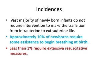
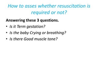
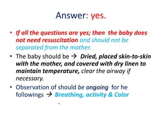
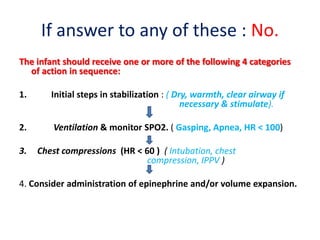
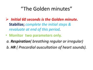
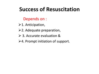
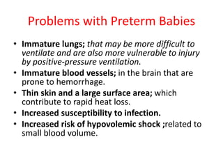
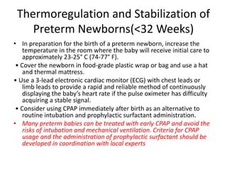
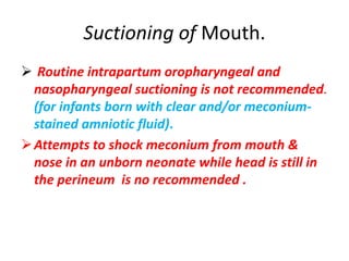
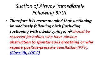
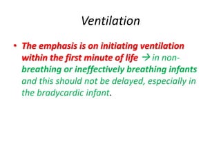
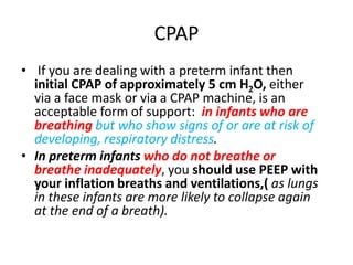
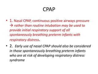
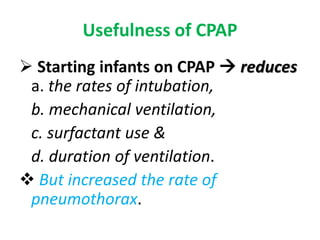
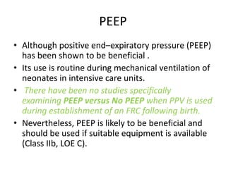
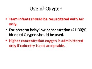
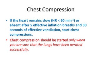
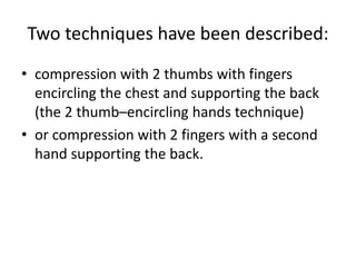
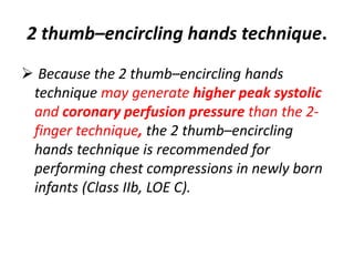
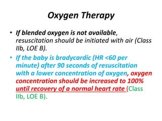
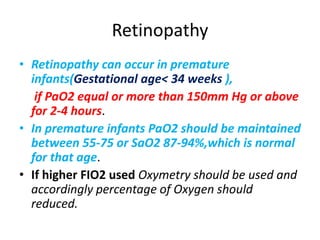
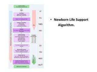
Neonatal Resuscitation.
Dr. P K Maharana.
Incidences
• Vast majority of newly born infants do not
require intervention to make the transition
from intrauterine to extrauterine life.
• Approximately 10% of newborns require
some assistance to begin breathing at birth.
• Less than 1% require extensive resuscitative
measures.
How to asses whether resuscitation is
required or not?
Answering these 3 questions.
• Is it Term gestation?
• Is the baby Crying or breathing?
• Is there Good muscle tone?
Answer: yes.
• If all the questions are yes; then the baby does
not need resuscitation and should not be
separated from the mother.
• The baby should be  Dried, placed skin-to-skin
with the mother, and covered with dry linen to
maintain temperature, clear the airway if
necessary.
• Observation of should be ongoing for he
followings  Breathing, activity & Color
.
If answer to any of these : No.
The infant should receive one or more of the following 4 categories
of action in sequence:
1. Initial steps in stabilization : ( Dry, warmth, clear airway if
necessary & stimulate).
2. Ventilation & monitor SPO2. ( Gasping, Apnea, HR < 100)
3. Chest compressions (HR < 60 ) ( Intubation, chest
compression, IPPV )
4. Consider administration of epinephrine and/or volume expansion.
“The Golden minutes”
 Initial 60 seconds is the Golden minute.
Stabilize; complete the initial steps &
revaluate at end of this period.
• Monitor two parameters only.
a. Respiration( breathing regular or irregular)
b. HR ( Precordial auscultation of heart sounds).
Success of Resuscitation
Depends on :
1. Anticipation,
2. Adequate preparation,
 3. Accurate evaluation &
4. Prompt initiation of support.
Problems with Preterm Babies
• Immature lungs; that may be more difficult to
ventilate and are also more vulnerable to injury
by positive-pressure ventilation.
• Immature blood vessels; in the brain that are
prone to hemorrhage.
• Thin skin and a large surface area; which
contribute to rapid heat loss.
• Increased susceptibility to infection.
• Increased risk of hypovolemic shock ;related to
small blood volume.
Thermoregulation and Stabilization of
Preterm Newborns(<32 Weeks)
• In preparation for the birth of a preterm newborn, increase the
temperature in the room where the baby will receive initial care to
approximately 23-25° C (74-77° F).
• Cover the newborn in food-grade plastic wrap or bag and use a hat
and thermal mattress.
• Use a 3-lead electronic cardiac monitor (ECG) with chest leads or
limb leads to provide a rapid and reliable method of continuously
displaying the baby’s heart rate if the pulse oximeter has difficulty
acquiring a stable signal.
• Consider using CPAP immediately after birth as an alternative to
routine intubation and prophylactic surfactant administration.
• Many preterm babies can be treated with early CPAP and avoid the
risks of intubation and mechanical ventilation. Criteria for CPAP
usage and the administration of prophylactic surfactant should be
developed in coordination with local experts
Suctioning of Mouth.
 Routine intrapartum oropharyngeal and
nasopharyngeal suctioning is not recommended.
(for infants born with clear and/or meconium-
stained amniotic fluid).
Attempts to shock meconium from mouth &
nose in an unborn neonate while head is still in
the perineum is no recommended .
Suction of Airway immediately
Following Birth.
• Therefore it is recommended that suctioning
immediately following birth (including
suctioning with a bulb syringe)  should be
reserved for babies who have obvious
obstruction to spontaneous breathing or who
require positive-pressure ventilation (PPV).
(Class IIb, LOE C)
When should consider Suctioning of
Oroppharynx.
• The presence of thick viscous meconium in a non-
vigorous infant is the only indication
for considering visualising the oropharynx and
suctioning material, which might obstruct the
airway.
• If an infant born through meconium-stained
amniotic fluid and is also floppy and makes no
immediate respiratory effort,  then it is
reasonable to rapidly inspect the oropharynx
with a view to removing any particulate matter
that might obstruct the airway.
Criteria to identify a healthy infant?
Heart Rate Tone Color Respiration
Healthy
Infant
120- 150 Good Blue Good
respiration
Unhealthy
infant
90-120 Less good tone Blue No good
respiration
Sick infant
( unwell)
< 100
not acceptable
Floppy Blue No respiration
Recent changes in the guidelines.
• Cord clamping is delayed up to 1 minute after
delivery of the neonate unless immediate
resuscitation is required.
• Milking of chord is no more recommended .
• The temperature is maintained between 36.5-
37.5 C.
• The emphasis should be on initiating lung
inflation within the first minute of life and
this should not be delayed  (in non-breathing
or ineffectively breathing infants ).
Cord Clamping
 1. Current evidence suggests that cord clamping
should be delayed for at least 30 to 60
seconds; for most vigorous term and
preterm newborns.
2. If the placental circulation is not intact the
cord should be clamped immediately after
birth; (such as after a placental abruption,
bleeding placenta previa, bleeding vasa
previa, or cord avulsion),
 There is insufficient evidence to recommend an
approach to cord clamping for newborns
who require resuscitation at birth.
Drying , Covering & Maintaining
Temperature
• In all cases; the term or near-term infants,
whether intervention is required or not; dry,
remove the wet towels, & cover the with dry
towels.
• Significantly preterm infants are best placed,
without drying, into polyethylene wrapping
under a radiant heater.
In infants of all gestations:  the head should be
covered with an appropriately sized hat.
Whatever the situation it is important that the
infant does not get cold.
How to maintain body Temperature
 Body temperature should be maintained between
36.5°C and 37.5°C after delivery. through the following
measures 
 Radiant warmer.
 Use of Plastic rap with a cap.
 Increased room temperature.
 Warmed humidified respiratory gases.
 Thermal mattress alone.
 A combination of increased room temperature with
plastic wrapping of head and body with thermal
mattress goes a long way.
Delivery Room Temperature.
 The delivery room temperature
should be at least 26°C for the most
immature infants.
Breath
• Most infants have a good heart rate
after birth and establish breathing by
about 90s.
• If the infant is not breathing
adequately aerate the lungs by giving 5
inflation breaths, preferably using air.
• Until now the infant's lungs will have
been filled with fluid.
Inflation breaths
Aeration of the lungs in these
circumstances is likely to require
sustained application of pressures
of about 30 cm H2O for 2–3 s.
Begin with lower pressures (20–25 cm
H2O) in preterm infants.
If the lungs have not been aerated
then consider:
1.Checking again that the infant’s head is in the neutral
position?
2. Is there a problem with face mask leak?
3. Do you need jaw thrust or a two-person approach to mask
inflation?
4. Do you need a longer inflation time? were the inspiratory
phases of your inflation breaths really of 2–3 s duration?
5. Is there an obstruction in the oropharynx ?(laryngoscope
and suction).
6. Will an oropharyngeal (Guedel’s airway) HELP?
7. Is there a tracheal obstruction?
Monitoring
• Respiration
• Heart rate
• Color
• Tone
• Reflexes
• Temperature
• Pulse Oxymetry
• Temperature
• ECG
Monitoring Heart Rate.
• During resuscitation of term and preterm
newborns, the use of 3-lead ECG for the rapid
and accurate measurement of the newborn’s
heart rate may be reasonable. (Class IIb, LOE
C-LD)
Methods to asses Heart Rate
• Auscultation of heart sound.
• Pulse Oximetry
• ECG
Preductal & Postductal Oximetry.
 SPO2%
 1 minute - 60-65%
• 2 minute – 65-70%
• 3 minute – 70-75%
• 4 minute – 75 80%
• 5 minute – 80-85%
• 10 minutes – 85-95%
Oxymetry in a full term vigorous
neonate at birth.
Ventilation
• The emphasis is on initiating ventilation
within the first minute of life  in non-
breathing or ineffectively breathing infants
and this should not be delayed, especially in
the bradycardic infant.
Adequacy of ventilation
• Assisted ventilation: 40 to 60 breaths per
minute to promptly, to achieve or maintain a
heart rate >100.
• An initial inflation pressure of 20 cm H2O may
be effective, but ≥30 to 40 cm H2O may be required in
some term babies without spontaneous ventilation (Class IIb,
LOE C).
• Inflation pressure should be monitored;
Effectiveness of ventilation.
 The primary measure of adequate initial
ventilation is prompt improvement in heart
rate.
Tracheal Intubation
Tracheal intubation should not be
routine.
It is only in the presence of thick
meconium, and is performed only for
suspected tracheal obstruction or
where IPPV is necessary.
Indications for Intubation
1. Meconium stained non vigorous newborn.
2. Bag mask ventilation is ineffective or for a
prolonged period.
3. Newborn without detectable HR.
4. Expected to need prolonged ventilation.
5. Special situations( Congenital diaphragmatic
hernia, Extreme low birth weight).
6. When chest compression is required.
Intubation
• In the absence of randomized, controlled trials,
there is insufficient evidence to recommend a
change in the current practice of;  Performing
endotracheal suctioning of nonvigorous babies
with meconium-stained amniotic fluid (Class IIb,
LOE C).
• However, if attempted intubation is prolonged
and unsuccessful,  bag-mask ventilation should
be considered, particularly if there is persistent
bradycardia.
Indicators of correct endotracheal
tube placement
After endotracheal intubation and administration of
intermittent positive pressure, a prompt increase in
heart rate is the best indicator that the tube is in
the tracheobronchial tree and providing effective
ventilation
 Moisture condensation in the endotracheal tube,
 Chest movement,
 Presence of equal breath sounds bilaterally,
 Capnography ( Exhaled Carbon dioxide ETCO2.)
 Other clinical indicators have not been systematically
evaluated in neonates (Class 11b, LOE C).
Size of ET tube
• A small leak should be resent between the ET
tube and tracheal wall when a breath is
delivered at a pressure of 25cm of water.
• 1 kg ------------2.5mm ID
• 1.5-2.5 kg ---- 3.0 mm ID
• 2.5 – 3.5 kg and above 3.5 mm ID
Ideal Length of Tube
• When a tube is placed the tip should 1- 2 cm
below the cords.
• 1 kg ----------- 7cm
• 2 kg ----------- 8cm
• 3 kg ------------ 9 cm
• 4 kg ------------- 10 cm
CPAP
• If you are dealing with a preterm infant then
initial CPAP of approximately 5 cm H2O, either
via a face mask or via a CPAP machine, is an
acceptable form of support: in infants who are
breathing but who show signs of or are at risk of
developing, respiratory distress.
• In preterm infants who do not breathe or
breathe inadequately, you should use PEEP with
your inflation breaths and ventilations,( as lungs
in these infants are more likely to collapse again
at the end of a breath).
CPAP
• 1. Nasal CPAP, continuous positive airways pressure
 rather than routine intubation may be used to
provide initial respiratory support of all
spontaneously breathing preterm infants with
respiratory distress.
• 2. Early use of nasal CPAP should also be considered
in those spontaneously breathing preterm infants
who are at risk of developing respiratory distress
syndrome
Usefulness of CPAP
 Starting infants on CPAP  reduces
a. the rates of intubation,
b. mechanical ventilation,
c. surfactant use &
d. duration of ventilation.
 But increased the rate of
pneumothorax.
PEEP
• Although positive end–expiratory pressure (PEEP)
has been shown to be beneficial .
• Its use is routine during mechanical ventilation of
neonates in intensive care units.
• There have been no studies specifically
examining PEEP versus No PEEP when PPV is used
during establishment of an FRC following birth.
• Nevertheless, PEEP is likely to be beneficial and
should be used if suitable equipment is available
(Class IIb, LOE C).
Use of Oxygen
• Term infants should be resuscitated with Air
only.
• For preterm baby low concentration (21-30)%
blended Oxygen should be used.
• Higher concentration oxygen is administered
only if oximetry is not acceptable.
Chest Compression
• If the heart remains slow (HR < 60 min-1) or
absent after 5 effective inflation breaths and 30
seconds of effective ventilation, start chest
compressions.
• Chest compression should be started only when
you are sure that the lungs have been aerated
successfully.
Two techniques have been described:
• compression with 2 thumbs with fingers
encircling the chest and supporting the back
(the 2 thumb–encircling hands technique)
• or compression with 2 fingers with a second
hand supporting the back.
2 thumb–encircling hands technique.
 Because the 2 thumb–encircling hands
technique may generate higher peak systolic
and coronary perfusion pressure than the 2-
finger technique, the 2 thumb–encircling
hands technique is recommended for
performing chest compressions in newly born
infants (Class IIb, LOE C).
Technique of 2 thumb–encircling
hands technique.
 In infants, the most efficient method of delivering chest
compression is to 
 Grip the chest in both hands in such a way that the two
thumbs can press on the lower third of the sternum,
just below an imaginary line joining the nipples, with
the fingers over the spine at the back.
 Compress the chest quickly and firmly, reducing the
antero-posterior diameter of the chest by about one
third.
 The ratio of compressions to inflations in new born
resuscitation is 3:1.
The 2-finger technique
• The 2-finger technique may be preferable
when access to the umbilicus is required
during insertion of an umbilical catheter,
although it is possible to administer the 2
thumb–encircling hands technique in
intubated infants with the rescuer standing at
the baby's head, thus permitting adequate
access to the umbilicus (Class IIb, LOE).
CPR(Compression: ventilation ratio)
• The recommended Compression: ventilation
ratio for CPR  remains at 3:1 for newborn
resuscitation.
• It should be synchronous, asynchronous
compressions are not recommended.
Compressions : Ventilations
• There is evidence from animals and non-
neonatal studies that sustained compressions
or a compression ratio of 15:2 or even 30:2
may be more effective when the arrest is of
primary cardiac etiology.
• One study in children suggests that CPR with
rescue breathing is preferable to chest
compressions alone when the arrest is of
noncardiac etiology
Important facts (Chest Compression)
• Intubation is strongly recommended prior to
beginning chest compressions.
• If intubation is not successful or not feasible, a
laryngeal mask may be used.
• Chest compressions are administered with the
two-thumb technique.
• Once the endotracheal tube or laryngeal mask is
secured, the compressor administers chest
compressions from the head of the newborn.
• Chest compressions continue for 60 seconds prior
to checking a heart rate.
Oxygen Requirement?
• Optimal management of oxygen during
neonatal resuscitation becomes particularly
important because of the evidence that either
insufficient or excessive oxygenation can be
harmful to the newborn infant.
 Hypoxia and ischemia are known to result in
injury to multiple organs.
 that adverse outcomes may result from even
brief exposure to excessive oxygen during and
following resuscitation.
How safe is the oxygen therapy?
• In healthy term infants, oxygen saturation increases
gradually from approximately 60% soon after birth to
over 90% at 10 min.
• In preterm infants hyperoxaemia is particularly
damaging and if oxygen is being used and the
saturation is above 95% the risk of hyperoxaemia is
high.
 Therefore the rate of rise in oxygen saturation after
birth in preterm infants should not exceed that seen in
term infants, although some supplemental oxygen may
be required to achieve this.
Oxygen Therapy
• If blended oxygen is not available,
resuscitation should be initiated with air (Class
IIb, LOE B).
• If the baby is bradycardic (HR <60 per
minute) after 90 seconds of resuscitation
with a lower concentration of oxygen, oxygen
concentration should be increased to 100%
until recovery of a normal heart rate (Class
IIb, LOE B).
Retinopathy
• Retinopathy can occur in premature
infants(Gestational age< 34 weeks ),
if PaO2 equal or more than 150mm Hg or above
for 2-4 hours.
• In premature infants PaO2 should be maintained
between 55-75 or SaO2 87-94%,which is normal
for that age.
• If higher FIO2 used Oxymetry should be used and
accordingly percentage of Oxygen should
reduced.
Apgar Score
Virginia Apgar.
Anesthesiologist at NewYork–Presbyterian Hospital
• Developed a score in 1952
to quantify the effects
of obstetric anesthesia on
babies.
• In 1953, she introduced the
first test, called the Apgar
score, to assess the health
of newborn babies.
Interpretation of scores
• The test is generally done: at 1 and 5 minutes
after birth and may be repeated later if the score
is and remains low.
Scores of 7 and above are normal;(nothing is
required)
 4 to 6, fairly low; ( responds to Vigorous
stimulation & blowing of Oxygen over face)
 3 and below are critically low
( immediate resuscitative efforts are recommended,
mostly responds to bag mask ventilation).
2 and below ( Vigorous resuscitation)
Implementation of scores.
 In cases where a newborn needs resuscitation, it should be
initiated before the Apgar score is assigned at the 1-minute
mark.
 Apgar score is not used to determine if initial resuscitation is
needed or not, rather it is used to determine if resuscitation
efforts should be continued.
 Variation between the 1-minute and 5-minute used to assess
an infant's response to resuscitation.
 If the score is below 7 at the 5-minute mark, then Apgar
score should be reassessed at 5-minute intervals for up to 20
minutes.
 To reduce the risk of negative outcomes, it is recommended
umbilical artery blood gas to be done when Apgar score of 5
or less at the 5-minute mark.
Limitations.
• There are numerous factors that contribute to the
Apgar Score, several of which are subjective, includes
but not limited to  color, tone, and reflex irritability.
• Preterm infants may receive a lower score in these
categories due to lack of maturity rather than
asphyxia.
• Other factors that may contribute to variability
among infants are  birth defects, sedation of the
mother during labor, gestational age or trauma.
• Inappropriately using the Apgar Score has led to
errors in diagnosing asphyxia.
Drugs
 The drugs used include:-
 1. Adrenaline
(1:10,000),
 2. Glucose (10%).
 3.Occasionally sodium
Bicarbonate (ideally 4.2%),
Drugs(Adrenaline )
The recommended intravenous dose for is 10
microgram kg-1 (0.1 mL kg-1 of 1:10,000
solution).
• If this is not effective, a dose of up to 30
microgram kg-1 (0.3 mL kg-1 of 1:10,000
solution) may be tried.
Drugs(Sodium bicarbonate)
 Is not recommended during brief resuscitation.
• If it is used during prolonged arrests unresponsive to
other therapy, it should be given only after adequate
ventilation and circulation (with chest compressions)
is established.
The dose for sodium bicarbonate is: 1 and 2 mmol of
bicarbonate kg-1 (2–4 mL kg-1 of 4.2% bicarbonate
solution). A diluted solution is used.
Drugs( Glucose)
• GLUCOSE : Intravenous
glucose infusion should
be considered as soon
as practical after
resuscitation, with the
goal of avoiding
hypoglycemia (Class IIb,
LOE C).
• The dose for glucose
10% : 2.5 mL kg-1 (250
mg kg-1.)
• (and should be considered if
there has been no response
to other drugs delivered
through a central venous
catheter).
Role of Glucose
• New born at increase risk of brain injury if
hypoglycemia.
• BG should be maintained at 2.5 mmol/kg or
more.
• Start with 0.5 -1ml/kg bolus, maintain 5-
7ml/kg/min.
Crystalloid Infusion
• Required very rarely : the heart rate cannot
increase because the infant has lost significant
blood volume.
• If this is the case, there is often a clear history of
blood loss from the infant, but not always.
• Use of isotonic crystalloid rather than albumin is
preferred for emergency volume replacement.
• In the presence of hypovolaemia: a bolus
of 10 mL kg-1 of 0.9% sodium chloride or similar
given over 10–20 s will often produce a rapid response
and can be repeated safely if needed.
Blood or Crystalloids
• If > 50% of Blood volume is lost( placenta
abrupted or placenta is transected).
• 10-20ml/kg transfusion restores arterial
pressure to normal.
• It should be titrated and slow infusion
10ml/kg over 10 minutes.
Therapeutic Hypothermia
• Infants born ≤ 36 weeks Gestations involving
with moderate to severe ischemic
encephalopathy at 6 hours of birth should be
offered therapeutic hypothermia.
• Temperature maintained between ( 33.5 –
34.5⁰C).
• Begins at 6hours & continues up to 72 hours.
• Rewarming should be slow, (at 0.2- 0.5 °C per
hour till 36.5⁰C).
Indications of Hypothermia
 1. Neonates ≥ 36 weeks gestational age and less than 6 hours of age
 2. Any one of the following:
• sentinel event prior to delivery, such as uterine rupture, profound fetal
bradycardia, or cord prolapse.
• low Apgar scores ➔ ≤ 5 at 10 minutes of life.
• prolonged resuscitation at birth ➔ chest compressions and/or
intubation and/or mask ventilation at 10 minutes.
• severe acidosis ➔ pH < 7.0 from cord or neonate blood gas
within 60 minutes of birth.
• abnormal base excess ➔ ≤ -16 mEq/L from cord gas or neonate blood
gas within 60 minutes of birth.
 3. Any one of the following: 
• clinical event concerning for seizure
• neonatal encephalopathy
Communication to Parents.
• It is important that the team caring for the
newborn baby informs the parents of the baby’s
progress.
• At delivery, adhere to the routine local plan and, if
possible, hand the baby to the mother at the
earliest opportunity.
• If resuscitation is required inform the parents of
the procedures undertaken and why they were
required.
• Record all discussions and decisions in the baby’s
records as soon as possible after birth.
When to stop resuscitation
 In a newly-born infant with no detectable
cardiac activity, and with cardiac activity that
remains undetectable for 10 min, it is
appropriate to consider stopping
resuscitation.
Decision to continue resuscitation
efforts beyond 10 min
• The decision to continue resuscitation efforts beyond
10 min with no cardiac activity is often complex and
may be influenced by issues such as:
• the availability of therapeutic hypothermia and
• intensive care facilities,
• the presumed aetiology of the arrest,
• the gestation of the infant,
• the presence or absence of complications,
• and the parents’ previous expressed feelings about
acceptable risk of morbidity.
Apnea
• Infant apnea is defined: by the American
Academy of Pediatrics as "an unexplained
episode of cessation of breathing for 20
seconds or longer, or a shorter respiratory
pause associated with bradycardia, cyanosis,
pallor, and/or marked hypotonia."
• Apnea is more common in preterm
infants.(Feb 13, 2018)
Primary Apnea.
• When a fetus or infant is deprived of oxygen, an
initial period of rapid breathing occurs.
• If the asphyxia continues, the respiratory
movements cease,  the heart rate begins to
fall, and the infant enters a period
of apnea known as primary apnea.
This is the first sign of oxygen deprivation, usually
related to labor and delivery events.
 When in primary apnea, the infant responds to -
o Tactile stimulation such as drying or slapping
the infant's feet o Free flow oxygen.
Primary Apnea
• A self-limited condition characterized by an absence of respiration.
• It may follow a blow to the head and is common
immediately after birth in the newborn who breathes spontaneousl
y when the carbon dioxide level in the circulation
reaches a certain value.
• Reflexes are present and the heart is beating, but the skin may be p
ale or blue and muscle tone is diminished.
• No treatment is necessary, but careful observation, maintenance of
body temperature, and oral pharyngeal
aspiration are usually performed.
• Within seconds the newborn usually begins breathing, becomes pin
ker, moves the arms and legs, and cries.
Secondary apnea
• Occurs when oxygen deprivation continues
following several gasps.
• Breathing stops.
• Bradycardia will progress to asystole.
• The BP falls as secondary apnea begins.
• When in secondary apnea –
o Tactile stimulation will not help.
Points to note
• If an infant begins breathing with stimulation, he is in
primary apnea.
• If an infant does not begin breathing with stimulation, he
has secondary apnea and will require positive-pressure
ventilation (PPV).
• The longer a baby remains in secondary apnea, the longer
it will take for spontaneous breathing to occur.
• Initiation of PPV results in rapid improvement in the
compromised infant--don't waste time continuing to
stimulate the infant.
• Knowledgeable and skilled caregivers capable of
responding to events surrounding birth should be present
at every delivery.
• Newborn Life Support
Algorithm.
Neonatal resuscitation

Neonatal resuscitation

  • 1.
  • 2.
    Incidences • Vast majorityof newly born infants do not require intervention to make the transition from intrauterine to extrauterine life. • Approximately 10% of newborns require some assistance to begin breathing at birth. • Less than 1% require extensive resuscitative measures.
  • 3.
    How to asseswhether resuscitation is required or not? Answering these 3 questions. • Is it Term gestation? • Is the baby Crying or breathing? • Is there Good muscle tone?
  • 4.
    Answer: yes. • Ifall the questions are yes; then the baby does not need resuscitation and should not be separated from the mother. • The baby should be  Dried, placed skin-to-skin with the mother, and covered with dry linen to maintain temperature, clear the airway if necessary. • Observation of should be ongoing for he followings  Breathing, activity & Color .
  • 5.
    If answer toany of these : No. The infant should receive one or more of the following 4 categories of action in sequence: 1. Initial steps in stabilization : ( Dry, warmth, clear airway if necessary & stimulate). 2. Ventilation & monitor SPO2. ( Gasping, Apnea, HR < 100) 3. Chest compressions (HR < 60 ) ( Intubation, chest compression, IPPV ) 4. Consider administration of epinephrine and/or volume expansion.
  • 6.
    “The Golden minutes” Initial 60 seconds is the Golden minute. Stabilize; complete the initial steps & revaluate at end of this period. • Monitor two parameters only. a. Respiration( breathing regular or irregular) b. HR ( Precordial auscultation of heart sounds).
  • 7.
    Success of Resuscitation Dependson : 1. Anticipation, 2. Adequate preparation,  3. Accurate evaluation & 4. Prompt initiation of support.
  • 8.
    Problems with PretermBabies • Immature lungs; that may be more difficult to ventilate and are also more vulnerable to injury by positive-pressure ventilation. • Immature blood vessels; in the brain that are prone to hemorrhage. • Thin skin and a large surface area; which contribute to rapid heat loss. • Increased susceptibility to infection. • Increased risk of hypovolemic shock ;related to small blood volume.
  • 9.
    Thermoregulation and Stabilizationof Preterm Newborns(<32 Weeks) • In preparation for the birth of a preterm newborn, increase the temperature in the room where the baby will receive initial care to approximately 23-25° C (74-77° F). • Cover the newborn in food-grade plastic wrap or bag and use a hat and thermal mattress. • Use a 3-lead electronic cardiac monitor (ECG) with chest leads or limb leads to provide a rapid and reliable method of continuously displaying the baby’s heart rate if the pulse oximeter has difficulty acquiring a stable signal. • Consider using CPAP immediately after birth as an alternative to routine intubation and prophylactic surfactant administration. • Many preterm babies can be treated with early CPAP and avoid the risks of intubation and mechanical ventilation. Criteria for CPAP usage and the administration of prophylactic surfactant should be developed in coordination with local experts
  • 10.
    Suctioning of Mouth. Routine intrapartum oropharyngeal and nasopharyngeal suctioning is not recommended. (for infants born with clear and/or meconium- stained amniotic fluid). Attempts to shock meconium from mouth & nose in an unborn neonate while head is still in the perineum is no recommended .
  • 11.
    Suction of Airwayimmediately Following Birth. • Therefore it is recommended that suctioning immediately following birth (including suctioning with a bulb syringe)  should be reserved for babies who have obvious obstruction to spontaneous breathing or who require positive-pressure ventilation (PPV). (Class IIb, LOE C)
  • 12.
    When should considerSuctioning of Oroppharynx. • The presence of thick viscous meconium in a non- vigorous infant is the only indication for considering visualising the oropharynx and suctioning material, which might obstruct the airway. • If an infant born through meconium-stained amniotic fluid and is also floppy and makes no immediate respiratory effort,  then it is reasonable to rapidly inspect the oropharynx with a view to removing any particulate matter that might obstruct the airway.
  • 13.
    Criteria to identifya healthy infant? Heart Rate Tone Color Respiration Healthy Infant 120- 150 Good Blue Good respiration Unhealthy infant 90-120 Less good tone Blue No good respiration Sick infant ( unwell) < 100 not acceptable Floppy Blue No respiration
  • 14.
    Recent changes inthe guidelines. • Cord clamping is delayed up to 1 minute after delivery of the neonate unless immediate resuscitation is required. • Milking of chord is no more recommended . • The temperature is maintained between 36.5- 37.5 C. • The emphasis should be on initiating lung inflation within the first minute of life and this should not be delayed  (in non-breathing or ineffectively breathing infants ).
  • 15.
    Cord Clamping  1.Current evidence suggests that cord clamping should be delayed for at least 30 to 60 seconds; for most vigorous term and preterm newborns. 2. If the placental circulation is not intact the cord should be clamped immediately after birth; (such as after a placental abruption, bleeding placenta previa, bleeding vasa previa, or cord avulsion),  There is insufficient evidence to recommend an approach to cord clamping for newborns who require resuscitation at birth.
  • 16.
    Drying , Covering& Maintaining Temperature • In all cases; the term or near-term infants, whether intervention is required or not; dry, remove the wet towels, & cover the with dry towels. • Significantly preterm infants are best placed, without drying, into polyethylene wrapping under a radiant heater. In infants of all gestations:  the head should be covered with an appropriately sized hat. Whatever the situation it is important that the infant does not get cold.
  • 17.
    How to maintainbody Temperature  Body temperature should be maintained between 36.5°C and 37.5°C after delivery. through the following measures   Radiant warmer.  Use of Plastic rap with a cap.  Increased room temperature.  Warmed humidified respiratory gases.  Thermal mattress alone.  A combination of increased room temperature with plastic wrapping of head and body with thermal mattress goes a long way.
  • 18.
    Delivery Room Temperature. The delivery room temperature should be at least 26°C for the most immature infants.
  • 19.
    Breath • Most infantshave a good heart rate after birth and establish breathing by about 90s. • If the infant is not breathing adequately aerate the lungs by giving 5 inflation breaths, preferably using air. • Until now the infant's lungs will have been filled with fluid.
  • 20.
    Inflation breaths Aeration ofthe lungs in these circumstances is likely to require sustained application of pressures of about 30 cm H2O for 2–3 s. Begin with lower pressures (20–25 cm H2O) in preterm infants.
  • 21.
    If the lungshave not been aerated then consider: 1.Checking again that the infant’s head is in the neutral position? 2. Is there a problem with face mask leak? 3. Do you need jaw thrust or a two-person approach to mask inflation? 4. Do you need a longer inflation time? were the inspiratory phases of your inflation breaths really of 2–3 s duration? 5. Is there an obstruction in the oropharynx ?(laryngoscope and suction). 6. Will an oropharyngeal (Guedel’s airway) HELP? 7. Is there a tracheal obstruction?
  • 22.
    Monitoring • Respiration • Heartrate • Color • Tone • Reflexes • Temperature • Pulse Oxymetry • Temperature • ECG
  • 23.
    Monitoring Heart Rate. •During resuscitation of term and preterm newborns, the use of 3-lead ECG for the rapid and accurate measurement of the newborn’s heart rate may be reasonable. (Class IIb, LOE C-LD)
  • 24.
    Methods to assesHeart Rate • Auscultation of heart sound. • Pulse Oximetry • ECG
  • 25.
  • 26.
     SPO2%  1minute - 60-65% • 2 minute – 65-70% • 3 minute – 70-75% • 4 minute – 75 80% • 5 minute – 80-85% • 10 minutes – 85-95% Oxymetry in a full term vigorous neonate at birth.
  • 27.
    Ventilation • The emphasisis on initiating ventilation within the first minute of life  in non- breathing or ineffectively breathing infants and this should not be delayed, especially in the bradycardic infant.
  • 28.
    Adequacy of ventilation •Assisted ventilation: 40 to 60 breaths per minute to promptly, to achieve or maintain a heart rate >100. • An initial inflation pressure of 20 cm H2O may be effective, but ≥30 to 40 cm H2O may be required in some term babies without spontaneous ventilation (Class IIb, LOE C). • Inflation pressure should be monitored;
  • 29.
    Effectiveness of ventilation. The primary measure of adequate initial ventilation is prompt improvement in heart rate.
  • 30.
    Tracheal Intubation Tracheal intubationshould not be routine. It is only in the presence of thick meconium, and is performed only for suspected tracheal obstruction or where IPPV is necessary.
  • 31.
    Indications for Intubation 1.Meconium stained non vigorous newborn. 2. Bag mask ventilation is ineffective or for a prolonged period. 3. Newborn without detectable HR. 4. Expected to need prolonged ventilation. 5. Special situations( Congenital diaphragmatic hernia, Extreme low birth weight). 6. When chest compression is required.
  • 32.
    Intubation • In theabsence of randomized, controlled trials, there is insufficient evidence to recommend a change in the current practice of;  Performing endotracheal suctioning of nonvigorous babies with meconium-stained amniotic fluid (Class IIb, LOE C). • However, if attempted intubation is prolonged and unsuccessful,  bag-mask ventilation should be considered, particularly if there is persistent bradycardia.
  • 33.
    Indicators of correctendotracheal tube placement After endotracheal intubation and administration of intermittent positive pressure, a prompt increase in heart rate is the best indicator that the tube is in the tracheobronchial tree and providing effective ventilation  Moisture condensation in the endotracheal tube,  Chest movement,  Presence of equal breath sounds bilaterally,  Capnography ( Exhaled Carbon dioxide ETCO2.)  Other clinical indicators have not been systematically evaluated in neonates (Class 11b, LOE C).
  • 34.
    Size of ETtube • A small leak should be resent between the ET tube and tracheal wall when a breath is delivered at a pressure of 25cm of water. • 1 kg ------------2.5mm ID • 1.5-2.5 kg ---- 3.0 mm ID • 2.5 – 3.5 kg and above 3.5 mm ID
  • 35.
    Ideal Length ofTube • When a tube is placed the tip should 1- 2 cm below the cords. • 1 kg ----------- 7cm • 2 kg ----------- 8cm • 3 kg ------------ 9 cm • 4 kg ------------- 10 cm
  • 36.
    CPAP • If youare dealing with a preterm infant then initial CPAP of approximately 5 cm H2O, either via a face mask or via a CPAP machine, is an acceptable form of support: in infants who are breathing but who show signs of or are at risk of developing, respiratory distress. • In preterm infants who do not breathe or breathe inadequately, you should use PEEP with your inflation breaths and ventilations,( as lungs in these infants are more likely to collapse again at the end of a breath).
  • 37.
    CPAP • 1. NasalCPAP, continuous positive airways pressure  rather than routine intubation may be used to provide initial respiratory support of all spontaneously breathing preterm infants with respiratory distress. • 2. Early use of nasal CPAP should also be considered in those spontaneously breathing preterm infants who are at risk of developing respiratory distress syndrome
  • 38.
    Usefulness of CPAP Starting infants on CPAP  reduces a. the rates of intubation, b. mechanical ventilation, c. surfactant use & d. duration of ventilation.  But increased the rate of pneumothorax.
  • 39.
    PEEP • Although positiveend–expiratory pressure (PEEP) has been shown to be beneficial . • Its use is routine during mechanical ventilation of neonates in intensive care units. • There have been no studies specifically examining PEEP versus No PEEP when PPV is used during establishment of an FRC following birth. • Nevertheless, PEEP is likely to be beneficial and should be used if suitable equipment is available (Class IIb, LOE C).
  • 40.
    Use of Oxygen •Term infants should be resuscitated with Air only. • For preterm baby low concentration (21-30)% blended Oxygen should be used. • Higher concentration oxygen is administered only if oximetry is not acceptable.
  • 41.
    Chest Compression • Ifthe heart remains slow (HR < 60 min-1) or absent after 5 effective inflation breaths and 30 seconds of effective ventilation, start chest compressions. • Chest compression should be started only when you are sure that the lungs have been aerated successfully.
  • 42.
    Two techniques havebeen described: • compression with 2 thumbs with fingers encircling the chest and supporting the back (the 2 thumb–encircling hands technique) • or compression with 2 fingers with a second hand supporting the back.
  • 43.
    2 thumb–encircling handstechnique.  Because the 2 thumb–encircling hands technique may generate higher peak systolic and coronary perfusion pressure than the 2- finger technique, the 2 thumb–encircling hands technique is recommended for performing chest compressions in newly born infants (Class IIb, LOE C).
  • 44.
    Technique of 2thumb–encircling hands technique.  In infants, the most efficient method of delivering chest compression is to   Grip the chest in both hands in such a way that the two thumbs can press on the lower third of the sternum, just below an imaginary line joining the nipples, with the fingers over the spine at the back.  Compress the chest quickly and firmly, reducing the antero-posterior diameter of the chest by about one third.  The ratio of compressions to inflations in new born resuscitation is 3:1.
  • 46.
    The 2-finger technique •The 2-finger technique may be preferable when access to the umbilicus is required during insertion of an umbilical catheter, although it is possible to administer the 2 thumb–encircling hands technique in intubated infants with the rescuer standing at the baby's head, thus permitting adequate access to the umbilicus (Class IIb, LOE).
  • 47.
    CPR(Compression: ventilation ratio) •The recommended Compression: ventilation ratio for CPR  remains at 3:1 for newborn resuscitation. • It should be synchronous, asynchronous compressions are not recommended.
  • 48.
    Compressions : Ventilations •There is evidence from animals and non- neonatal studies that sustained compressions or a compression ratio of 15:2 or even 30:2 may be more effective when the arrest is of primary cardiac etiology. • One study in children suggests that CPR with rescue breathing is preferable to chest compressions alone when the arrest is of noncardiac etiology
  • 49.
    Important facts (ChestCompression) • Intubation is strongly recommended prior to beginning chest compressions. • If intubation is not successful or not feasible, a laryngeal mask may be used. • Chest compressions are administered with the two-thumb technique. • Once the endotracheal tube or laryngeal mask is secured, the compressor administers chest compressions from the head of the newborn. • Chest compressions continue for 60 seconds prior to checking a heart rate.
  • 50.
    Oxygen Requirement? • Optimalmanagement of oxygen during neonatal resuscitation becomes particularly important because of the evidence that either insufficient or excessive oxygenation can be harmful to the newborn infant.  Hypoxia and ischemia are known to result in injury to multiple organs.  that adverse outcomes may result from even brief exposure to excessive oxygen during and following resuscitation.
  • 51.
    How safe isthe oxygen therapy? • In healthy term infants, oxygen saturation increases gradually from approximately 60% soon after birth to over 90% at 10 min. • In preterm infants hyperoxaemia is particularly damaging and if oxygen is being used and the saturation is above 95% the risk of hyperoxaemia is high.  Therefore the rate of rise in oxygen saturation after birth in preterm infants should not exceed that seen in term infants, although some supplemental oxygen may be required to achieve this.
  • 52.
    Oxygen Therapy • Ifblended oxygen is not available, resuscitation should be initiated with air (Class IIb, LOE B). • If the baby is bradycardic (HR <60 per minute) after 90 seconds of resuscitation with a lower concentration of oxygen, oxygen concentration should be increased to 100% until recovery of a normal heart rate (Class IIb, LOE B).
  • 53.
    Retinopathy • Retinopathy canoccur in premature infants(Gestational age< 34 weeks ), if PaO2 equal or more than 150mm Hg or above for 2-4 hours. • In premature infants PaO2 should be maintained between 55-75 or SaO2 87-94%,which is normal for that age. • If higher FIO2 used Oxymetry should be used and accordingly percentage of Oxygen should reduced.
  • 54.
    Apgar Score Virginia Apgar. Anesthesiologistat NewYork–Presbyterian Hospital • Developed a score in 1952 to quantify the effects of obstetric anesthesia on babies. • In 1953, she introduced the first test, called the Apgar score, to assess the health of newborn babies.
  • 56.
    Interpretation of scores •The test is generally done: at 1 and 5 minutes after birth and may be repeated later if the score is and remains low. Scores of 7 and above are normal;(nothing is required)  4 to 6, fairly low; ( responds to Vigorous stimulation & blowing of Oxygen over face)  3 and below are critically low ( immediate resuscitative efforts are recommended, mostly responds to bag mask ventilation). 2 and below ( Vigorous resuscitation)
  • 61.
    Implementation of scores. In cases where a newborn needs resuscitation, it should be initiated before the Apgar score is assigned at the 1-minute mark.  Apgar score is not used to determine if initial resuscitation is needed or not, rather it is used to determine if resuscitation efforts should be continued.  Variation between the 1-minute and 5-minute used to assess an infant's response to resuscitation.  If the score is below 7 at the 5-minute mark, then Apgar score should be reassessed at 5-minute intervals for up to 20 minutes.  To reduce the risk of negative outcomes, it is recommended umbilical artery blood gas to be done when Apgar score of 5 or less at the 5-minute mark.
  • 62.
    Limitations. • There arenumerous factors that contribute to the Apgar Score, several of which are subjective, includes but not limited to  color, tone, and reflex irritability. • Preterm infants may receive a lower score in these categories due to lack of maturity rather than asphyxia. • Other factors that may contribute to variability among infants are  birth defects, sedation of the mother during labor, gestational age or trauma. • Inappropriately using the Apgar Score has led to errors in diagnosing asphyxia.
  • 63.
    Drugs  The drugsused include:-  1. Adrenaline (1:10,000),  2. Glucose (10%).  3.Occasionally sodium Bicarbonate (ideally 4.2%),
  • 64.
    Drugs(Adrenaline ) The recommendedintravenous dose for is 10 microgram kg-1 (0.1 mL kg-1 of 1:10,000 solution). • If this is not effective, a dose of up to 30 microgram kg-1 (0.3 mL kg-1 of 1:10,000 solution) may be tried.
  • 65.
    Drugs(Sodium bicarbonate)  Isnot recommended during brief resuscitation. • If it is used during prolonged arrests unresponsive to other therapy, it should be given only after adequate ventilation and circulation (with chest compressions) is established. The dose for sodium bicarbonate is: 1 and 2 mmol of bicarbonate kg-1 (2–4 mL kg-1 of 4.2% bicarbonate solution). A diluted solution is used.
  • 66.
    Drugs( Glucose) • GLUCOSE: Intravenous glucose infusion should be considered as soon as practical after resuscitation, with the goal of avoiding hypoglycemia (Class IIb, LOE C). • The dose for glucose 10% : 2.5 mL kg-1 (250 mg kg-1.) • (and should be considered if there has been no response to other drugs delivered through a central venous catheter).
  • 67.
    Role of Glucose •New born at increase risk of brain injury if hypoglycemia. • BG should be maintained at 2.5 mmol/kg or more. • Start with 0.5 -1ml/kg bolus, maintain 5- 7ml/kg/min.
  • 68.
    Crystalloid Infusion • Requiredvery rarely : the heart rate cannot increase because the infant has lost significant blood volume. • If this is the case, there is often a clear history of blood loss from the infant, but not always. • Use of isotonic crystalloid rather than albumin is preferred for emergency volume replacement. • In the presence of hypovolaemia: a bolus of 10 mL kg-1 of 0.9% sodium chloride or similar given over 10–20 s will often produce a rapid response and can be repeated safely if needed.
  • 69.
    Blood or Crystalloids •If > 50% of Blood volume is lost( placenta abrupted or placenta is transected). • 10-20ml/kg transfusion restores arterial pressure to normal. • It should be titrated and slow infusion 10ml/kg over 10 minutes.
  • 70.
    Therapeutic Hypothermia • Infantsborn ≤ 36 weeks Gestations involving with moderate to severe ischemic encephalopathy at 6 hours of birth should be offered therapeutic hypothermia. • Temperature maintained between ( 33.5 – 34.5⁰C). • Begins at 6hours & continues up to 72 hours. • Rewarming should be slow, (at 0.2- 0.5 °C per hour till 36.5⁰C).
  • 71.
    Indications of Hypothermia 1. Neonates ≥ 36 weeks gestational age and less than 6 hours of age  2. Any one of the following: • sentinel event prior to delivery, such as uterine rupture, profound fetal bradycardia, or cord prolapse. • low Apgar scores ➔ ≤ 5 at 10 minutes of life. • prolonged resuscitation at birth ➔ chest compressions and/or intubation and/or mask ventilation at 10 minutes. • severe acidosis ➔ pH < 7.0 from cord or neonate blood gas within 60 minutes of birth. • abnormal base excess ➔ ≤ -16 mEq/L from cord gas or neonate blood gas within 60 minutes of birth.  3. Any one of the following:  • clinical event concerning for seizure • neonatal encephalopathy
  • 72.
    Communication to Parents. •It is important that the team caring for the newborn baby informs the parents of the baby’s progress. • At delivery, adhere to the routine local plan and, if possible, hand the baby to the mother at the earliest opportunity. • If resuscitation is required inform the parents of the procedures undertaken and why they were required. • Record all discussions and decisions in the baby’s records as soon as possible after birth.
  • 73.
    When to stopresuscitation  In a newly-born infant with no detectable cardiac activity, and with cardiac activity that remains undetectable for 10 min, it is appropriate to consider stopping resuscitation.
  • 74.
    Decision to continueresuscitation efforts beyond 10 min • The decision to continue resuscitation efforts beyond 10 min with no cardiac activity is often complex and may be influenced by issues such as: • the availability of therapeutic hypothermia and • intensive care facilities, • the presumed aetiology of the arrest, • the gestation of the infant, • the presence or absence of complications, • and the parents’ previous expressed feelings about acceptable risk of morbidity.
  • 75.
    Apnea • Infant apneais defined: by the American Academy of Pediatrics as "an unexplained episode of cessation of breathing for 20 seconds or longer, or a shorter respiratory pause associated with bradycardia, cyanosis, pallor, and/or marked hypotonia." • Apnea is more common in preterm infants.(Feb 13, 2018)
  • 76.
    Primary Apnea. • Whena fetus or infant is deprived of oxygen, an initial period of rapid breathing occurs. • If the asphyxia continues, the respiratory movements cease,  the heart rate begins to fall, and the infant enters a period of apnea known as primary apnea. This is the first sign of oxygen deprivation, usually related to labor and delivery events.  When in primary apnea, the infant responds to - o Tactile stimulation such as drying or slapping the infant's feet o Free flow oxygen.
  • 77.
    Primary Apnea • Aself-limited condition characterized by an absence of respiration. • It may follow a blow to the head and is common immediately after birth in the newborn who breathes spontaneousl y when the carbon dioxide level in the circulation reaches a certain value. • Reflexes are present and the heart is beating, but the skin may be p ale or blue and muscle tone is diminished. • No treatment is necessary, but careful observation, maintenance of body temperature, and oral pharyngeal aspiration are usually performed. • Within seconds the newborn usually begins breathing, becomes pin ker, moves the arms and legs, and cries.
  • 78.
    Secondary apnea • Occurswhen oxygen deprivation continues following several gasps. • Breathing stops. • Bradycardia will progress to asystole. • The BP falls as secondary apnea begins. • When in secondary apnea – o Tactile stimulation will not help.
  • 79.
    Points to note •If an infant begins breathing with stimulation, he is in primary apnea. • If an infant does not begin breathing with stimulation, he has secondary apnea and will require positive-pressure ventilation (PPV). • The longer a baby remains in secondary apnea, the longer it will take for spontaneous breathing to occur. • Initiation of PPV results in rapid improvement in the compromised infant--don't waste time continuing to stimulate the infant. • Knowledgeable and skilled caregivers capable of responding to events surrounding birth should be present at every delivery.
  • 80.
    • Newborn LifeSupport Algorithm.

Editor's Notes

  • #4 Pre term <37 weeks Term between 37-39 weeks.
  • #8 All these are critical for successful neonatal resuscitation
  • #11 There is no evidence to support suctioning of the mouth and nose of infants born through clear amniotic fluid. A multicenteric, multinational randomized clinical trial provided evidence that newborns born through meconium-stained amniotic fluid who were vigorous at birth did not benefit from intervention and could avoid the risk of intubation.
  • #12  Deterioration of pulmonary compliance, oxygenation, and cerebral blood flow velocity shown to accompany tracheal suction in intubated infants in the neonatal intensive care unit also suggests the need for caution in the use of suction immediately after birth.103–
  • #15 The importance of achieving this has been highlighted and reinforced because of the strong association with mortality and morbidity. Even the mild hypothermia that was once felt to be inevitable and therefore clinically acceptable carries a risk. The admission temperature should be recorded as a predictor of outcomes as well as a quality indicator.
  • #18 The temperature must be actively maintained between 36.5°C and 37.5°C after birth unless a decision has been taken to start therapeutic hypothermia. The admission temperature should always be recorded as a predictor of outcomes as well as a quality indicator. Hyperthermia >38 Celsius is harmful.
  • #20  If the heart rate does not increase, and the chest does not passively move with each inflation breath, then you have not aerated the lungs. 
  • #23  A note should be made of the colour and tone, although these are of lesser importance in determining the immediate approach to be taken they can point towards the severely acidaemic baby (potentially requiring substantial resuscitation) or anaemic baby (potentially requiring urgent transfusion). 
  • #24 The use of ECG does not replace the need for pulse oximetry to evaluate the newborn’s oxygenation.
  • #42 Because ventilation is the most effective action in neonatal resuscitation and because chest compressions are likely to compete with effective ventilation, rescuers should ensure that assisted ventilation is being delivered optimally before starting chest compressions
  • #45 Do not use asynchronous compressions, even if the infant has a tracheal tube placed, as maintaining air entry into the lung remains as important now as it was during the initial aeration. Compressing the chest during a ventilation breath may reduce air entry, which may be harmful. 
  • #48 ( as respiratory failure is the primary cause)
  • #50 To determine tip-to-lip depth of the endotracheal tube after insertion, use the endotracheal tube initial insertion depth table or measure the nasal-tragus length (NTL). •
  • #51 Conversely there is growing experimental evidence, as well as evidence from studies of babies receiving resuscitation
  • #55  Apgar scoring remains the accepted method of assessment and is endorsed by both the American College of Obstetricians and Gynecologists and the American Academy of Pediatrics
  • #56 Maximum Score 10 Minimum 0.
  • #62 A systematic review that analyzed the relationship between umbilical cord pH and neonatal outcomes found that low cord pH is strongly correlated with mortality, morbidity and cerebral palsy in childhood.
  • #64 All resuscitation drugs are best delivered via an umbilical venous catheter or if this is not possible through an intraosseous needle
  • #67 Higher IV doses are not recommended because animal86,87 and pediatric88,89 studies show exaggerated hypertension, decreased myocardial function, and worse neurological function after administration
  • #71 There is now compelling clinical evidence that mild induced hypothermia significantly improves survival and disability, including cerebral palsy and neuro-cognitive outcomes, Infants should have serial neurological examinations during and at the end of cooling and at discharge.
  • #75 The difficulty of this decision-making emphasises the need for senior help to be sought as soon as possible
  • #77 It is difficult to differentiate between primary and secondary apnea. Bradycardia develops at the same time the infant enters primary apnea. BP is maintained.