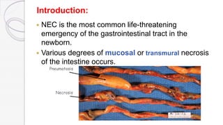
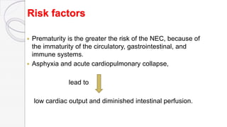
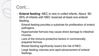
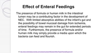
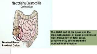
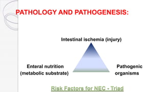
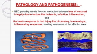
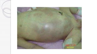
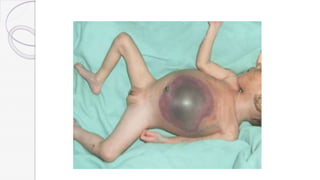
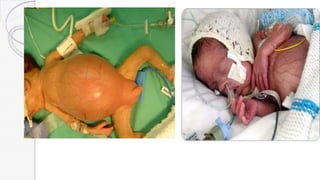
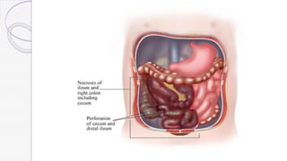
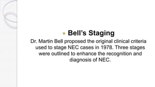
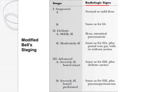
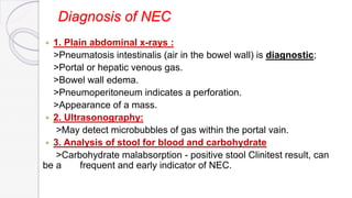
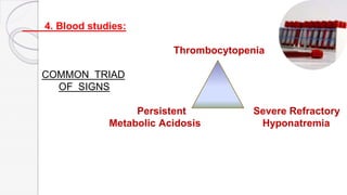
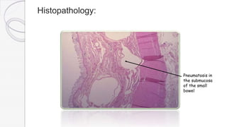
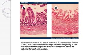
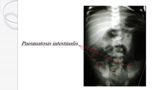
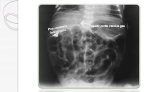
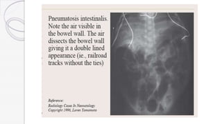
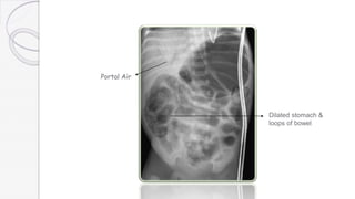
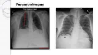
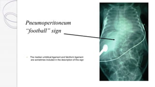
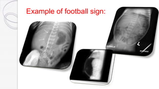
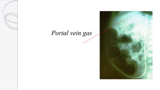
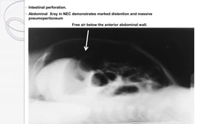
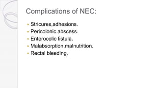
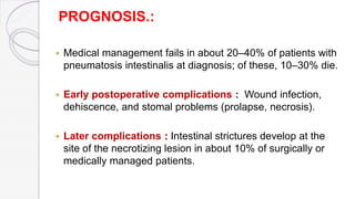
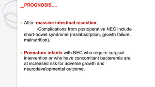
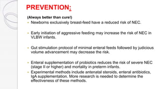
morning
and
Welcom
e to
our
morning
Neonatal Necrotizing Enterocolitis
Dr.Sumona chowdhury keya
Assistant register
Department of Pediatrics
SWMCH,Sylhet.
Introduction:
 NEC is the most common life-threatening
emergency of the gastrointestinal tract in the
newborn.
 Various degrees of mucosal or transmural necrosis
of the intestine occurs.
Pneumatosis
Necrosis
Incidence:
 The incidence of NEC is 1% to 7% of all admitted neonats
in NICU or 1 to 3 per 1,000 live births.
 It occurs equally in males and females. Sex, race, geography, climate
has no role in determining the incidence of NEC
 The mortality rate is 11.5 to 12.3 per 100,000 infant deaths.
 In 2001, the National Institute of Child Health and Human
Development (NICHD) Neonatal Research Network reported (from a
data set of 14 centers) an overall incidence of proven NEC of 7% in
VLBW .
Birth weight
Incidence, Fatality
Gestational age
(less than 28 weeks of gestation)
Ref…http://www.medscape.com/viewarticle/458205_2
Cont…
 The incidence of NEC varies from different NICUs in the
same country. Research by the National Institute of Child
Health and Human Development observed variations from
4 to 20% in the prevalence of NEC at centres across
northern America, suggesting that iatrogenic factors may
play a large role.
ref..
http://www.hindawi.com/journals/isrn/2012/628317/
Risk factors
 Prematurity is the greater the risk of the NEC, because of
the immaturity of the circulatory, gastrointestinal, and
immune systems.
 Asphyxia and acute cardiopulmonary collapse,
lead to
low cardiac output and diminished intestinal perfusion.
Cont…
 Enteral feeding: NEC is rare in unfed infants. About 90-
95% of infants with NEC received at least one enteral
feeding.
◦ Enteral feeding provides a substrate for proliferation of enteric
pathogens.
◦ Hyperosmolar formula may cause direct damage to intestinal
mucosa.
◦ Lack of the immuno-protective factors in commercially
prepared formula.
◦ Breast-feeding significantly lowers the risk of NEC .
◦ Large feeding volumes and rapid advancement of enteral
feedings.
Cont…
 Enteric pathogenic organisms comprising bacterial
and viral pathogens. These include E.coli,
Klebsiella, salmonella, rotaviruses, and
enteroviruses.
 Maternal cocaine use during pregnancy may result
in vasoconstriction and GI ischemia from
cocaine's -adrenergic action.
Cont..
In Term Babies,
the disease can occur in full-term neonates. NEC in term
infants is often a “secondary” disease, seen more frequently
in infants with
> History of birth asphyxia,
>Down syndrome,
>Congenital heart disease,
>Rotavirus infections, and
>Hirschsprung disease.
Effect of Enteral Feedings
The presence of formula or human milk in the intestinal
lumen may be a contributing factor in the development of
NEC. With limited absorptive abilities of the infant's gut and
the possibility of bowel mucosal damage from ischemia,
enteral feedings may remain in the gut for extended periods
of time. Furthermore, the presence of formula and/or
human milk may simply provide a media upon which the
bacteria can feed and flourish.
ref..http://www.motherbabyuniversity.com/outreach/outreach/Modules/GINec/Pages/ParentalSupport.htm
Terminal ileum/
Proximal Colon
The distal part of the ileum and the
proximal segment of colon are involved
most frequently; in fatal cases,
gangrene may extend from the
stomach to the rectum.
PATHOLOGY AND PATHOGENESIS:
Intestinal ischemia (injury)
Enteral nutrition Pathogenic
(metabolic substrate) organisms
PATHOLOGY AND PATHOGENESIS: ..
 NEC probably results from an interaction between loss of mucosal
integrity due to factors like ischemia, infection, inflammation,
and
the host's response to that injury like circulatory, immunologic,
inflammatory responses resulting in necrosis of the affected area.
Cont…
 Various bacterial and viral agents, including
Escherichia coli, Klebsiella, Clostridium perfringens,
Staphylococcus epidermidis, and rotavirus, have
been recovered from culture.
 Coagulation necrosis is the characteristic
histologic finding of intestinal specimens.
Cont…
These factors may contribute to the development of a
necrotic segment of intestine,
gas accumulation in the submucosa of the bowel wall
(pneumatosis intestinalis),
and progression of the necrosis to perforation, peritonitis,
sepsis, and death.
Why the Preterm Gut is Different
 Decreased IgA
 Decreased Intestinal T-lymphocytes
 Poor Antibody response
 Higher Membrane Permeability of GI epithelial
lining
 Lower Gastric motility
 More scant and more permeable Mucin Blanket
Intestinal Archetecture
small slice of an
enormous organ that has
multiple layers of complexity…
necrosis of right colon and ileum from a neonate with NEC
CLINICAL MANIFESTATIONS:
 Onset is insidious or sudden catastrophic onset .The onset of NEC is
usually in the 2nd or 3rd week of life but can be as late as 3 months in
VLBW infants. Age of onset is inversely related to gestational age.
CLINICAL MANIFESTATIONS:
 The 1st signs of impending disease may be
-Nonspecific including lethargy and temperature
instability
or
-Related to gastrointestinal pathology such as
abdominal
distention and gastric retention.
 Obvious bloody stools are seen in 25% of patients.
 The spectrum of illness is broad and ranges from
-Mild disease with only guaiac-positive stools
to
-Severe illness with bowel perforation, peritonitis,
Cont..
 Bell’s Staging
Dr. Martin Bell proposed the original clinical criteria
used to stage NEC cases in 1978. Three stages
were outlined to enhance the recognition and
diagnosis of NEC.
Based on:
1. Systemic Signs
2. Intestinal Signs
3. Radiological Signs
Classified into:
I. Suspected:
II. Definite :
A (Mildly ill) ,
B (Moderately ill)
III. Advanced:
A (Severely ill,bowel intact),
B (Severely ill,bowel perforated)
Modified
Bell’s
Staging
Modified
Bell’s
Staging
Modified
Bell’s
Staging
Diagnosis of NEC
 1. Plain abdominal x-rays :
>Pneumatosis intestinalis (air in the bowel wall) is diagnostic;
>Portal or hepatic venous gas.
>Bowel wall edema.
>Pneumoperitoneum indicates a perforation.
>Appearance of a mass.
 2. Ultrasonography:
>May detect microbubbles of gas within the portal vain.
 3. Analysis of stool for blood and carbohydrate
>Carbohydrate malabsorption - positive stool Clinitest result, can
be a frequent and early indicator of NEC.
4. Blood studies:
Thrombocytopenia
COMMON TRIAD
OF SIGNS
Persistent Severe Refractory
Metabolic Acidosis Hyponatremia
Histopathology:
Pneumatosis in
the submucosa
of the small
bowel
Microscopic images of (A) normal bowel and (B) characteristic findings
of NEC, which illustrates hemorrhagic necrosis, beginning in the
mucosa and extending to the muscular bowel wall, where the
potential for perforation exists.
Portal Air
Dilated stomach &
loops of bowel
Pneumoperitoneum
Pneumoperitoneum
“football” sign
 The median umbilical ligament and falciform ligament
are sometimes included in the description of this sign
Example of football sign:
Portal vein gas
◦ Intestinal perforation.
◦ Abdominal Xray in NEC demonstrates marked distention and massive
pneumoperitoneum
Free air below the anterior abdominal wall.
 3 principles of management:
1.Place gut at rest.
2.Prevent continuing injury.
3.Correct or modify the systemic response.
Treatment:
Cont…
 1.Gut rest:NPO until the condition is stable.
 2.GI decompression through NG tube for suction
and free drainage to stomach.
 3.Broad spectrum antibiotics for 10-14 days.
>Ampicillin
>Gentamycin
>Metronidazol.
Cont..
4.Monitoring:
 Vital signs.
 Serial abdominal x-rays to detect intestinal perforation;
 Serial determination of hematologic status,
 Serial determination of electrolyte status, and
 Serial determination of acid-base status.
Cont…..
 5.Parenteral nutrition should be maintain.Afterthat,feeds
should be started slowly with EBM or with hypoallergenic
formula feeds.i.e. lactose free soya based formulas as
lactose and cow milk protien intolerance are often seen in
recovery phase.
 6.Treatment of shock,acidosis,hyponatremia,
thrombocytopenia or DIC.
 7.Surgical intervention.
Indications for surgery :
Absolute indications:
 Evidence of perforation on abdominal roentgenograms
(pneumoperitoneum) or
 Positive abdominal paracentesis (stool or organism on Gram stain
from peritoneal fluid).
Relative indications:
 Failure of medical management,
 Single fixed bowel loop on roentgenograms,
 Abdominal wall erythema, or
 A palpable mass.
 Portal vein gas.
Cont..
 Ideally, surgery should be performed after intestinal
necrosis develops, but before perforation and peritonitis
occurs.
 Peritoneal drainage may be helpful for patients with
peritonitis who are too unstable to undergo surgery.
Peritoneal drainage is more successful in patients with
isolated intestinal perforation.
Complications of NEC:
 Stricures,adhesions.
 Pericolonic abscess.
 Enterocolic fistula.
 Malabsorption,malnutrition.
 Rectal bleeding.
PROGNOSIS.:
 Medical management fails in about 20–40% of patients with
pneumatosis intestinalis at diagnosis; of these, 10–30% die.
 Early postoperative complications : Wound infection,
dehiscence, and stomal problems (prolapse, necrosis).
 Later complications : Intestinal strictures develop at the
site of the necrotizing lesion in about 10% of surgically or
medically managed patients.
PROGNOSIS….
 After massive intestinal resection,
-Complications from postoperative NEC include
short-bowel syndrome (malabsorption, growth failure,
malnutrition),
 Premature infants with NEC who require surgical
intervention or who have concomitant bacteremia are
at increased risk for adverse growth and
neurodevelopmental outcome.
PREVENTION:
(Always better than cure!)
 Newborns exclusively breast-feed have a reduced risk of NEC.
 Early initiation of aggressive feeding may increase the risk of NEC in
VLBW infants.
 Gut stimulation protocol of minimal enteral feeds followed by judicious
volume advancement may decrease the risk.
 Enteral supplementation of probiotics reduces the risk of severe NEC
(stage II or higher) and mortality in preterm infants.
 Experimental methods include antenatal steroids, enteral antibiotics,
IgA supplementation. More research is needed to determine the
effectiveness of these methods.
References:
 Nelson Textbook of Pediatrics.
(19th Edition)
 Concise Textbook of Pediatrics.
(Dr.M.R.Molla)
 Internet
http://www.ncbi.nlm.nih.gov/pmc/articles/PMC2709096/
emedicine.medscape.com/article/977956-overview
https://en.wikipedia.org/wiki/Necrotizing_enterocolitis
http://www.ncbi.nlm.nih.gov/pubmed/18547133
..http://www.motherbabyuniversity.com/outreach/outreach/Modules/GINec/Pages/ParentalSupport.htm
http://www.hindawi.com/journals/isrn/2012/628317/
a

Neonatal Necrotizing Enterocolitis

  • 1.
  • 2.
    Neonatal Necrotizing Enterocolitis Dr.Sumonachowdhury keya Assistant register Department of Pediatrics SWMCH,Sylhet.
  • 3.
    Introduction:  NEC isthe most common life-threatening emergency of the gastrointestinal tract in the newborn.  Various degrees of mucosal or transmural necrosis of the intestine occurs. Pneumatosis Necrosis
  • 4.
    Incidence:  The incidenceof NEC is 1% to 7% of all admitted neonats in NICU or 1 to 3 per 1,000 live births.  It occurs equally in males and females. Sex, race, geography, climate has no role in determining the incidence of NEC  The mortality rate is 11.5 to 12.3 per 100,000 infant deaths.  In 2001, the National Institute of Child Health and Human Development (NICHD) Neonatal Research Network reported (from a data set of 14 centers) an overall incidence of proven NEC of 7% in VLBW . Birth weight Incidence, Fatality Gestational age (less than 28 weeks of gestation) Ref…http://www.medscape.com/viewarticle/458205_2
  • 5.
    Cont…  The incidenceof NEC varies from different NICUs in the same country. Research by the National Institute of Child Health and Human Development observed variations from 4 to 20% in the prevalence of NEC at centres across northern America, suggesting that iatrogenic factors may play a large role. ref.. http://www.hindawi.com/journals/isrn/2012/628317/
  • 6.
    Risk factors  Prematurityis the greater the risk of the NEC, because of the immaturity of the circulatory, gastrointestinal, and immune systems.  Asphyxia and acute cardiopulmonary collapse, lead to low cardiac output and diminished intestinal perfusion.
  • 7.
    Cont…  Enteral feeding:NEC is rare in unfed infants. About 90- 95% of infants with NEC received at least one enteral feeding. ◦ Enteral feeding provides a substrate for proliferation of enteric pathogens. ◦ Hyperosmolar formula may cause direct damage to intestinal mucosa. ◦ Lack of the immuno-protective factors in commercially prepared formula. ◦ Breast-feeding significantly lowers the risk of NEC . ◦ Large feeding volumes and rapid advancement of enteral feedings.
  • 8.
    Cont…  Enteric pathogenicorganisms comprising bacterial and viral pathogens. These include E.coli, Klebsiella, salmonella, rotaviruses, and enteroviruses.  Maternal cocaine use during pregnancy may result in vasoconstriction and GI ischemia from cocaine's -adrenergic action.
  • 9.
    Cont.. In Term Babies, thedisease can occur in full-term neonates. NEC in term infants is often a “secondary” disease, seen more frequently in infants with > History of birth asphyxia, >Down syndrome, >Congenital heart disease, >Rotavirus infections, and >Hirschsprung disease.
  • 10.
    Effect of EnteralFeedings The presence of formula or human milk in the intestinal lumen may be a contributing factor in the development of NEC. With limited absorptive abilities of the infant's gut and the possibility of bowel mucosal damage from ischemia, enteral feedings may remain in the gut for extended periods of time. Furthermore, the presence of formula and/or human milk may simply provide a media upon which the bacteria can feed and flourish. ref..http://www.motherbabyuniversity.com/outreach/outreach/Modules/GINec/Pages/ParentalSupport.htm
  • 11.
    Terminal ileum/ Proximal Colon Thedistal part of the ileum and the proximal segment of colon are involved most frequently; in fatal cases, gangrene may extend from the stomach to the rectum.
  • 12.
    PATHOLOGY AND PATHOGENESIS: Intestinalischemia (injury) Enteral nutrition Pathogenic (metabolic substrate) organisms
  • 13.
    PATHOLOGY AND PATHOGENESIS:..  NEC probably results from an interaction between loss of mucosal integrity due to factors like ischemia, infection, inflammation, and the host's response to that injury like circulatory, immunologic, inflammatory responses resulting in necrosis of the affected area.
  • 14.
    Cont…  Various bacterialand viral agents, including Escherichia coli, Klebsiella, Clostridium perfringens, Staphylococcus epidermidis, and rotavirus, have been recovered from culture.  Coagulation necrosis is the characteristic histologic finding of intestinal specimens.
  • 15.
    Cont… These factors maycontribute to the development of a necrotic segment of intestine, gas accumulation in the submucosa of the bowel wall (pneumatosis intestinalis), and progression of the necrosis to perforation, peritonitis, sepsis, and death.
  • 16.
    Why the PretermGut is Different  Decreased IgA  Decreased Intestinal T-lymphocytes  Poor Antibody response  Higher Membrane Permeability of GI epithelial lining  Lower Gastric motility  More scant and more permeable Mucin Blanket
  • 17.
    Intestinal Archetecture small sliceof an enormous organ that has multiple layers of complexity…
  • 18.
    necrosis of rightcolon and ileum from a neonate with NEC
  • 19.
    CLINICAL MANIFESTATIONS:  Onsetis insidious or sudden catastrophic onset .The onset of NEC is usually in the 2nd or 3rd week of life but can be as late as 3 months in VLBW infants. Age of onset is inversely related to gestational age.
  • 20.
    CLINICAL MANIFESTATIONS:  The1st signs of impending disease may be -Nonspecific including lethargy and temperature instability or -Related to gastrointestinal pathology such as abdominal distention and gastric retention.  Obvious bloody stools are seen in 25% of patients.  The spectrum of illness is broad and ranges from -Mild disease with only guaiac-positive stools to -Severe illness with bowel perforation, peritonitis,
  • 21.
  • 26.
     Bell’s Staging Dr.Martin Bell proposed the original clinical criteria used to stage NEC cases in 1978. Three stages were outlined to enhance the recognition and diagnosis of NEC.
  • 27.
    Based on: 1. SystemicSigns 2. Intestinal Signs 3. Radiological Signs Classified into: I. Suspected: II. Definite : A (Mildly ill) , B (Moderately ill) III. Advanced: A (Severely ill,bowel intact), B (Severely ill,bowel perforated)
  • 28.
  • 29.
  • 30.
  • 31.
    Diagnosis of NEC 1. Plain abdominal x-rays : >Pneumatosis intestinalis (air in the bowel wall) is diagnostic; >Portal or hepatic venous gas. >Bowel wall edema. >Pneumoperitoneum indicates a perforation. >Appearance of a mass.  2. Ultrasonography: >May detect microbubbles of gas within the portal vain.  3. Analysis of stool for blood and carbohydrate >Carbohydrate malabsorption - positive stool Clinitest result, can be a frequent and early indicator of NEC.
  • 32.
    4. Blood studies: Thrombocytopenia COMMONTRIAD OF SIGNS Persistent Severe Refractory Metabolic Acidosis Hyponatremia
  • 33.
  • 34.
    Microscopic images of(A) normal bowel and (B) characteristic findings of NEC, which illustrates hemorrhagic necrosis, beginning in the mucosa and extending to the muscular bowel wall, where the potential for perforation exists.
  • 38.
  • 39.
  • 40.
    Pneumoperitoneum “football” sign  Themedian umbilical ligament and falciform ligament are sometimes included in the description of this sign
  • 41.
  • 42.
  • 43.
    ◦ Intestinal perforation. ◦Abdominal Xray in NEC demonstrates marked distention and massive pneumoperitoneum Free air below the anterior abdominal wall.
  • 44.
     3 principlesof management: 1.Place gut at rest. 2.Prevent continuing injury. 3.Correct or modify the systemic response. Treatment:
  • 45.
    Cont…  1.Gut rest:NPOuntil the condition is stable.  2.GI decompression through NG tube for suction and free drainage to stomach.  3.Broad spectrum antibiotics for 10-14 days. >Ampicillin >Gentamycin >Metronidazol.
  • 46.
    Cont.. 4.Monitoring:  Vital signs. Serial abdominal x-rays to detect intestinal perforation;  Serial determination of hematologic status,  Serial determination of electrolyte status, and  Serial determination of acid-base status.
  • 47.
    Cont…..  5.Parenteral nutritionshould be maintain.Afterthat,feeds should be started slowly with EBM or with hypoallergenic formula feeds.i.e. lactose free soya based formulas as lactose and cow milk protien intolerance are often seen in recovery phase.  6.Treatment of shock,acidosis,hyponatremia, thrombocytopenia or DIC.  7.Surgical intervention.
  • 48.
    Indications for surgery: Absolute indications:  Evidence of perforation on abdominal roentgenograms (pneumoperitoneum) or  Positive abdominal paracentesis (stool or organism on Gram stain from peritoneal fluid). Relative indications:  Failure of medical management,  Single fixed bowel loop on roentgenograms,  Abdominal wall erythema, or  A palpable mass.  Portal vein gas.
  • 49.
    Cont..  Ideally, surgeryshould be performed after intestinal necrosis develops, but before perforation and peritonitis occurs.  Peritoneal drainage may be helpful for patients with peritonitis who are too unstable to undergo surgery. Peritoneal drainage is more successful in patients with isolated intestinal perforation.
  • 50.
    Complications of NEC: Stricures,adhesions.  Pericolonic abscess.  Enterocolic fistula.  Malabsorption,malnutrition.  Rectal bleeding.
  • 51.
    PROGNOSIS.:  Medical managementfails in about 20–40% of patients with pneumatosis intestinalis at diagnosis; of these, 10–30% die.  Early postoperative complications : Wound infection, dehiscence, and stomal problems (prolapse, necrosis).  Later complications : Intestinal strictures develop at the site of the necrotizing lesion in about 10% of surgically or medically managed patients.
  • 52.
    PROGNOSIS….  After massiveintestinal resection, -Complications from postoperative NEC include short-bowel syndrome (malabsorption, growth failure, malnutrition),  Premature infants with NEC who require surgical intervention or who have concomitant bacteremia are at increased risk for adverse growth and neurodevelopmental outcome.
  • 53.
    PREVENTION: (Always better thancure!)  Newborns exclusively breast-feed have a reduced risk of NEC.  Early initiation of aggressive feeding may increase the risk of NEC in VLBW infants.  Gut stimulation protocol of minimal enteral feeds followed by judicious volume advancement may decrease the risk.  Enteral supplementation of probiotics reduces the risk of severe NEC (stage II or higher) and mortality in preterm infants.  Experimental methods include antenatal steroids, enteral antibiotics, IgA supplementation. More research is needed to determine the effectiveness of these methods.
  • 54.
    References:  Nelson Textbookof Pediatrics. (19th Edition)  Concise Textbook of Pediatrics. (Dr.M.R.Molla)  Internet http://www.ncbi.nlm.nih.gov/pmc/articles/PMC2709096/ emedicine.medscape.com/article/977956-overview https://en.wikipedia.org/wiki/Necrotizing_enterocolitis http://www.ncbi.nlm.nih.gov/pubmed/18547133 ..http://www.motherbabyuniversity.com/outreach/outreach/Modules/GINec/Pages/ParentalSupport.htm http://www.hindawi.com/journals/isrn/2012/628317/
  • 55.

Editor's Notes

  • #5 NEC is predominantly a disorder of preterm infants, with an incidence of 6-10% in infants weighing <1.5 kg. The incidence increases with decreasing gestational age. Seventy to 90% of cases occur in high-risk low birth weight infants, whereas 10-25% occur in full-term newborns. Infants with NEC represent 2-5% of neonatal intensive care unit (NICU) admissions. (clinical manual ) It has been estimated that 90% of cases occur in premature infants and that NEC may develop in 1% to 10% of infants hospitalized in neonatal intensive care units (84). Significant intercenter differences in the prevalence of NEC have been reported (79). The mortality rates vary from 10% to 50%. The age of onset of NEC is related to birth weight and gestational age. Smaller, more immature infants (less than 28 weeks of gestation) tend to have NEC at an older age than larger, more mature (greater than 31 weeks of age) infants . ( avery )