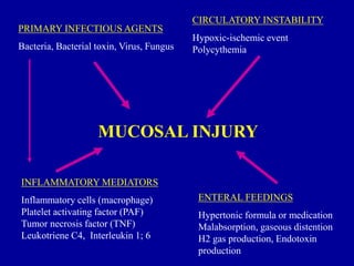
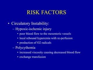
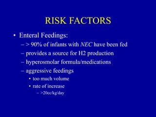
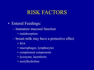
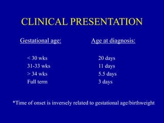
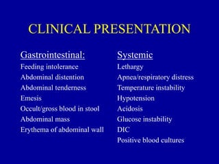
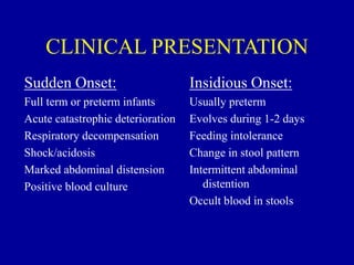
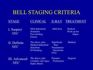
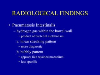
NECROTIZING
ENTEROCOLITIS
Janice Nicklay Catalan M.D.
OBJECTIVES
• Ability to diagnose and treat the signs and
symptoms of NEC
• Ability to evaluate radiographs for the
classic findings of NEC
• List several long-term complications
associated with NEC
NECROTIZING
ENTEROCOLITIS
• Epidemiology:
– most commonly occurring gastrointestinal
emergency in preterm infants
– leading cause of emergency surgery in neonates
– overall incidence: 1-5% in most NICU’s
– most common in VLBW preterm infants
• 10% of all cases occur in term infants
NECROTIZING
ENTEROCOLITIS
• Epidemiology:
– 10x more likely to occur in infants who have
been fed
– males = females
– blacks > whites
– mortality rate: 25-30%
– 50% of survivors experience long-term
sequelae
NECROTIZING
ENTEROCOLITIS
• Pathology:
– most commonly involved areas: terminal ileum
and proximal colon
– GROSS:
• bowel appears irregularly dilated with hemorrhagic
or ischemic areas of frank necrosis
– focal or diffuse
– MICROSCOPIC:
• mucosal edema, hemorrhage and ulceration
NECROTIZING
ENTEROCOLITIS
• MICROSCOPIC:
– minimal inflammation during the acute phase
• increases during revascularization
– granulation tissue and fibrosis develop
• stricture formation
– microthrombi in mesenteric arterioles and
venules
NECROTIZING
ENTEROCOLITIS
• Pathophysiology:
UNKNOWN
CAUSE…….
PRIMARY INFECTIOUS AGENTS
Bacteria, Bacterial toxin, Virus, Fungus
CIRCULATORY INSTABILITY
Hypoxic-ischemic event
Polycythemia
MUCOSAL INJURY
ENTERAL FEEDINGS
Hypertonic formula or medication
Malabsorption, gaseous distention
H2 gas production, Endotoxin
production
INFLAMMATORY MEDIATORS
Inflammatory cells (macrophage)
Platelet activating factor (PAF)
Tumor necrosis factor (TNF)
Leukotriene C4, Interleukin 1; 6
RISK FACTORS
• Prematurity:
* primary risk factor
– 90% of cases are premature infants
– immature gastrointestinal system
• mucosal barrier
• poor motility
– immature immune response
– impaired circulatory dynamics
RISK FACTORS
• Infectious Agents:
– usually occurs in clustered epidemics
– normal intestinal flora
• E. coli
• Klebsiella spp.
• Pseudomonas spp.
• Clostridium difficile
• Staph. Epi
• Viruses
RISK FACTORS
• Inflammatory Mediators:
– involved in the development of intestinal injury
and systemic side effects
• neutropenia, thrombocytopenia, acidosis,
hypotension
– primary factors
• Tumor necrosis factor (TNF)
• Platelet activating factor (PAF)
• LTC4
• Interleukin 1& 6
RISK FACTORS
• Circulatory Instability:
– Hypoxic-ischemic injury
• poor blood flow to the mesenteric vessels
• local rebound hyperemia with re-perfusion
• production of O2 radicals
– Polycythemia
• increased viscosity causing decreased blood flow
• exchange transfusion
RISK FACTORS
• Enteral Feedings:
– > 90% of infants with NEC have been fed
– provides a source for H2 production
– hyperosmolar formula/medications
– aggressive feedings
• too much volume
• rate of increase
– >20cc/kg/day
RISK FACTORS
• Enteral Feedings:
– immature mucosal function
• malabsorption
– breast milk may have a protective effect
• IGA
• macrophages, lymphocytes
• complement components
• lysozyme, lactoferrin
• acetylhydrolase
CLINICAL PRESENTATION
Gestational age:
< 30 wks
31-33 wks
> 34 wks
Full term
Age at diagnosis:
20 days
11 days
5.5 days
3 days
*Time of onset is inversely related to gestational age/birthweight
CLINICAL PRESENTATION
Gastrointestinal:
Feeding intolerance
Abdominal distention
Abdominal tenderness
Emesis
Occult/gross blood in stool
Abdominal mass
Erythema of abdominal wall
Systemic
Lethargy
Apnea/respiratory distress
Temperature instability
Hypotension
Acidosis
Glucose instability
DIC
Positive blood cultures
CLINICAL PRESENTATION
Sudden Onset:
Full term or preterm infants
Acute catastrophic deterioration
Respiratory decompensation
Shock/acidosis
Marked abdominal distension
Positive blood culture
Insidious Onset:
Usually preterm
Evolves during 1-2 days
Feeding intolerance
Change in stool pattern
Intermittent abdominal
distention
Occult blood in stools
BELL STAGING CRITERIA
STAGE CLINICAL X-RAY TREATMENT
I. Suspect
NEC
Mild abdominal
distention
Poor feeding
Emesis
Mild ileus Medical
Work up for
Sepsis
II. Definite
NEC
The above, plus
Marked abdominal
distention
GI bleeding
Significant
Ileus
Pneumatosis
Intestinalis
PVG
Medical
III. Advanced
NEC
The above, plus
Unstable vital signs
Septic Shock
Pneumo-
Peritoneum
Surgical
RADIOLOGICAL FINDINGS
• Pneumatosis Intestinalis
– hydrogen gas within the bowel wall
• product of bacterial metabolism
a. linear streaking pattern
• more diagnostic
b. bubbly pattern
• appears like retained meconium
• less specific
RADIOLOGICAL FINDINGS
• Portal Venous Gas
– extension of pneumatosis intestinalis into the
portal venous circulation
• linear branching lucencies overlying the liver and
extending to the periphery
• associated with severe disease and high mortality
RADIOLOGICAL FINDINGS
• Pneumoperitoneum
– free air in the peritoneal cavity secondary to
perforation
• falciform ligament may be outlined
– “football” sign
– surgical emergency
LABORATORY FINDINGS
• CBC
– neutropenia/elevated WBC
– thrombocytopenia
• Acidosis
– metabolic
• Hyperkalemia
– increased secondary to release from necrotic
tissue
LABORATORY FINDINGS
• DIC
• Positive cultures
– blood
– CSF
– urine
– stool
TREATMENT
• Stop enteral feeds
– re-start or increase IVF
• Nasogastric decompression
– low intermittent suction
• Antibiotics
– Amp/Gent; Vanc/Cefotaxime
– Clindamycin
• suspected or proven perforation
TREATMENT
• Surgical Consult
– suspected or proven NEC
– indications for surgery:
• portal venous gas; pneumoperitoneum
• clinical deterioration
– despite medical management
• positive paracentesis
• fixed intestinal loop on serial x-rays
• erythema of abdominal wall
TREATMENT
• Labs: q6-8hrs
– CBC, electrolytes, DIC panel, blood gases
• X-rays: q6-8hrs
– AP, left lateral decubitus or cross-table lateral
• Supportive Therapy
– fluids, blood products, pressors, mechanical
ventilation
PROGNOSIS
• Depends on the severity of the illness
• Associated with late complications
* strictures
– short-gut syndrome
– malabsorption
– fistulas
– abscess
* MOST COMMON

2. NEC.ppt

  • 1.
  • 2.
    OBJECTIVES • Ability todiagnose and treat the signs and symptoms of NEC • Ability to evaluate radiographs for the classic findings of NEC • List several long-term complications associated with NEC
  • 3.
    NECROTIZING ENTEROCOLITIS • Epidemiology: – mostcommonly occurring gastrointestinal emergency in preterm infants – leading cause of emergency surgery in neonates – overall incidence: 1-5% in most NICU’s – most common in VLBW preterm infants • 10% of all cases occur in term infants
  • 4.
    NECROTIZING ENTEROCOLITIS • Epidemiology: – 10xmore likely to occur in infants who have been fed – males = females – blacks > whites – mortality rate: 25-30% – 50% of survivors experience long-term sequelae
  • 5.
    NECROTIZING ENTEROCOLITIS • Pathology: – mostcommonly involved areas: terminal ileum and proximal colon – GROSS: • bowel appears irregularly dilated with hemorrhagic or ischemic areas of frank necrosis – focal or diffuse – MICROSCOPIC: • mucosal edema, hemorrhage and ulceration
  • 6.
    NECROTIZING ENTEROCOLITIS • MICROSCOPIC: – minimalinflammation during the acute phase • increases during revascularization – granulation tissue and fibrosis develop • stricture formation – microthrombi in mesenteric arterioles and venules
  • 7.
  • 8.
    PRIMARY INFECTIOUS AGENTS Bacteria,Bacterial toxin, Virus, Fungus CIRCULATORY INSTABILITY Hypoxic-ischemic event Polycythemia MUCOSAL INJURY ENTERAL FEEDINGS Hypertonic formula or medication Malabsorption, gaseous distention H2 gas production, Endotoxin production INFLAMMATORY MEDIATORS Inflammatory cells (macrophage) Platelet activating factor (PAF) Tumor necrosis factor (TNF) Leukotriene C4, Interleukin 1; 6
  • 9.
    RISK FACTORS • Prematurity: *primary risk factor – 90% of cases are premature infants – immature gastrointestinal system • mucosal barrier • poor motility – immature immune response – impaired circulatory dynamics
  • 10.
    RISK FACTORS • InfectiousAgents: – usually occurs in clustered epidemics – normal intestinal flora • E. coli • Klebsiella spp. • Pseudomonas spp. • Clostridium difficile • Staph. Epi • Viruses
  • 11.
    RISK FACTORS • InflammatoryMediators: – involved in the development of intestinal injury and systemic side effects • neutropenia, thrombocytopenia, acidosis, hypotension – primary factors • Tumor necrosis factor (TNF) • Platelet activating factor (PAF) • LTC4 • Interleukin 1& 6
  • 12.
    RISK FACTORS • CirculatoryInstability: – Hypoxic-ischemic injury • poor blood flow to the mesenteric vessels • local rebound hyperemia with re-perfusion • production of O2 radicals – Polycythemia • increased viscosity causing decreased blood flow • exchange transfusion
  • 13.
    RISK FACTORS • EnteralFeedings: – > 90% of infants with NEC have been fed – provides a source for H2 production – hyperosmolar formula/medications – aggressive feedings • too much volume • rate of increase – >20cc/kg/day
  • 14.
    RISK FACTORS • EnteralFeedings: – immature mucosal function • malabsorption – breast milk may have a protective effect • IGA • macrophages, lymphocytes • complement components • lysozyme, lactoferrin • acetylhydrolase
  • 15.
    CLINICAL PRESENTATION Gestational age: <30 wks 31-33 wks > 34 wks Full term Age at diagnosis: 20 days 11 days 5.5 days 3 days *Time of onset is inversely related to gestational age/birthweight
  • 16.
    CLINICAL PRESENTATION Gastrointestinal: Feeding intolerance Abdominaldistention Abdominal tenderness Emesis Occult/gross blood in stool Abdominal mass Erythema of abdominal wall Systemic Lethargy Apnea/respiratory distress Temperature instability Hypotension Acidosis Glucose instability DIC Positive blood cultures
  • 17.
    CLINICAL PRESENTATION Sudden Onset: Fullterm or preterm infants Acute catastrophic deterioration Respiratory decompensation Shock/acidosis Marked abdominal distension Positive blood culture Insidious Onset: Usually preterm Evolves during 1-2 days Feeding intolerance Change in stool pattern Intermittent abdominal distention Occult blood in stools
  • 18.
    BELL STAGING CRITERIA STAGECLINICAL X-RAY TREATMENT I. Suspect NEC Mild abdominal distention Poor feeding Emesis Mild ileus Medical Work up for Sepsis II. Definite NEC The above, plus Marked abdominal distention GI bleeding Significant Ileus Pneumatosis Intestinalis PVG Medical III. Advanced NEC The above, plus Unstable vital signs Septic Shock Pneumo- Peritoneum Surgical
  • 19.
    RADIOLOGICAL FINDINGS • PneumatosisIntestinalis – hydrogen gas within the bowel wall • product of bacterial metabolism a. linear streaking pattern • more diagnostic b. bubbly pattern • appears like retained meconium • less specific
  • 20.
    RADIOLOGICAL FINDINGS • PortalVenous Gas – extension of pneumatosis intestinalis into the portal venous circulation • linear branching lucencies overlying the liver and extending to the periphery • associated with severe disease and high mortality
  • 21.
    RADIOLOGICAL FINDINGS • Pneumoperitoneum –free air in the peritoneal cavity secondary to perforation • falciform ligament may be outlined – “football” sign – surgical emergency
  • 22.
    LABORATORY FINDINGS • CBC –neutropenia/elevated WBC – thrombocytopenia • Acidosis – metabolic • Hyperkalemia – increased secondary to release from necrotic tissue
  • 23.
    LABORATORY FINDINGS • DIC •Positive cultures – blood – CSF – urine – stool
  • 24.
    TREATMENT • Stop enteralfeeds – re-start or increase IVF • Nasogastric decompression – low intermittent suction • Antibiotics – Amp/Gent; Vanc/Cefotaxime – Clindamycin • suspected or proven perforation
  • 25.
    TREATMENT • Surgical Consult –suspected or proven NEC – indications for surgery: • portal venous gas; pneumoperitoneum • clinical deterioration – despite medical management • positive paracentesis • fixed intestinal loop on serial x-rays • erythema of abdominal wall
  • 26.
    TREATMENT • Labs: q6-8hrs –CBC, electrolytes, DIC panel, blood gases • X-rays: q6-8hrs – AP, left lateral decubitus or cross-table lateral • Supportive Therapy – fluids, blood products, pressors, mechanical ventilation
  • 27.
    PROGNOSIS • Depends onthe severity of the illness • Associated with late complications * strictures – short-gut syndrome – malabsorption – fistulas – abscess * MOST COMMON