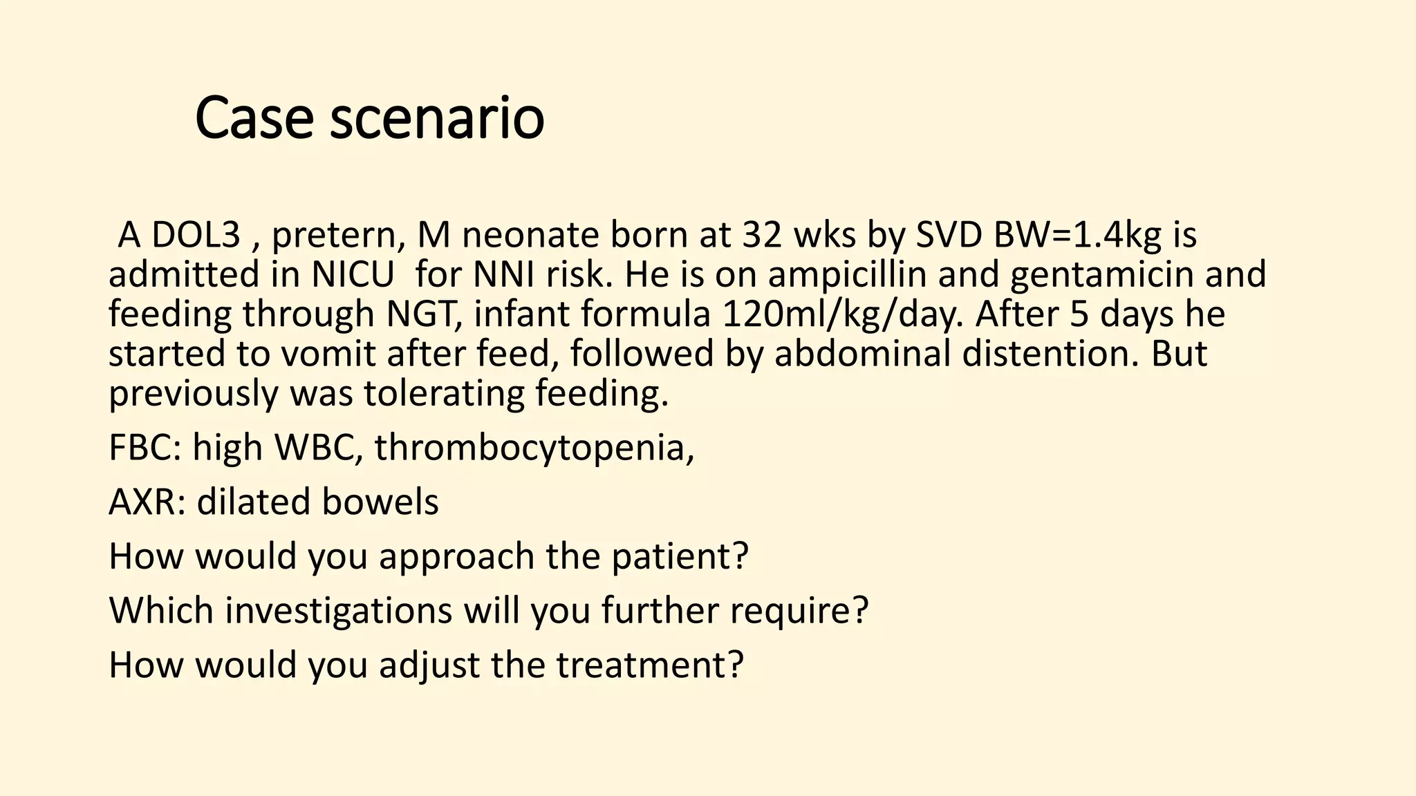
1) A premature neonate presented with vomiting after feeds and abdominal distention after previously tolerating feeding. Examination found high white blood cell count, thrombocytopenia, and dilated bowels on x-ray.
2) Necrotizing enterocolitis is a disease of premature infants characterized by ischemic necrosis of the intestinal mucosa caused by immature gut and immune system, enteral nutrition, and bacterial overgrowth. Risk factors include prematurity, formula feeding, and low birth weight.
3) Treatment involves bowel rest, antibiotics, intravenous fluids and nutrition. Surgical intervention with resection may be needed for perforation or failure to improve. Outcomes depend on severity but include short bowel syndrome,
this presentation includes anatomy physiology function of peritoneum ,also includes cause of peritonitis its severity ,various scoring system investigation and treatment.It includes the recent advancement and latest articles from latest books of surgery.
Tuberculosis (TB) is a life threatening disease which can virtually affect any organ system.
Abdominal tuberculosis is a most common type of extra-pulmonary tuberculosis, comprising of tuberculosis of gastrointestinal tract, peritoneum, omentum, mysentery and its lymph nodes and other abdominal organs such as liver, spleen and pancreas.
Abdominal tuberculosis can occur primarily or it can be secondary to a tubercular focus elsewhere in the body.
Gastrointestinal tuberculosis occurring due to ingestion of milk or food infected with Mycobacterium bovis can result in primary intestinal tuberculosis, but it is now-a days rare.
Firstly, the tubercle bacilli may enter the intestinal tract through the ingestion of infected milk or sputum. The mucosal layer of the GI tract can be infected with the bacilli with formation of epithelioid tubercles in the lymphoid tissue of the submucosa.
After 2-4 weeks, caseous necrosis of the tubercles leads to ulceration of the overlying mucosa which can later spread into the deeper layers and into the adjacent lymph nodes and into peritoneum.
Information about Abdominal sepsis and peritonitis final by Dr Dhaval Mangukiya.
Details of Anatomy, intra abdominal infections, physiology, peritonitis, risks for failure of source control, management of critical issues.
https://drdhavalmangukiya.com/
http://www.youtube.com/c/DrDhavalMangukiyaGastrosurgeonSurat
https://gastrosurgerysurat.blogspot.com/
Management of Typhoid Intestinal Perforation which is a common and the most dreaded surgical complication of Typhoid fever.
This menace is still on the rise in low and medium income countries where we still battle with lack of potable water and open defecation.
This presentation is especially targeted at trainee surgeons in Nigeria and Medical Students also who may find it worthwhile.
Necrotizing enterocolitis (NEC) is a life-threatening emergency of the gastrointestinal tract in the newborn period.
The most common gastrointestinal condition in premature neonates.
It is characterized by inflammation, ischemia, and permeability of the neonatal bowel wall to bacteria.
It is potentially life-threatening with significant associated morbidity.
The disease is characterized by various degrees of mucosal or transmural necrosis of the intestine.
Neonatal necrotizing enterocolitis
NEC is the most common life-threatening emergency of the gastrointestinal tract in the newborn period. The disease is characterized by various degrees of mucosal or transmural necrosis of the intestine. The cause of NEC remains unclear but is most likely multifactorial. The incidence of NEC is 1-5% of infants in neonatal intensive care units (NICUs). Both incidence and case fatality rates increase with decreasing birthweight and gestational age. Because very small, ill preterm infants are particularly susceptible to NEC, a rising incidence may reflect improved survival of this high-risk group of patients.
Clinical Manifestations
Infants with NEC have a variety of signs and symptoms and may have an insidious or sudden catastrophic onset (Table 96-1). The onset of NEC is usually in the 2nd or 3rd week of life but can be as late as 3 mo in VLBW infants. Age of onset is inversely related to gestational age. The 1st signs of impending disease may be nonspecific, including lethargy and temperature instability, or related to gastrointestinal pathology, such as abdominal distention and gastric retention. Obvious bloody stools are seen in 25% of patients. Because of nonspecific signs, sepsis may be suspected before NEC. The spectrum of illness is broad, ranging from mild disease with only guaiac-positive stools to severe illness with bowel perforation, peritonitis, systemic inflammatory response syndrome, shock, and death. Progression may be rapid, but it is unusual for the disease to progress from mild to severe after 72 hr.
Diagnosis
A very high index of suspicion in treating preterm at-risk infants is crucial. Plain abdominal radiographs are essential to make a diagnosis of NEC. The finding of pneumatosis intestinalis (air in the bowel wall) confirms the clinical suspicion of NEC and is diagnostic; 50-75% of patients have pneumatosis when treatment is started (Fig. 96-4). Portal venous gas is a sign of severe disease, and pneumoperitoneum indicates a perforation (Figs. 96-4 and 96-5). Hepatic ultrasonography may detect portal venous gas despite normal abdominal roentgenograms .
this ppt will explain the problem of Acute Appendicitis in Children, its etiology, pathophysiology, clinical manifestation, diagnostic evaluation, therapeutic management and nursing consideration.
These simplified slides by Dr. Sidra Arshad present an overview of the non-respiratory functions of the respiratory tract.
Learning objectives:
1. Enlist the non-respiratory functions of the respiratory tract
2. Briefly explain how these functions are carried out
3. Discuss the significance of dead space
4. Differentiate between minute ventilation and alveolar ventilation
5. Describe the cough and sneeze reflexes
Study Resources:
1. Chapter 39, Guyton and Hall Textbook of Medical Physiology, 14th edition
2. Chapter 34, Ganong’s Review of Medical Physiology, 26th edition
3. Chapter 17, Human Physiology by Lauralee Sherwood, 9th edition
4. Non-respiratory functions of the lungs https://academic.oup.com/bjaed/article/13/3/98/278874
Ozempic: Preoperative Management of Patients on GLP-1 Receptor Agonists Saeid Safari
Preoperative Management of Patients on GLP-1 Receptor Agonists like Ozempic and Semiglutide
ASA GUIDELINE
NYSORA Guideline
2 Case Reports of Gastric Ultrasound
- Video recording of this lecture in English language: https://youtu.be/lK81BzxMqdo
- Video recording of this lecture in Arabic language: https://youtu.be/Ve4P0COk9OI
- Link to download the book free: https://nephrotube.blogspot.com/p/nephrotube-nephrology-books.html
- Link to NephroTube website: www.NephroTube.com
- Link to NephroTube social media accounts: https://nephrotube.blogspot.com/p/join-nephrotube-on-social-media.html
MANAGEMENT OF ATRIOVENTRICULAR CONDUCTION BLOCK.pdfJim Jacob Roy
Cardiac conduction defects can occur due to various causes.
Atrioventricular conduction blocks ( AV blocks ) are classified into 3 types.
This document describes the acute management of AV block.
Couples presenting to the infertility clinic- Do they really have infertility...Sujoy Dasgupta
Dr Sujoy Dasgupta presented the study on "Couples presenting to the infertility clinic- Do they really have infertility? – The unexplored stories of non-consummation" in the 13th Congress of the Asia Pacific Initiative on Reproduction (ASPIRE 2024) at Manila on 24 May, 2024.
HOT NEW PRODUCT! BIG SALES FAST SHIPPING NOW FROM CHINA!! EU KU DB BK substit...GL Anaacs
Contact us if you are interested:
Email / Skype : kefaya1771@gmail.com
Threema: PXHY5PDH
New BATCH Ku !!! MUCH IN DEMAND FAST SALE EVERY BATCH HAPPY GOOD EFFECT BIG BATCH !
Contact me on Threema or skype to start big business!!
Hot-sale products:
NEW HOT EUTYLONE WHITE CRYSTAL!!
5cl-adba precursor (semi finished )
5cl-adba raw materials
ADBB precursor (semi finished )
ADBB raw materials
APVP powder
5fadb/4f-adb
Jwh018 / Jwh210
Eutylone crystal
Protonitazene (hydrochloride) CAS: 119276-01-6
Flubrotizolam CAS: 57801-95-3
Metonitazene CAS: 14680-51-4
Payment terms: Western Union,MoneyGram,Bitcoin or USDT.
Deliver Time: Usually 7-15days
Shipping method: FedEx, TNT, DHL,UPS etc.Our deliveries are 100% safe, fast, reliable and discreet.
Samples will be sent for your evaluation!If you are interested in, please contact me, let's talk details.
We specializes in exporting high quality Research chemical, medical intermediate, Pharmaceutical chemicals and so on. Products are exported to USA, Canada, France, Korea, Japan,Russia, Southeast Asia and other countries.
TEST BANK for Operations Management, 14th Edition by William J. Stevenson, Ve...kevinkariuki227
TEST BANK for Operations Management, 14th Edition by William J. Stevenson, Verified Chapters 1 - 19, Complete Newest Version.pdf
Title: Sense of Taste
Presenter: Dr. Faiza, Assistant Professor of Physiology
Qualifications:
MBBS (Best Graduate, AIMC Lahore)
FCPS Physiology
ICMT, CHPE, DHPE (STMU)
MPH (GC University, Faisalabad)
MBA (Virtual University of Pakistan)
Learning Objectives:
Describe the structure and function of taste buds.
Describe the relationship between the taste threshold and taste index of common substances.
Explain the chemical basis and signal transduction of taste perception for each type of primary taste sensation.
Recognize different abnormalities of taste perception and their causes.
Key Topics:
Significance of Taste Sensation:
Differentiation between pleasant and harmful food
Influence on behavior
Selection of food based on metabolic needs
Receptors of Taste:
Taste buds on the tongue
Influence of sense of smell, texture of food, and pain stimulation (e.g., by pepper)
Primary and Secondary Taste Sensations:
Primary taste sensations: Sweet, Sour, Salty, Bitter, Umami
Chemical basis and signal transduction mechanisms for each taste
Taste Threshold and Index:
Taste threshold values for Sweet (sucrose), Salty (NaCl), Sour (HCl), and Bitter (Quinine)
Taste index relationship: Inversely proportional to taste threshold
Taste Blindness:
Inability to taste certain substances, particularly thiourea compounds
Example: Phenylthiocarbamide
Structure and Function of Taste Buds:
Composition: Epithelial cells, Sustentacular/Supporting cells, Taste cells, Basal cells
Features: Taste pores, Taste hairs/microvilli, and Taste nerve fibers
Location of Taste Buds:
Found in papillae of the tongue (Fungiform, Circumvallate, Foliate)
Also present on the palate, tonsillar pillars, epiglottis, and proximal esophagus
Mechanism of Taste Stimulation:
Interaction of taste substances with receptors on microvilli
Signal transduction pathways for Umami, Sweet, Bitter, Sour, and Salty tastes
Taste Sensitivity and Adaptation:
Decrease in sensitivity with age
Rapid adaptation of taste sensation
Role of Saliva in Taste:
Dissolution of tastants to reach receptors
Washing away the stimulus
Taste Preferences and Aversions:
Mechanisms behind taste preference and aversion
Influence of receptors and neural pathways
Impact of Sensory Nerve Damage:
Degeneration of taste buds if the sensory nerve fiber is cut
Abnormalities of Taste Detection:
Conditions: Ageusia, Hypogeusia, Dysgeusia (parageusia)
Causes: Nerve damage, neurological disorders, infections, poor oral hygiene, adverse drug effects, deficiencies, aging, tobacco use, altered neurotransmitter levels
Neurotransmitters and Taste Threshold:
Effects of serotonin (5-HT) and norepinephrine (NE) on taste sensitivity
Supertasters:
25% of the population with heightened sensitivity to taste, especially bitterness
Increased number of fungiform papillae
2. Case scenario
A DOL3 , pretern, M neonate born at 32 wks by SVD BW=1.4kg is
admitted in NICU for NNI risk. He is on ampicillin and gentamicin and
feeding through NGT, infant formula 120ml/kg/day. After 5 days he
started to vomit after feed, followed by abdominal distention. But
previously was tolerating feeding.
FBC: high WBC, thrombocytopenia,
AXR: dilated bowels
How would you approach the patient?
Which investigations will you further require?
How would you adjust the treatment?
3. Introduction
• Is one of the most common gastrointestinal emergencies in the
newborn infant.
• It is a disorder characterized by ischemic necrosis of the intestinal
mucosa, which is associated with inflammation, invasion of enteric
gas forming organisms, transmural involvement and perforation
• Necrotizing enterocolitis (NEC) is an inflammatory intestinal disease
that affects 5–7% of preterm neonates.
• The terminal ileum and proximal colon are the sections of bowel
affected most commonly, although more extensive disease is possible
from the stomach to the rectum.
5. Why the Preterm Gut is Different?
◦Decreased IgA
◦Decreased Intestinal T lymphocytes
◦Poor Antibody Response
◦Higher Membrane Permeability of GI Epithelial Lining
◦Lower Gastric Motility
6. Pathophysiology
Is based on 3 main problems:
1. immature gut an immune system
2. enteral nutrition and
3. the presence of bacteria
9. Pathophysiology
• Bacteria reported to be associated with NEC include
Escherichia coli, Klebsiella, Enterobacter, Pseudomonas,
Salmonella, Clostridium, coagulase-negative
Staphylococcus, and Enterococcus sp.
• Gram-positive organisms are more common in stages I and
II disease, and enteric organisms predominate in more severe
cases.
10. Pathophysiology
• Viral pathogens, namely, rotavirus, coronavirus, and enteroviruses
also have been described in association with NEC.
• The type of nutrition that a baby receives appears to have an impact
on subsequent colonization patterns, with formula-fed infants
showing an early increase in Enterobacteriaceae colonization and the
guts of breastfed infants colonized early with Enterobacteriaceae and
Bifidobacterium.
11. Clinical presentation
The majority of preterm infants who develop NEC are
generally healthy, feeding well, and growing prior to
developing NEC.
The most frequent sign of NEC is a sudden change in feeding
tolerance, which can be manifest by numerous clinical signs
listed below.
12. SPECIFIC GASTROINTESTINAL SIGNS
Early presenting signs:
• Abdominal distention (70% to 98%)
• Feeding intolerance with increased gastric residuals (70%)
emesis (70%)
• Gross blood per rectum (25% to 63%)
• Occult gastrointestinal bleeding (22% to 59%)
diarrhea (4% to 26%).
14. As the disease progresses
abdominal findings become more severe.
marked abdominal distention due to increased intestinal dilation and
ascites.
Abdominal wall erythema may be caused by necrotic bowel loops
abutting the thin abdominal wall.
When the intestine is perforated, the abdomen may develop a bluish
cast as intraperitoneal meconium is seen through the abdominal wall
18. Pathology
• Histologically, NEC is characterized by mucosal edema, inflammation,
hemorrhage, coagulation necrosis, and mucosal ulceration.
19. IMAGING
ABDOMINAL X RAY
• Fixed, dilated loop of bowel
• The characteristic radiologic finding is pneumatosis intestinalis,
which Is believed to be intraluminal gas produced by bacterial
fermentation
• Pneumatosis intestinalis is found in 70% to 80% of confirmed cases of
NEC.
• Pneumoperitoneum: CONFIRM PERFORATION
20. STAGING: bell’s staging
Stage I – Normal dilatation, mild ileus
Stage II
• Stage II A – Intestinal dilatation, ileus, pneumatosis intestinalis
• Stage II B – Same as II A plus ascites
Stage III
• Stage III A – Same as stage II A plus ascites
• Stage III B – Same as II A plus ascites and pneumoperitoneum
21. Ddx
• Sepsis with ileus
• Isolated gastric perforation
• Iatrogenic gastric perforation
• Hirschsprung enterocolitis
• Severe gastroenteritis
22. 1. Medical management
Rapid initiation of therapy is required for suspected as well as proven
NEC cases this include:
Supportive care
Empirical antibiotic therapy
Serial examinations and close laboratory and radiologic monitoring
23. SUPPORTIVE
Bowel rest: Cessation of feeding (NIL PER OS)
Gastric decompression using intermittent nasogastric suction
Total parenteral nutrition
Assessment and support the cardiovascular system (eg, inotropic support
in addition to fluid replacement)
Assessment and support respiratory systems (supplemental oxygen and
mechanical ventilation as needed)
Correction of hematologic (eg, DIC) and metabolic abnormalities (eg,
metabolic acidosis).
24. ANTIBIOTHERAPY
• 20- 30% NEC cases have positive bacteremia
• Empiric antimicrobial regimens are used to provide coverage for
pathogens that cause late-onset bacteremia.
• Anaerobic coverage should be considered when intestinal perforation
is suspected (based on the presence of signs of peritonitis or
radiologic evidence of pneumoperitoneum)
25. Empirical broad spectrum antibiotics of choice
include:
Ampicillin gentamicin (or amikacin), and metronidazole
• Ampicillin, gentamicin (or amikacin), and clindamycin
• Ampicillin, cefotaxime (if available), and metronidazole (ceftazidime is
an alternative choice for cefotaxime)
Piperacillin-tazobactam and gentamicin (or amikacin)
Vancomycin, piperacillin-tazobactam, and gentamicin
Meropenem
26. 2. SURGICAL MANAGEMENT
1. Exploratory laparotomy with resection of the affected
intestinal region(s)
2. Primary peritoneal drainage (PPD)
27. INDICATIONS
1 Absolute indications:
• Evidence of perforation on abdominal roentgenograms
(pneumoperitoneum)
2 ) Relative indications:
• Failure of medical management,
• Single fixed bowel loop on roentgenograms,
• Abdominal wall erythema,
• A palpable mass.