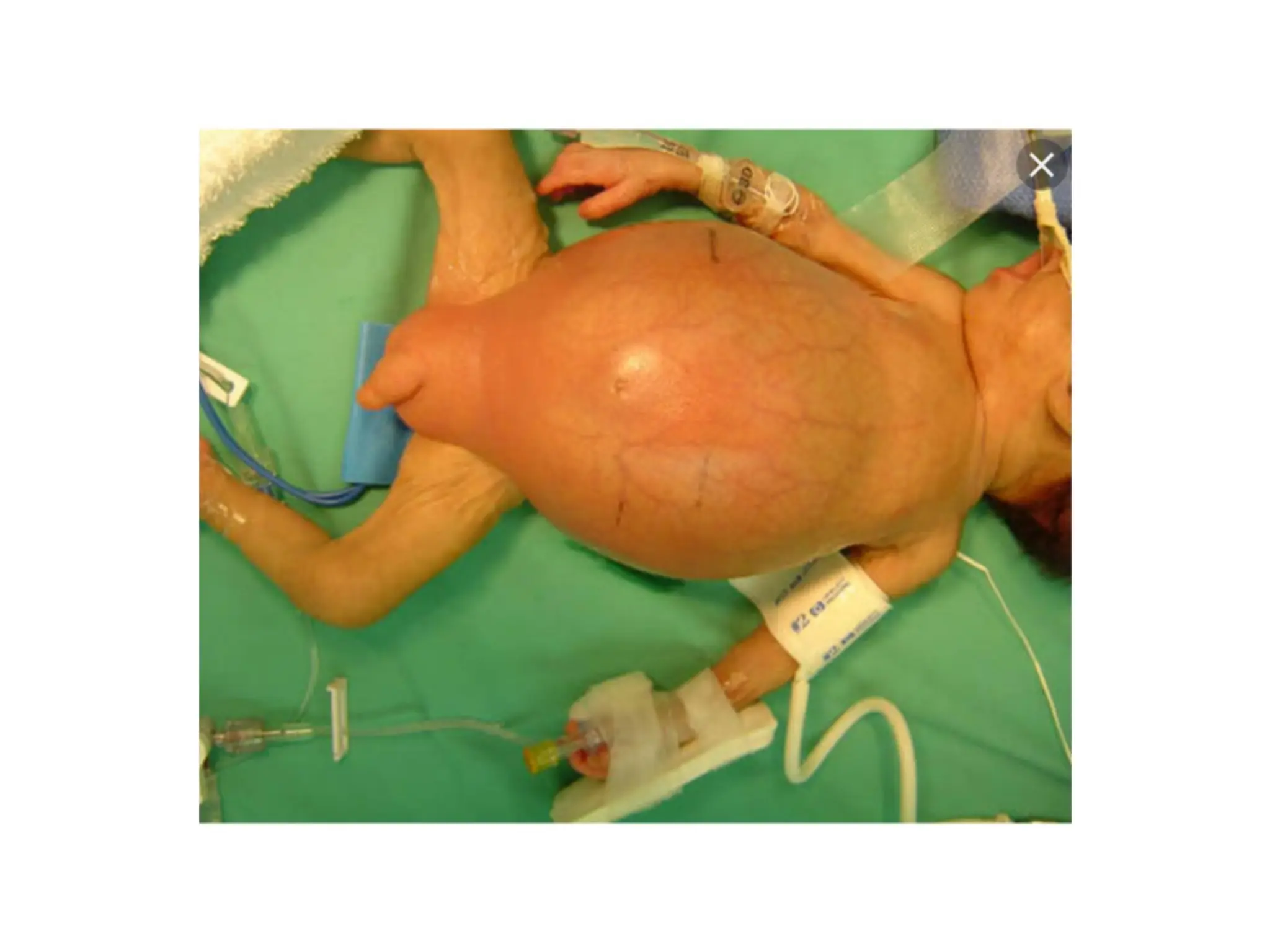
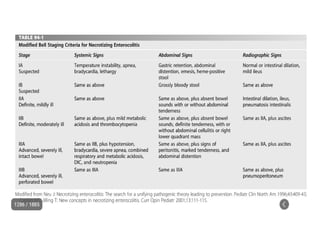
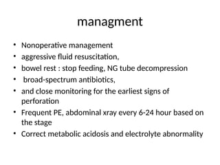
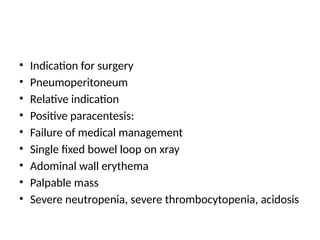
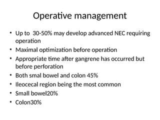
INTRODUCTION
• Is an acquired inflammatory condition of the
GI tract with variable degree of tissue necrosis
• Occur in 3/ 1000 births
• Disease first described in 1960
• Still difficulty in diagnosis and treatment
approach
• Leading cause of mortality and morbidity in NICU
• Most common newborn surgical emergency
• Incidence is 5-10% among newborn infants in NICU
• The incidence widely vary among different
geographic areas
• About 90% of cases occur in preterm
• 7-13 % cases may occur in term
Risk factor
• Prematurity is the greatest risk factor
• The lowest the birth weight the highest
incidence of NEC
• Indomethacin
• Umblical artery cathetherization
• Maternal cocaine use
• If it occurs in term neonate usually occurs with
Down syndrome, HSD, CHD, asphyxia
prematurity
• the mean gestational age of NEC was 31
weeks and average birth weight, 1460 g)
• Greatest risk before 28week and below 1kg
• Age of onset is within 1st
and 2nd
week usually
• inverse relationship between the age at onset
of NEC and gestational age
• Declining risk after 35-36week
Physiology of premature gut
• Decrease peristalsis leading to less clearance
of bacteria and stasis
• Fewer B lymphocytes so less IgA
• Invasion of mucosa by bacteria
• Less pepsin, gastric acid, mucous resulting in
increased bacterial proliferation
Role of feeding
• Composition and quantity of feeding
• Highly concentrated formulas,,,, increased PAF
• High osmolarity…. Damage to villi
• Fatty acids ….mucosal injury
• Large carbohdrate…. Hydrogen production
• No clear guideline
• 90% occur after feeding
• Minimal volume feeding 20ml/kg before going to
full feeding for about 10days found to decrease NEC
pathophysiology
• Maladaptive response of the gut to feeding
and bacterial colonization
hypoxic ischemic insult; due to diminished
splanchnic perfusion
Enteral feeding
Bacterial overgrowth
Exaggerated inflammatory response
Ischemia….
• Ischemia and reperfusion injury
• Cellular hypoxia leading to increase xanthine
oxidase production
• XO result in increased production of xanthine and
uric acid during reperfusion
• Release of reactive oxygen species
• Damage of cell membrane
• Membrane permeability and bacterial
translocation will be the effect
Exaggerated inflammatory response
• Key mediators of inflammation being NO, PAF,
TNFa
• Maladaptive vasoregulatory reponses
• leading to vasoconstriction
• Increased level of inflammaory mediators
enteral feeding
• Breast-fed infants colonized predominantly
with gram positive bacteria
• In contrast, formula-fed infants colonized
predominantly by coliforms, enterococci, and
Bacteroides species
• Affected symbiotic gut microbial axis
Role of infectious agent
• Affection of symbiotic gut-microbe relationship
• gut colonization during the newborn period
• In 2/3rd of cases bacteria isolated
• E. coli, Klebsiella, Enterobacter, Pseudomonas,
Clostridium, and coagulase-negative
Staphylococci
• Pneumatosis intestinalis:the accumulation of
hydrogen gas in the intestinal wall due to
fermentation of carbohydrates by intestinal flora
The agreed hypothesis
• the interaction among the enterocyte,immune
effector cells, and resident microbiota initiates
an inflammatory cascade that becomes
unbalanced
pathology
• Involves single( 50% ) or multiple segment of
inestine
• About 20 % of surgically treated NEC have feature
of pan necrosis
• bowel is markedly distended with patchy areas of
thinning
• serosal surfaces are typically red to gray
• and may be covered by a fibrinous exudate
• Frank gangrene, hemorrhagic fluid
Histopathologic changes
• Acute on chronic inflammation
• coagulation necrosis of the superficial mucosa
• Edema and hemorrhage of the submucosa
• In resolving cases epithelial regeneration and
fibrosis
• Small vessels thrombosis
• Full and partial thickness necrosis
• Perforation
• Pneumatosis intestinalis
diagnosis
• Physiologic instability
that may mimic sepsis
• Hypo/hyperthermia
• Bradycardia
• Apnea
• shock
• Feeding intolerance
• Gi bleeding(79-86%)
• Gastric residuals
• Abdominal distension(70-
98%
• Diarrhea(26%
• Vomiting(70%
• Progressing sepsis
• Fixed abdominal mass
• Abdominal wall erythema
Physical examination
• Abdominal distension
• Fixed palpable mass
• Palpable bowel loops
• Edema/erythema abdominal wall
laboratory
• neutropenia, thrombocytopenia, and metabolic
acidosis
• Paracentesis: When there is high suspcion with
no radiologic evidence of perforation
• Positive paracentesis: aspiration of more than 0.5
mL of brown fluid that contains bacteria on Gram
stain
• Is done for extensive pneumatosis intestinalis
and failed medical therapy
diffferntial
• Ileus with sepsi
• Focal intestinal perforation
imaging
• Multiple gas filled loops of intestine
• Pneumatosis intestinalisc: pathognomonic for
NEC
• a collection of cystic or linear lucencies in the
bowel wall,
• Frequency may reach 19-98%but may be
absent in upto 14% of cases of NEC
• Portal venous gas: linear branching radiolucencies
overlying the liver and often extending to its
periphery
• incidence of 10% to 30%.poor prognosis
• Pneumoperitoneum: in 12-30% of patients
• Best seen on lateral decubitus film
• Football sign, double wall sign,
• Only 63% of patients with perforation show the
xray sign
• Intraperitoneal fluid: gasless abdomen, flank
opacity, separation of bowel loops
• Persistent dilated bowel loop: dilated bowel
loop remained unchanged for 24 to 36 hours
managment
• Nonoperative management
• aggressive fluid resuscitation,
• bowel rest : stop feeding, NG tube decompression
• broad-spectrum antibiotics,
• and close monitoring for the earliest signs of
perforation
• Frequent PE, abdominal xray every 6-24 hour based on
the stage
• Correct metabolic acidosis and electrolyte abnormality
• Indication for surgery
• Pneumoperitoneum
• Relative indication
• Positive paracentesis:
• Failure of medical management
• Single fixed bowel loop on xray
• Adominal wall erythema
• Palpable mass
• Severe neutropenia, severe thrombocytopenia, acidosis
Operative management
• Up to 30-50% may develop advanced NEC requiring
operation
• Maximal optimization before operation
• Appropriate time after gangrene has occurred but
before perforation
• Both smal bowel and colon 45%
• Ileocecal region being the most common
• Small bowel20%
• Colon30%
laparatomy
• May be focal, multifocal, paninvolvement
• Focal disease : resection and stoma
• Multifocal disease: resection and multiple
anastomosis
• Clip and drop back technique
• Depend on the condition of the patient and
the extent of the bowel involved
• Resect gangreneous part so as to reduce
sepsis
• Early intervention to reduce conatamination
• Prevent short bowel syndrome
paracentesis
• Peritoneal drainage vs exploraory laparatomy
• Primary peritoneal drainage
• In ELBW/VLBW infant as a treatement
• In other neonates as away of stabilizing before the
definitive laparatomy
• From studies from those PD done 40% doesn’t
require additional laparatomy and other 40%
laparatomy was needed
• Recent studies no difference in outcome
• Peritoneal drainage
• making a right lower
quadrant incision,
• irrigating the peritoneal
cavity,
• placing a small rubber
drain in the abdomen
• enterostomy versus primary anastomosis
• The traditional trend was resection and stoma
• In a localized ,focal disease with a good bowel
condition and stable neonate primary
anastomosis could be considered
• For multifocal and pan NEC proximal diverting
jejunostomy
• Clip and drop technique
• Gangreneous bowel resected out and the end
tied of and left in the abdomen
• 24-48hour relaparatomy for possible stoma or
anastomosis
• Prognosis
• Medical management fails in approximately
20-40% of patients with
• pneumatosis intestinalis at diagnosis; of these,
10-30% die
Postop complication
• Wound infection
• Stoma complication
• Sepsis
• Neuro developemtal problems
• Intestinal stricture
• Short bowel syndrome
• Prevention
• Breast feeding
• Following feeding guidelines
• Trophic feedings are advised
• ?prophylactic antibiotic
NEC.pptx necrotizing enterocolitis in neonates

NEC.pptx necrotizing enterocolitis in neonates

  • 2.
    INTRODUCTION • Is anacquired inflammatory condition of the GI tract with variable degree of tissue necrosis • Occur in 3/ 1000 births • Disease first described in 1960 • Still difficulty in diagnosis and treatment approach
  • 3.
    • Leading causeof mortality and morbidity in NICU • Most common newborn surgical emergency • Incidence is 5-10% among newborn infants in NICU • The incidence widely vary among different geographic areas • About 90% of cases occur in preterm • 7-13 % cases may occur in term
  • 4.
    Risk factor • Prematurityis the greatest risk factor • The lowest the birth weight the highest incidence of NEC • Indomethacin • Umblical artery cathetherization • Maternal cocaine use • If it occurs in term neonate usually occurs with Down syndrome, HSD, CHD, asphyxia
  • 5.
    prematurity • the meangestational age of NEC was 31 weeks and average birth weight, 1460 g) • Greatest risk before 28week and below 1kg • Age of onset is within 1st and 2nd week usually • inverse relationship between the age at onset of NEC and gestational age • Declining risk after 35-36week
  • 6.
    Physiology of prematuregut • Decrease peristalsis leading to less clearance of bacteria and stasis • Fewer B lymphocytes so less IgA • Invasion of mucosa by bacteria • Less pepsin, gastric acid, mucous resulting in increased bacterial proliferation
  • 7.
    Role of feeding •Composition and quantity of feeding • Highly concentrated formulas,,,, increased PAF • High osmolarity…. Damage to villi • Fatty acids ….mucosal injury • Large carbohdrate…. Hydrogen production • No clear guideline • 90% occur after feeding • Minimal volume feeding 20ml/kg before going to full feeding for about 10days found to decrease NEC
  • 8.
    pathophysiology • Maladaptive responseof the gut to feeding and bacterial colonization hypoxic ischemic insult; due to diminished splanchnic perfusion Enteral feeding Bacterial overgrowth Exaggerated inflammatory response
  • 9.
    Ischemia…. • Ischemia andreperfusion injury • Cellular hypoxia leading to increase xanthine oxidase production • XO result in increased production of xanthine and uric acid during reperfusion • Release of reactive oxygen species • Damage of cell membrane • Membrane permeability and bacterial translocation will be the effect
  • 10.
    Exaggerated inflammatory response •Key mediators of inflammation being NO, PAF, TNFa • Maladaptive vasoregulatory reponses • leading to vasoconstriction • Increased level of inflammaory mediators
  • 11.
    enteral feeding • Breast-fedinfants colonized predominantly with gram positive bacteria • In contrast, formula-fed infants colonized predominantly by coliforms, enterococci, and Bacteroides species • Affected symbiotic gut microbial axis
  • 12.
    Role of infectiousagent • Affection of symbiotic gut-microbe relationship • gut colonization during the newborn period • In 2/3rd of cases bacteria isolated • E. coli, Klebsiella, Enterobacter, Pseudomonas, Clostridium, and coagulase-negative Staphylococci • Pneumatosis intestinalis:the accumulation of hydrogen gas in the intestinal wall due to fermentation of carbohydrates by intestinal flora
  • 13.
    The agreed hypothesis •the interaction among the enterocyte,immune effector cells, and resident microbiota initiates an inflammatory cascade that becomes unbalanced
  • 15.
    pathology • Involves single(50% ) or multiple segment of inestine • About 20 % of surgically treated NEC have feature of pan necrosis • bowel is markedly distended with patchy areas of thinning • serosal surfaces are typically red to gray • and may be covered by a fibrinous exudate • Frank gangrene, hemorrhagic fluid
  • 16.
    Histopathologic changes • Acuteon chronic inflammation • coagulation necrosis of the superficial mucosa • Edema and hemorrhage of the submucosa • In resolving cases epithelial regeneration and fibrosis • Small vessels thrombosis • Full and partial thickness necrosis • Perforation • Pneumatosis intestinalis
  • 17.
    diagnosis • Physiologic instability thatmay mimic sepsis • Hypo/hyperthermia • Bradycardia • Apnea • shock • Feeding intolerance • Gi bleeding(79-86%) • Gastric residuals • Abdominal distension(70- 98% • Diarrhea(26% • Vomiting(70% • Progressing sepsis • Fixed abdominal mass • Abdominal wall erythema
  • 18.
    Physical examination • Abdominaldistension • Fixed palpable mass • Palpable bowel loops • Edema/erythema abdominal wall
  • 19.
    laboratory • neutropenia, thrombocytopenia,and metabolic acidosis • Paracentesis: When there is high suspcion with no radiologic evidence of perforation • Positive paracentesis: aspiration of more than 0.5 mL of brown fluid that contains bacteria on Gram stain • Is done for extensive pneumatosis intestinalis and failed medical therapy
  • 20.
    diffferntial • Ileus withsepsi • Focal intestinal perforation
  • 21.
    imaging • Multiple gasfilled loops of intestine • Pneumatosis intestinalisc: pathognomonic for NEC • a collection of cystic or linear lucencies in the bowel wall, • Frequency may reach 19-98%but may be absent in upto 14% of cases of NEC
  • 25.
    • Portal venousgas: linear branching radiolucencies overlying the liver and often extending to its periphery • incidence of 10% to 30%.poor prognosis • Pneumoperitoneum: in 12-30% of patients • Best seen on lateral decubitus film • Football sign, double wall sign, • Only 63% of patients with perforation show the xray sign
  • 26.
    • Intraperitoneal fluid:gasless abdomen, flank opacity, separation of bowel loops • Persistent dilated bowel loop: dilated bowel loop remained unchanged for 24 to 36 hours
  • 30.
    managment • Nonoperative management •aggressive fluid resuscitation, • bowel rest : stop feeding, NG tube decompression • broad-spectrum antibiotics, • and close monitoring for the earliest signs of perforation • Frequent PE, abdominal xray every 6-24 hour based on the stage • Correct metabolic acidosis and electrolyte abnormality
  • 31.
    • Indication forsurgery • Pneumoperitoneum • Relative indication • Positive paracentesis: • Failure of medical management • Single fixed bowel loop on xray • Adominal wall erythema • Palpable mass • Severe neutropenia, severe thrombocytopenia, acidosis
  • 32.
    Operative management • Upto 30-50% may develop advanced NEC requiring operation • Maximal optimization before operation • Appropriate time after gangrene has occurred but before perforation • Both smal bowel and colon 45% • Ileocecal region being the most common • Small bowel20% • Colon30%
  • 33.
    laparatomy • May befocal, multifocal, paninvolvement • Focal disease : resection and stoma • Multifocal disease: resection and multiple anastomosis • Clip and drop back technique
  • 35.
    • Depend onthe condition of the patient and the extent of the bowel involved • Resect gangreneous part so as to reduce sepsis • Early intervention to reduce conatamination • Prevent short bowel syndrome
  • 36.
  • 37.
    • Peritoneal drainagevs exploraory laparatomy • Primary peritoneal drainage • In ELBW/VLBW infant as a treatement • In other neonates as away of stabilizing before the definitive laparatomy • From studies from those PD done 40% doesn’t require additional laparatomy and other 40% laparatomy was needed • Recent studies no difference in outcome
  • 38.
    • Peritoneal drainage •making a right lower quadrant incision, • irrigating the peritoneal cavity, • placing a small rubber drain in the abdomen
  • 39.
    • enterostomy versusprimary anastomosis • The traditional trend was resection and stoma • In a localized ,focal disease with a good bowel condition and stable neonate primary anastomosis could be considered • For multifocal and pan NEC proximal diverting jejunostomy
  • 40.
    • Clip anddrop technique • Gangreneous bowel resected out and the end tied of and left in the abdomen • 24-48hour relaparatomy for possible stoma or anastomosis
  • 41.
    • Prognosis • Medicalmanagement fails in approximately 20-40% of patients with • pneumatosis intestinalis at diagnosis; of these, 10-30% die
  • 42.
    Postop complication • Woundinfection • Stoma complication • Sepsis • Neuro developemtal problems • Intestinal stricture • Short bowel syndrome
  • 43.
    • Prevention • Breastfeeding • Following feeding guidelines • Trophic feedings are advised • ?prophylactic antibiotic