Downloaded 131 times

![LABORATORY FINDINGS
• A patient who has NEC can present with an abnormal white blood cell count.
• It may be elevated, but more commonly it is depressed.
• A severely low white blood cell count (,1.5 3 109/L [,1,500 cells/cu mm]) has been reported in
37% of cases.
• It results from both decreased production and increased utilization of leukocytes.
• Thrombocytopenia is also common, seen in up to 87% of patients.
• In addition, patients may develop other coagulation abnormalities, including prolongation of
prothrombin time and hypofibrinogenemia.
• Glucose instability (hypoglycemia or hyperglycemia), metabolic acidosis, and electrolyte
imbalance may occur.
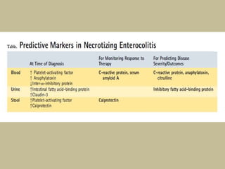
• Some patients have elevated C-reactive protein levels.
• Because no unique infectious agents have been incriminated in NEC, bacteriologic and fungal
cultures may prove helpful but not conclusive.](https://image.slidesharecdn.com/necrotizingenterocolitis-150421142343-conversion-gate02/85/Necrotizing-enterocolitis-14-320.jpg)
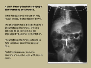

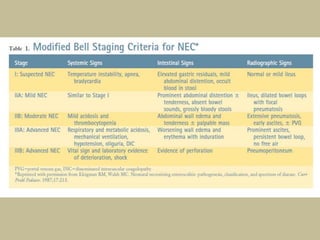


Necrotizing enterocolitis (NEC) is an inflammatory disease of the intestine affecting premature infants. It has a mortality rate of 10-50% and is the most common intestinal emergency in neonatal intensive care units. The disease results from an aberrant immune response of the immature gut to enteral feeding and bacterial colonization. Risk factors include prematurity, type of feeding, and hypoxic events. Clinical signs include abdominal distention and feeding intolerance. Diagnosis involves abdominal x-rays showing pneumatosis intestinalis or free air. Treatment involves bowel rest, antibiotics, and may require surgery for resection of necrotic intestine. Long term outcomes can include strictures, short bowel syndrome, and neurodevelopmental


