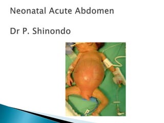
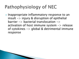
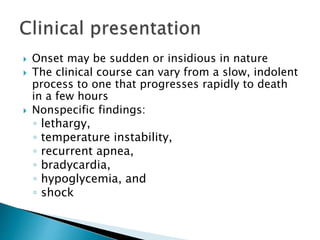
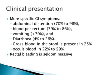
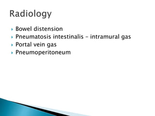
 One of surgical emergencies in neonates
 May include
- Intestinal obstruction
- Necrotising Enterocolitis
- Intussusception
- GI perforation in a newborn
 Sepsis & ileus – bilious vomitting & abdominal
distension (most important non-surgical
cause)
 Intracranial lesions – Hydrocephalus &
subdural hemorrhage
 Renal Dx with uremia - renal agenesis,
polycystic Dx.
 Bilious vomiting – always abnormal. NB! Until
proven otherwise it’s a malrotation and/or
volvulus
 Abdominal distension (scaphoid possible)
 Delayed, scanty, or no meconium
• Polyhydramnios in mother
• Downs Syndrome
• Family history (HSD, DM mother, jejunal
atresia)
 High Obstruction
• Gastric outlet
• Duodenal
• Jejunal
 Low obstruction
• Distal small bowel
• Colonic
 Congenital (bowel atresias & stenoses; HSD;
Meconium Ileus)
 Acquired (pyloric stenosis, malrotation &
midgut volvulus, hernias)
 Intrinsic developmental defects
 Insults acquired in utero, after the formation
of normal bowel
 Abnormalities of peristalsis and/or abnormal
intestinal contents.
1. Plain abdominal X-Rays.
 Complete high obstruction – no gas in distal
small bowel
 Low obstruction – many gas filled loops
(24hrs)
 May be non specific – malrotation
2. Contrast enema – differentiates types of low
IO. (meconium plug, HSD)
3. Upper GI series – procedure of choice to
diagnose malrotation
4. Rectal biopsy – HSD (suction biopsy & full
thickness)
 Diagnose early to prevent clinical
deterioration, aspiration pneumonia, sepsis &
biochem & hematological derangements.
 Surgery …… once preliminaries done
- Resuscitation
- NGT
- Baseline labs
- Discuss with parents & consent for OT.
 1:1000 live births
 Risk factors
– prematurity and low birth weight. 750-1000g
have highest incidence.
- Infant related (perinatal asphyxia, congenital
heart disease. Etc)
- Maternal related (pre-eclampsia, prolonged
PROM, drugs)
 Inappropriate inflammatory response to an
insult -> injury & disruption of epithelial
barrier -> bacterial translocation ->
activation of host immune system -> release
of cytokines -> global & detrimental immune
response
 NEC may be
- Focal (isolated) disease : single area of
the bowel is necrotic or perforated
-Multifocal disease : multisegmental
disease with > 50% viable
- NEC totalis : necrosis of at least 75% of
the gut
 NEC - mucosal disease
that extends well into the
normal intestine
 Terminal ileum,
ascending & transverse
colon commonly affected
 The disease can occur
anywhere from stomach
to rectum
 Onset may be sudden or insidious in nature
 The clinical course can vary from a slow, indolent
process to one that progresses rapidly to death
in a few hours
 Nonspecific findings:
◦ lethargy,
◦ temperature instability,
◦ recurrent apnea,
◦ bradycardia,
◦ hypoglycemia, and
◦ shock
 More specific GI symptoms:
◦ abdominal distention (70% to 98%),
◦ blood per rectum (79% to 86%),
◦ vomiting (>70%), and
◦ Diarrhoea (4% to 26%).
◦ Gross blood in the stool is present in 25%
◦ occult blood in 22% to 59%.
 Rectal bleeding is seldom massive
 Bowel distension
 Pneumatosis intestinalis – intramural gas
 Portal vein gas
 Pneumoperitoneum
 Pneumatosis
intestinalis with air
over the liver shadow
dispersed within the
radicles of the portal
venous system
 Portal venous gas and
pneumatosis
STAGE CLINICAL X-RAY TREATMENT
I. SUSPECT
NEC
-Mild
abdominal
distention
-Poor feeding
-Emesis
-Mild ileus -Medical
workup for
sepsis
II. DEFINITE
NEC
-The above plus
-Marked
abdominal
distention
-GI bleeding
-Significant ileus
-Pneumatosis
intestinalis
-PVG
Medical
III. ADVANCED
NEC
-The above plus
-Unstable vital
signs
-Septic shock
Pneumoperitoneum Surgical
 Nonoperative Therapy is the initial
management
 Commence immediately upon suspicion of
the diagnosis of NEC
 The goals of medical management
include restoration of tissue perfusion,
control of infection or sepsis
careful observation for evidence of
intestinal gangrene or perforation.
 NEC without necrosis or perforation –
Treatment is supportive
 Keep NPO – Discontinue feeding
 Gastric tube - decompression
 Commence IVF resuscitation
 Commence Antibiotics
 Consider Empirical antifungal if clinical
course is prolonged
Absolute indication
1. Pneumoperitoneum
Relative indication
2. Clinical deterioration despite adequate therapy
a. Erythema and oedema of abdominal wall
b. Abdominal mass
c. Signs of peritonitis
d. Increasing acidosis
e. Persistent and progressive thrombocytopenia
 Idiopathic :
- 10days after URTI or GE
- Change from breast to bottle feeds
- Increase in lymphoid tissue mass (Peyers
Patches) – most likely cause
- Seasonal variation, higher in spring &
summer
- Viruses may play a role (rotavirus; associated
with vaccination)
 Lead Point
- Seen in older children.
- E.g Meckels diverticulum, enlarged LN,
polyps.
- Lead points initiate intussusception on a
mechanical basis
 Postoperative
- 2-5days postop, painless, observation, NG
aspiration is done. May self limit.
 Well nourished/ fat children 3-18months.
 Sudden onset ‘colicky’ abdominal pain,
associated vomiting.
 ‘Red currant jelly’ stool
 Dehydration/shock
 Palpable mass ‘sausage like’ (may palpate in
rectum – DRE)
 Intussusceptum may prolapse out of rectum
1. Abdominal Xray :
- Signs of obstruction
- May see a mass
2. Contrast enema:
- Barium or gas may be used.
3. U/S scan.
Donut-like. “Target
lesion” “pseudo kidney”
Invagination seen. “
 Resuscitation !!! Massive third space loss
 NGT
 Antibiotics (Penicillin, Aminoglycoside,
metronidazole)
 Resuscitate before contrast enema, and prior
to reduction
 Sedation/analgesia. pethidine
 Reduction
- Enema reduction
- Pneumatic/ Hydrostatic reduction
Contra-indications:
Shock, septicemia, peritonitis, perforation,
Intestinal infarction, chronic obstruction.
 Surgical management
- When reduction unsuccessful
- Peritonitis
- Perforation
 THANKYOU FOR YOUR
ATTENTION!

Neonatal acute abdomen. 7th yr

  • 2.
     One ofsurgical emergencies in neonates  May include - Intestinal obstruction - Necrotising Enterocolitis - Intussusception - GI perforation in a newborn
  • 3.
     Sepsis &ileus – bilious vomitting & abdominal distension (most important non-surgical cause)  Intracranial lesions – Hydrocephalus & subdural hemorrhage  Renal Dx with uremia - renal agenesis, polycystic Dx.
  • 4.
     Bilious vomiting– always abnormal. NB! Until proven otherwise it’s a malrotation and/or volvulus  Abdominal distension (scaphoid possible)  Delayed, scanty, or no meconium • Polyhydramnios in mother • Downs Syndrome • Family history (HSD, DM mother, jejunal atresia)
  • 5.
     High Obstruction •Gastric outlet • Duodenal • Jejunal  Low obstruction • Distal small bowel • Colonic  Congenital (bowel atresias & stenoses; HSD; Meconium Ileus)  Acquired (pyloric stenosis, malrotation & midgut volvulus, hernias)
  • 6.
     Intrinsic developmentaldefects  Insults acquired in utero, after the formation of normal bowel  Abnormalities of peristalsis and/or abnormal intestinal contents.
  • 7.
    1. Plain abdominalX-Rays.  Complete high obstruction – no gas in distal small bowel  Low obstruction – many gas filled loops (24hrs)  May be non specific – malrotation
  • 11.
    2. Contrast enema– differentiates types of low IO. (meconium plug, HSD) 3. Upper GI series – procedure of choice to diagnose malrotation 4. Rectal biopsy – HSD (suction biopsy & full thickness)
  • 12.
     Diagnose earlyto prevent clinical deterioration, aspiration pneumonia, sepsis & biochem & hematological derangements.  Surgery …… once preliminaries done - Resuscitation - NGT - Baseline labs - Discuss with parents & consent for OT.
  • 14.
     1:1000 livebirths  Risk factors – prematurity and low birth weight. 750-1000g have highest incidence. - Infant related (perinatal asphyxia, congenital heart disease. Etc) - Maternal related (pre-eclampsia, prolonged PROM, drugs)
  • 16.
     Inappropriate inflammatoryresponse to an insult -> injury & disruption of epithelial barrier -> bacterial translocation -> activation of host immune system -> release of cytokines -> global & detrimental immune response
  • 17.
     NEC maybe - Focal (isolated) disease : single area of the bowel is necrotic or perforated -Multifocal disease : multisegmental disease with > 50% viable - NEC totalis : necrosis of at least 75% of the gut
  • 18.
     NEC -mucosal disease that extends well into the normal intestine  Terminal ileum, ascending & transverse colon commonly affected  The disease can occur anywhere from stomach to rectum
  • 19.
     Onset maybe sudden or insidious in nature  The clinical course can vary from a slow, indolent process to one that progresses rapidly to death in a few hours  Nonspecific findings: ◦ lethargy, ◦ temperature instability, ◦ recurrent apnea, ◦ bradycardia, ◦ hypoglycemia, and ◦ shock
  • 20.
     More specificGI symptoms: ◦ abdominal distention (70% to 98%), ◦ blood per rectum (79% to 86%), ◦ vomiting (>70%), and ◦ Diarrhoea (4% to 26%). ◦ Gross blood in the stool is present in 25% ◦ occult blood in 22% to 59%.  Rectal bleeding is seldom massive
  • 21.
     Bowel distension Pneumatosis intestinalis – intramural gas  Portal vein gas  Pneumoperitoneum
  • 22.
     Pneumatosis intestinalis withair over the liver shadow dispersed within the radicles of the portal venous system
  • 23.
     Portal venousgas and pneumatosis
  • 24.
    STAGE CLINICAL X-RAYTREATMENT I. SUSPECT NEC -Mild abdominal distention -Poor feeding -Emesis -Mild ileus -Medical workup for sepsis II. DEFINITE NEC -The above plus -Marked abdominal distention -GI bleeding -Significant ileus -Pneumatosis intestinalis -PVG Medical III. ADVANCED NEC -The above plus -Unstable vital signs -Septic shock Pneumoperitoneum Surgical
  • 25.
     Nonoperative Therapyis the initial management  Commence immediately upon suspicion of the diagnosis of NEC  The goals of medical management include restoration of tissue perfusion, control of infection or sepsis careful observation for evidence of intestinal gangrene or perforation.
  • 26.
     NEC withoutnecrosis or perforation – Treatment is supportive  Keep NPO – Discontinue feeding  Gastric tube - decompression  Commence IVF resuscitation  Commence Antibiotics  Consider Empirical antifungal if clinical course is prolonged
  • 27.
    Absolute indication 1. Pneumoperitoneum Relativeindication 2. Clinical deterioration despite adequate therapy a. Erythema and oedema of abdominal wall b. Abdominal mass c. Signs of peritonitis d. Increasing acidosis e. Persistent and progressive thrombocytopenia
  • 29.
     Idiopathic : -10days after URTI or GE - Change from breast to bottle feeds - Increase in lymphoid tissue mass (Peyers Patches) – most likely cause - Seasonal variation, higher in spring & summer - Viruses may play a role (rotavirus; associated with vaccination)
  • 30.
     Lead Point -Seen in older children. - E.g Meckels diverticulum, enlarged LN, polyps. - Lead points initiate intussusception on a mechanical basis  Postoperative - 2-5days postop, painless, observation, NG aspiration is done. May self limit.
  • 31.
     Well nourished/fat children 3-18months.  Sudden onset ‘colicky’ abdominal pain, associated vomiting.  ‘Red currant jelly’ stool  Dehydration/shock  Palpable mass ‘sausage like’ (may palpate in rectum – DRE)  Intussusceptum may prolapse out of rectum
  • 32.
    1. Abdominal Xray: - Signs of obstruction - May see a mass 2. Contrast enema: - Barium or gas may be used. 3. U/S scan.
  • 34.
    Donut-like. “Target lesion” “pseudokidney” Invagination seen. “
  • 35.
     Resuscitation !!!Massive third space loss  NGT  Antibiotics (Penicillin, Aminoglycoside, metronidazole)  Resuscitate before contrast enema, and prior to reduction  Sedation/analgesia. pethidine
  • 36.
     Reduction - Enemareduction - Pneumatic/ Hydrostatic reduction Contra-indications: Shock, septicemia, peritonitis, perforation, Intestinal infarction, chronic obstruction.
  • 37.
     Surgical management -When reduction unsuccessful - Peritonitis - Perforation
  • 39.
     THANKYOU FORYOUR ATTENTION!

Editor's Notes

  • #8 Double bubble- duodenal atresia; few gas filled loops beyond duodenum- jejunal atresia
  • #12 Malrotation – ceacum in upper mid abdomen, or left upper quadrant.
  • #15 Immature GI immunity (low mucin, low MALT), low motility, disrupted tight junctions. Enteral feedings.
  • #19 Involvement of both large and small intestines occur in 44% of NEC cases
  • #22 Lab findings- non specific- leucocytosis, thrombocytopenia, metabolic acidosis. AP or left lateral decubitus Xray. PVG- branching radiolucencies over the liver.
  • #29 Telescope!