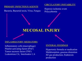
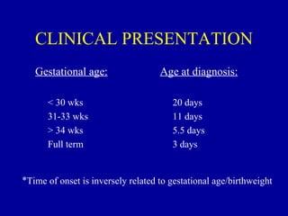
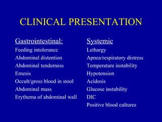
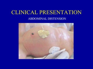
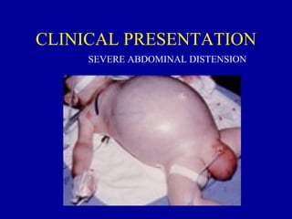
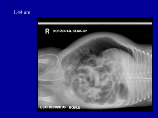
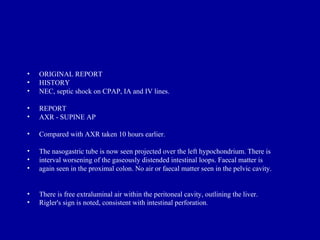
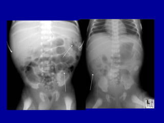
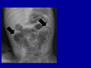
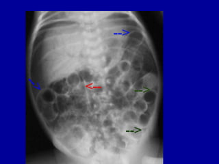
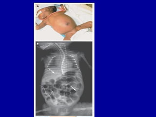
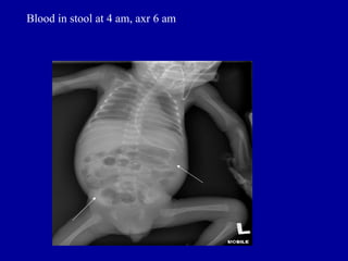
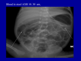
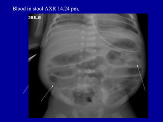
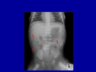
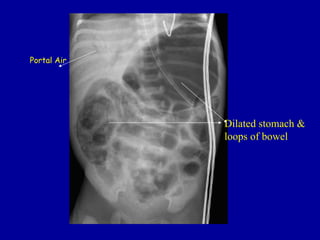
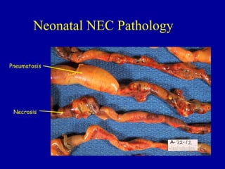
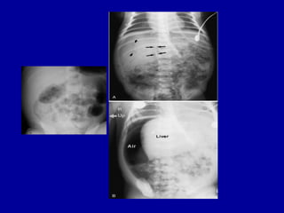
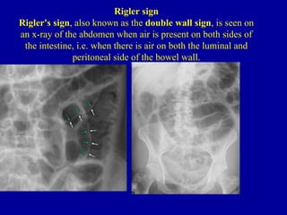
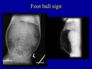
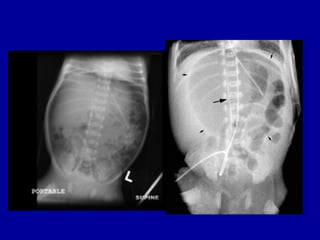
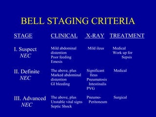
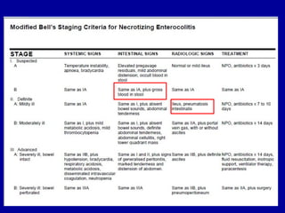
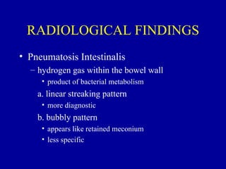
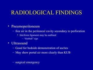
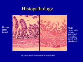
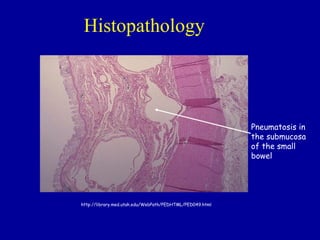
NECROTIZING
ENTEROCOLITIS
  Dr Varsha Shah
OBJECTIVES
• Ability to diagnose and treat the signs and
  symptoms of NEC

• Ability to evaluate radiographs for the
  classic findings of NEC

• List several long-term complications
  associated with NEC
NECROTIZING
          ENTEROCOLITIS
• Epidemiology:
  – most commonly occurring gastrointestinal
    emergency in preterm infants
  – leading cause of emergency surgery in neonates
  – overall incidence: 1-5% in most NICU’s
  – most common in VLBW preterm infants
     • 10% of all cases occur in term infants
NECROTIZING
          ENTEROCOLITIS
• Epidemiology:
  – 10x more likely to occur in infants who have
    been fed
  – males = females
  – blacks > whites
  – mortality rate: 25-30%
  – 50% of survivors experience long-term
    sequelae
NECROTIZING
          ENTEROCOLITIS
• Pathology:
  – most commonly involved areas: terminal ileum
    and proximal colon
  – GROSS:
     • bowel appears irregularly dilated with hemorrhagic
       or ischemic areas of frank necrosis
        – focal or diffuse
  – MICROSCOPIC:
     • mucosal edema, hemorrhage and ulceration
NECROTIZING
         ENTEROCOLITIS
• MICROSCOPIC:
 – minimal inflammation during the acute phase
    • increases during revascularization
 – granulation tissue and fibrosis develop
    • stricture formation
 – microthrombi in mesenteric arterioles and
   venules
NECROTIZING
         ENTEROCOLITIS

• Pathophysiology:


   UNKNOWN
   CAUSE…….
CIRCULATORY INSTABILITY
PRIMARY INFECTIOUS AGENTS
                                           Hypoxic-ischemic event
Bacteria, Bacterial toxin, Virus, Fungus   Polycythemia


                    MUCOSAL INJURY

INFLAMMATORY MEDIATORS
Inflammatory cells (macrophage)             ENTERAL FEEDINGS
Platelet activating factor (PAF)            Hypertonic formula or medication
Tumor necrosis factor (TNF)                 Malabsorption, gaseous distention
Leukotriene C4, Interleukin 1; 6            H2 gas production, Endotoxin
                                            production
RISK FACTORS
• Prematurity:
  * primary risk factor
  – 90% of cases are premature infants
  – immature gastrointestinal system
     • mucosal barrier
     • poor motility
  – immature immune response
  – impaired circulatory dynamics
RISK FACTORS
• Infectious Agents:
  – usually occurs in clustered epidemics
  – normal intestinal flora
     •   E. coli
     •   Klebsiella spp.
     •   Pseudomonas spp.
     •   Clostridium difficile
     •   Staph. Epi
     •   Viruses
RISK FACTORS
• Inflammatory Mediators:
  – involved in the development of intestinal injury
    and systemic side effects
     • neutropenia, thrombocytopenia, acidosis,
       hypotension
  – primary factors
     •   Tumor necrosis factor (TNF)
     •   Platelet activating factor (PAF)
     •   LTC4
     •   Interleukin 1& 6
RISK FACTORS
• Circulatory Instability:
  – Hypoxic-ischemic injury
     • poor blood flow to the mesenteric vessels
     • local rebound hyperemia with re-perfusion
     • production of O2 radicals
  – Polycythemia
     • increased viscosity causing decreased blood flow
     • exchange transfusion
RISK FACTORS
• Enteral Feedings:
  –   > 90% of infants with NEC have been fed
  –   provides a source for H2 production
  –   hyperosmolar formula/medications
  –   aggressive feedings
       • too much volume
       • rate of increase
          – >20cc/kg/day
RISK FACTORS
• Enteral Feedings:
  – immature mucosal function
     • malabsorption
  – breast milk may have a protective effect
     •   IGA
     •   macrophages, lymphocytes
     •   complement components
     •   lysozyme, lactoferrin
     •   acetylhydrolase
From UpToDate online, Adapted from Kliegman, RM, Pediatr Res 1993; 34:701.
CLINICAL PRESENTATION
   Gestational age:                 Age at diagnosis:

      < 30 wks                         20 days
      31-33 wks                        11 days
      > 34 wks                         5.5 days
      Full term                        3 days


*Time of onset is inversely related to gestational age/birthweight
CLINICAL PRESENTATION
Gastrointestinal:             Systemic
Feeding intolerance           Lethargy
Abdominal distention          Apnea/respiratory distress
Abdominal tenderness          Temperature instability
Emesis                        Hypotension
Occult/gross blood in stool   Acidosis
Abdominal mass                Glucose instability
Erythema of abdominal wall    DIC
                              Positive blood cultures
CLINICAL PRESENTATION
     ABDOMINAL DISTENSION
CLINICAL PRESENTATION
     SEVERE ABDOMINAL DISTENSION
CLINICAL PRESENTATION
Sudden Onset:                      Insidious Onset:
Full term or preterm infants       Usually preterm
Acute catastrophic deterioration   Evolves during 1-2 days
Respiratory decompensation         Increase desaturations
Shock/acidosis
                                   Feeding intolerance (regurgitation,
Marked abdominal distension
                                   high/green aspirates)
Positive blood culture
                                   Change in stool pattern
                                   Intermittent abdominal
                                   distention,loops,crepitus
                                   Occult blood in stools
                                   Poor perfusion, lethargy
12.52 pm AXR
•  ADDENDUM
•  Review of the images reveal air lucency noted in both hypochondrial regions
   which
• are worrisome for free gas. There is also suggestion of portal venous gas.
• ***FINAL ADDENDUM***
 Verified by: Dr PCC, Registrar,
• Amended Date/Time: 30-MAR-2005 09:27
• ORIGINAL REPORT
• HISTORY
• High NG aspirates

•   REPORT
•   The small bowel loops are dilated and there is faecal material noted in the large
•   colon but not much in the rectum. However, no definite pneumotosis intestinalis
•   is seen. Tip of the NG tube is in the distal oesophagus rather than the stomach
•   and should be adjusted.
•   CONCLUSION
•   The small bowel is dilated. The cause however is not visualised in the study.
15.52 pm AXR
•   ORIGINAL REPORT
•   HISTORY
•   ? sepsis. Growing prem. Abdominal distension.

•   REPORT
•   CXR - MOBILE SUPINE AP

•   Compared with CXR from 14/12/2004.

•   NG tube is again noted. There is now bilateral perihilar peribronchial thickening.
•   Cardiac size is unremarkable.

•   The intestinal loops remained gaseously distended though faecal matter is seen within
•   the colon. There is a lack of rectal air through a catheter is seen in the lower
•   pelvic cavity.
22.40 pm
•   ORIGINAL REPORT
•   HISTORY
•   Septicaemia. NEC. Intubated.

•   REPORT
•   MOBILE SUPINE (CHEST AND ABDOMEN)

•   The tip of the NG tube is projected over the distal esophagus. The tip of the ETT
•   is about 0.6cm from the carina.
•   A tube is projected over the rectal area.

•   There is diffuse haziness of both lungs. The heart size cannot be assessed.
•   There are multiple gas distended intestinal loops and intraperitoneal free gas present
•   which appears worse when compared to the last AXR.
1.44 am
1.44 am
•   ORIGINAL REPORT
•   HISTORY
•   NEC, septic shock on CPAP, IA and IV lines.

•   REPORT
•   AXR - SUPINE AP

•   Compared with AXR taken 10 hours earlier.

•   The nasogastric tube is now seen projected over the left hypochondrium. There is
•   interval worsening of the gaseously distended intestinal loops. Faecal matter is
•   again seen in the proximal colon. No air or faecal matter seen in the pelvic cavity.


•   There is free extraluminal air within the peritoneal cavity, outlining the liver.
•   Rigler's sign is noted, consistent with intestinal perforation.
Blood in stool at 4 am, axr 6 am
Blood in stool AXR 10. 30 am,
Blood in stool AXR 14.24 pm,
Portal Air


             Dilated stomach &
             loops of bowel
Neonatal NEC Pathology

Pneumatosis


 Necrosis
Rigler sign
Rigler's sign, also known as the double wall sign, is seen on
an x-ray of the abdomen when air is present on both sides of
 the intestine, i.e. when there is air on both the luminal and
              peritoneal side of the bowel wall.
Foot ball sign
BELL STAGING CRITERIA
STAGE           CLINICAL               X-RAY TREATMENT

I. Suspect      Mild abdominal         Mild ileus      Medical
                distention                             Work up for
   NEC          Poor feeding                               Sepsis
                Emesis

II. Definite    The above, plus        Significant     Medical
                Marked abdominal         Ileus
   NEC          distention             Pneumatosis
                GI bleeding             Intestinalis
                                       PVG

III. Advanced   The above, plus        Pneumo-         Surgical
                Unstable vital signs    Peritoneum
    NEC         Septic Shock
RADIOLOGICAL FINDINGS
• Pneumatosis Intestinalis
  – hydrogen gas within the bowel wall
     • product of bacterial metabolism
    a. linear streaking pattern
     • more diagnostic
    b. bubbly pattern
     • appears like retained meconium
     • less specific
RADIOLOGICAL FINDINGS

• Portal Venous Gas
  – extension of pneumatosis intestinalis into the
    portal venous circulation
     • linear branching lucencies overlying the liver and
       extending to the periphery
     • associated with severe disease and high mortality
RADIOLOGICAL FINDINGS

• Pneumoperitoneum
  – free air in the peritoneal cavity secondary to perforation
     • falciform ligament may be outlined
         – “football” sign

• Ultrasound
  – Good for bedside demonstration of ascites
  – May show portal air more clearly than KUB


  – surgical emergency
LABORATORY FINDINGS
• FBC
  – neutropenia/elevated WBC
  – thrombocytopenia
• Acidosis
  – metabolic
• Hyponatreamia
• Hyperkalemia
  – increased secondary to release from necrotic
    tissue
LABORATORY FINDINGS
• DIC
• Positive cultures
  –   Blood culture aerobic, anaerobic
  –   CSF
  –   urine
  –   Stool culture, aerobic, rotavirus
MEDICAL TREATMENT
• Stage Ia
   – NPO x 3 days
• Stage Ib - IIb/IIIa
   – NPOX 7 days since blood in stool
   – Broad spectrum antibiotics
      • Cover Gram +, Gram - & Anaerobes, Ampi/Amikacin/Flagyl
      • Ib = 3 days, IIa = 7-10 d, IIb & up = 14 d
   – Follow KUB for resolution
      • Resume enteral feeds 10-14 days after radiographic resolution
   – May require paracentesis if IIIa
SURGICAL CONSULT and
         TREATMENT
• Surgical Consult
  – suspected or proven NEC
  – indications for surgery:
     • portal venous gas; pneumoperitoneum
     • clinical deterioration
        – despite medical management
     • positive paracentesis
     • fixed intestinal loop on serial x-rays
     • erythema of abdominal wall
TREATMENT
• Labs: q6-8hrs
   – FBC PLT, Electrolytes, DIC panel (PT, PTT), blood
     gases
• X-rays: q6-8hrs
   – AP, left lateral decubitus or cross-table lateral
• Supportive Therapy
   – fluids, blood products (PCT< FFP< PLT), Inotropes,
     mechanical ventilation (May need long line, ETT,IA
     line)
Surgical treatment
• Stage IIIa - IIIb
   – Laparotomy
      • Resection of necrotic bowel
      • Ileostomy with mucous fistula
          – Subsequent re-anastamosis
      • May result in strictures requiring further surgery later
   – Peritoneal drain
      • Placement in NICU under local anesthetic
      • Used when infant is too clinically unstable for surgery
      • May help stabilize pt for subsequent surgery
Histopathology


Normal                                                             NEC:
small                                                              Hemorrhagic
bowel                                                              necrosis
                                                                   beginning at
                                                                   the mucosa
                                                                   and working
                                                                   its way down
                                                                   into the wall


         http://library.med.utah.edu/WebPath/PEDHTML/PED045.html
Histopathology


                                                          Pneumatosis in
                                                          the submucosa
                                                          of the small
                                                          bowel


http://library.med.utah.edu/WebPath/PEDHTML/PED049.html
PROGNOSIS
• Depends on the severity of the illness
• Associated with late complications
  *   strictures
  –   short-gut syndrome
  –   malabsorption
  –   fistulas
  –   abscess

                       * MOST COMMON
Outcomes
• Mortality varies with birth weight:
   – <1000 g = 40-100%
   – <1500 g = 10-44%
   – >2500 g = 0-20%
• Morbidity/Mortality vary with severity:
   – Resection -> Short gut -> FTT, malabsorbtion
   – Strictures -> further surgery in medical and surgical
     NEC
   – Prolonged NPO status on TPN -> cholestasis &
     metabolic abnormalities

Nec

  • 1.
  • 2.
    OBJECTIVES • Ability todiagnose and treat the signs and symptoms of NEC • Ability to evaluate radiographs for the classic findings of NEC • List several long-term complications associated with NEC
  • 3.
    NECROTIZING ENTEROCOLITIS • Epidemiology: – most commonly occurring gastrointestinal emergency in preterm infants – leading cause of emergency surgery in neonates – overall incidence: 1-5% in most NICU’s – most common in VLBW preterm infants • 10% of all cases occur in term infants
  • 4.
    NECROTIZING ENTEROCOLITIS • Epidemiology: – 10x more likely to occur in infants who have been fed – males = females – blacks > whites – mortality rate: 25-30% – 50% of survivors experience long-term sequelae
  • 5.
    NECROTIZING ENTEROCOLITIS • Pathology: – most commonly involved areas: terminal ileum and proximal colon – GROSS: • bowel appears irregularly dilated with hemorrhagic or ischemic areas of frank necrosis – focal or diffuse – MICROSCOPIC: • mucosal edema, hemorrhage and ulceration
  • 6.
    NECROTIZING ENTEROCOLITIS • MICROSCOPIC: – minimal inflammation during the acute phase • increases during revascularization – granulation tissue and fibrosis develop • stricture formation – microthrombi in mesenteric arterioles and venules
  • 7.
    NECROTIZING ENTEROCOLITIS • Pathophysiology: UNKNOWN CAUSE…….
  • 8.
    CIRCULATORY INSTABILITY PRIMARY INFECTIOUSAGENTS Hypoxic-ischemic event Bacteria, Bacterial toxin, Virus, Fungus Polycythemia MUCOSAL INJURY INFLAMMATORY MEDIATORS Inflammatory cells (macrophage) ENTERAL FEEDINGS Platelet activating factor (PAF) Hypertonic formula or medication Tumor necrosis factor (TNF) Malabsorption, gaseous distention Leukotriene C4, Interleukin 1; 6 H2 gas production, Endotoxin production
  • 9.
    RISK FACTORS • Prematurity: * primary risk factor – 90% of cases are premature infants – immature gastrointestinal system • mucosal barrier • poor motility – immature immune response – impaired circulatory dynamics
  • 10.
    RISK FACTORS • InfectiousAgents: – usually occurs in clustered epidemics – normal intestinal flora • E. coli • Klebsiella spp. • Pseudomonas spp. • Clostridium difficile • Staph. Epi • Viruses
  • 11.
    RISK FACTORS • InflammatoryMediators: – involved in the development of intestinal injury and systemic side effects • neutropenia, thrombocytopenia, acidosis, hypotension – primary factors • Tumor necrosis factor (TNF) • Platelet activating factor (PAF) • LTC4 • Interleukin 1& 6
  • 12.
    RISK FACTORS • CirculatoryInstability: – Hypoxic-ischemic injury • poor blood flow to the mesenteric vessels • local rebound hyperemia with re-perfusion • production of O2 radicals – Polycythemia • increased viscosity causing decreased blood flow • exchange transfusion
  • 13.
    RISK FACTORS • EnteralFeedings: – > 90% of infants with NEC have been fed – provides a source for H2 production – hyperosmolar formula/medications – aggressive feedings • too much volume • rate of increase – >20cc/kg/day
  • 14.
    RISK FACTORS • EnteralFeedings: – immature mucosal function • malabsorption – breast milk may have a protective effect • IGA • macrophages, lymphocytes • complement components • lysozyme, lactoferrin • acetylhydrolase
  • 15.
    From UpToDate online,Adapted from Kliegman, RM, Pediatr Res 1993; 34:701.
  • 16.
    CLINICAL PRESENTATION Gestational age: Age at diagnosis: < 30 wks 20 days 31-33 wks 11 days > 34 wks 5.5 days Full term 3 days *Time of onset is inversely related to gestational age/birthweight
  • 17.
    CLINICAL PRESENTATION Gastrointestinal: Systemic Feeding intolerance Lethargy Abdominal distention Apnea/respiratory distress Abdominal tenderness Temperature instability Emesis Hypotension Occult/gross blood in stool Acidosis Abdominal mass Glucose instability Erythema of abdominal wall DIC Positive blood cultures
  • 18.
    CLINICAL PRESENTATION ABDOMINAL DISTENSION
  • 19.
    CLINICAL PRESENTATION SEVERE ABDOMINAL DISTENSION
  • 20.
    CLINICAL PRESENTATION Sudden Onset: Insidious Onset: Full term or preterm infants Usually preterm Acute catastrophic deterioration Evolves during 1-2 days Respiratory decompensation Increase desaturations Shock/acidosis Feeding intolerance (regurgitation, Marked abdominal distension high/green aspirates) Positive blood culture Change in stool pattern Intermittent abdominal distention,loops,crepitus Occult blood in stools Poor perfusion, lethargy
  • 21.
  • 22.
    • ADDENDUM • Review of the images reveal air lucency noted in both hypochondrial regions which • are worrisome for free gas. There is also suggestion of portal venous gas. • ***FINAL ADDENDUM*** Verified by: Dr PCC, Registrar, • Amended Date/Time: 30-MAR-2005 09:27 • ORIGINAL REPORT • HISTORY • High NG aspirates • REPORT • The small bowel loops are dilated and there is faecal material noted in the large • colon but not much in the rectum. However, no definite pneumotosis intestinalis • is seen. Tip of the NG tube is in the distal oesophagus rather than the stomach • and should be adjusted. • CONCLUSION • The small bowel is dilated. The cause however is not visualised in the study.
  • 23.
  • 24.
    ORIGINAL REPORT • HISTORY • ? sepsis. Growing prem. Abdominal distension. • REPORT • CXR - MOBILE SUPINE AP • Compared with CXR from 14/12/2004. • NG tube is again noted. There is now bilateral perihilar peribronchial thickening. • Cardiac size is unremarkable. • The intestinal loops remained gaseously distended though faecal matter is seen within • the colon. There is a lack of rectal air through a catheter is seen in the lower • pelvic cavity.
  • 25.
  • 26.
    ORIGINAL REPORT • HISTORY • Septicaemia. NEC. Intubated. • REPORT • MOBILE SUPINE (CHEST AND ABDOMEN) • The tip of the NG tube is projected over the distal esophagus. The tip of the ETT • is about 0.6cm from the carina. • A tube is projected over the rectal area. • There is diffuse haziness of both lungs. The heart size cannot be assessed. • There are multiple gas distended intestinal loops and intraperitoneal free gas present • which appears worse when compared to the last AXR.
  • 27.
  • 28.
  • 29.
    ORIGINAL REPORT • HISTORY • NEC, septic shock on CPAP, IA and IV lines. • REPORT • AXR - SUPINE AP • Compared with AXR taken 10 hours earlier. • The nasogastric tube is now seen projected over the left hypochondrium. There is • interval worsening of the gaseously distended intestinal loops. Faecal matter is • again seen in the proximal colon. No air or faecal matter seen in the pelvic cavity. • There is free extraluminal air within the peritoneal cavity, outlining the liver. • Rigler's sign is noted, consistent with intestinal perforation.
  • 34.
    Blood in stoolat 4 am, axr 6 am
  • 35.
    Blood in stoolAXR 10. 30 am,
  • 36.
    Blood in stoolAXR 14.24 pm,
  • 38.
    Portal Air Dilated stomach & loops of bowel
  • 39.
  • 41.
    Rigler sign Rigler's sign,also known as the double wall sign, is seen on an x-ray of the abdomen when air is present on both sides of the intestine, i.e. when there is air on both the luminal and peritoneal side of the bowel wall.
  • 42.
  • 44.
    BELL STAGING CRITERIA STAGE CLINICAL X-RAY TREATMENT I. Suspect Mild abdominal Mild ileus Medical distention Work up for NEC Poor feeding Sepsis Emesis II. Definite The above, plus Significant Medical Marked abdominal Ileus NEC distention Pneumatosis GI bleeding Intestinalis PVG III. Advanced The above, plus Pneumo- Surgical Unstable vital signs Peritoneum NEC Septic Shock
  • 46.
    RADIOLOGICAL FINDINGS • PneumatosisIntestinalis – hydrogen gas within the bowel wall • product of bacterial metabolism a. linear streaking pattern • more diagnostic b. bubbly pattern • appears like retained meconium • less specific
  • 47.
    RADIOLOGICAL FINDINGS • PortalVenous Gas – extension of pneumatosis intestinalis into the portal venous circulation • linear branching lucencies overlying the liver and extending to the periphery • associated with severe disease and high mortality
  • 48.
    RADIOLOGICAL FINDINGS • Pneumoperitoneum – free air in the peritoneal cavity secondary to perforation • falciform ligament may be outlined – “football” sign • Ultrasound – Good for bedside demonstration of ascites – May show portal air more clearly than KUB – surgical emergency
  • 49.
    LABORATORY FINDINGS • FBC – neutropenia/elevated WBC – thrombocytopenia • Acidosis – metabolic • Hyponatreamia • Hyperkalemia – increased secondary to release from necrotic tissue
  • 50.
    LABORATORY FINDINGS • DIC •Positive cultures – Blood culture aerobic, anaerobic – CSF – urine – Stool culture, aerobic, rotavirus
  • 51.
    MEDICAL TREATMENT • StageIa – NPO x 3 days • Stage Ib - IIb/IIIa – NPOX 7 days since blood in stool – Broad spectrum antibiotics • Cover Gram +, Gram - & Anaerobes, Ampi/Amikacin/Flagyl • Ib = 3 days, IIa = 7-10 d, IIb & up = 14 d – Follow KUB for resolution • Resume enteral feeds 10-14 days after radiographic resolution – May require paracentesis if IIIa
  • 52.
    SURGICAL CONSULT and TREATMENT • Surgical Consult – suspected or proven NEC – indications for surgery: • portal venous gas; pneumoperitoneum • clinical deterioration – despite medical management • positive paracentesis • fixed intestinal loop on serial x-rays • erythema of abdominal wall
  • 53.
    TREATMENT • Labs: q6-8hrs – FBC PLT, Electrolytes, DIC panel (PT, PTT), blood gases • X-rays: q6-8hrs – AP, left lateral decubitus or cross-table lateral • Supportive Therapy – fluids, blood products (PCT< FFP< PLT), Inotropes, mechanical ventilation (May need long line, ETT,IA line)
  • 54.
    Surgical treatment • StageIIIa - IIIb – Laparotomy • Resection of necrotic bowel • Ileostomy with mucous fistula – Subsequent re-anastamosis • May result in strictures requiring further surgery later – Peritoneal drain • Placement in NICU under local anesthetic • Used when infant is too clinically unstable for surgery • May help stabilize pt for subsequent surgery
  • 55.
    Histopathology Normal NEC: small Hemorrhagic bowel necrosis beginning at the mucosa and working its way down into the wall http://library.med.utah.edu/WebPath/PEDHTML/PED045.html
  • 56.
    Histopathology Pneumatosis in the submucosa of the small bowel http://library.med.utah.edu/WebPath/PEDHTML/PED049.html
  • 57.
    PROGNOSIS • Depends onthe severity of the illness • Associated with late complications * strictures – short-gut syndrome – malabsorption – fistulas – abscess * MOST COMMON
  • 58.
    Outcomes • Mortality varieswith birth weight: – <1000 g = 40-100% – <1500 g = 10-44% – >2500 g = 0-20% • Morbidity/Mortality vary with severity: – Resection -> Short gut -> FTT, malabsorbtion – Strictures -> further surgery in medical and surgical NEC – Prolonged NPO status on TPN -> cholestasis & metabolic abnormalities