SlideShare a Scribd company logo
Zoo 211: Lower invertebrate
General Characteristics,
Biology and Economic
Importance of Phylum
Nematoda
Mr Ibrahim A. G
1
Introduction
•Nematoda (nematos-thread), about 25,000 species
have been named
•They live in the sea, freshwater and in good top soils
from polar to the tropic region
•They parasitized virtually every type of animal and
many plants
•Virtually every species of vertebrate and invertebrates
serve as host for one or more types of parasitic
nematodes
•Nematodes have numerous pathological impact on
human and domestic animals
Mr Ibrahim A. G
2
Characteristics
•Bilaterally symmetrical, unsegmented worms
•Triploblastic and pseudocoelomate animals
•Body wall with cuticle, cellular or syncytial epidermis and
longitudinal muscles in four bands
•No cilia, no circulatory and respiratory systems
•Excretory system of glandular organ or canal or both
•Digestive system complete with muscular pharynx and glands
•Sense organs are in the form of pits, papillae, bristles and
eye spots
•Dioecious; reproductive organs relatively simple and male is
smaller than female
•Fertilization is internal
•Aquatic or terrestrial, free-living or parasitic, enlongated
roundworms
Mr Ibrahim A. G
3
Mr Ibrahim A. G
Ascaris Hookworm
Wucheraria
whipworm
Guinea worm
Brugia 4
Morphology
•Nematode body is cylindrical in shape, flexible and
made up of nonliving cuticle
•Most are less than 5cm long but some parasitic
forms are more than 1m in length
•Covering is relatively thick, noncellular cuticle,
secreted by underlying epidermis (hypodermis)
•Cuticle is shed during juvenile growth stages
•Several layers of the cuticle are primarily collagen
•Fluid-filled pseudocoel constitute a hydrostatic
skeleton
•The interaction of their muscles, cuticle and fluid-
filled pseudocoel aid the movement of nematodes
Mr Ibrahim A. G
5
Digestive system
• The alimentary canal of nematodes consist of:
-mouth
-muscular pharynx
-a long nonmuscular intestine
-a short rectum and
-a terminal anus
•Food is sucked into the pharynx when muscle in its anterior portion
contract rapidly and open the lumen
•Relaxation of the muscle anterior to the food mass closes the lumen
of the pharynx, forcing the food posteriorly towards the intestine (one
cell-layer thick)
•Food matter moves posteriorly by body movements and by additional
food being passed into the intestine from the pharynx
•Defecation is accomplished by muscles that simply pull anus open,
and expulsive force is provided by the high pseudocoelomic pressure.
Mr Ibrahim A. G
6
Nervous system
•A ring of nerve tissue and ganglia around the pharynx gives rise to small nerves to the
anterior end and to two nerve cords, one dorsal and one ventral
•Sensory papillae are concentrated around the head and tail
•The amphidial opening leads into a deep cuticular pit with sensory endings of modified cilia
*The Amphids are a pair of somewhat more complex sensory organs that open on each side of the head at about the same level as the cephalic circle of papillae
•Most nematodes bear a bilateral pair of phasmids (similar in structure to amphids) near the
posterior end.
Mr Ibrahim A. G
7
Reproduction
•Most nematodes are dioecious
•Males are smaller than females and their posterior
end usually bears a pair of copulatory spicules
•Fertilization is internal, and eggs are usually stored
in the uterus until deposition
•Development among free-living forms is typically
direct
•Four juvenile stages are each separated by a molt,
or shedding, of the cuticle
•Many parasitic nematodes have free-living juvenile
stages
•Others require intermediate host to complete their
life cycles
Mr Ibrahim A. G
8
Classification of the Phylum Nematoda
The Phylum Nematoda is divided into two classes:
Class Adenophorea (Aphasmidia)
Class Secernentea (Phasmidia)
Class Adenophorea (Aphasmidia)
• They lack phasmids (caudal sensory organs).
• Excretory system is rudimentary or poorly developed.
• Males have one spicule.
• E.g. Trichuris sp. etc.
Class Secernentea (Phasmidia)
•They have phasmids
• Excretory system is present and well developed.
• Males have two spicules.
• E.g. Rhabditis, Wuchereria, Onchocerca spp., Ascaris, etc.
Mr Ibrahim A. G
9
Mr Ibrahim A. G
Ascaris Lumbricoides
Ascaris lumbricoides , (askaris-intestinal worm) common
saying “round worm of man”, is the largest of the
intestinal nematodes parasitizing humans. It is the most
common worm found in human. It is worldwide in
distribution and most prevalent through out the tropics,
sub-tropics and more prevalent in the countryside than
in the city
10
Mr Ibrahim A. G
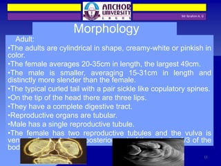
Morphology
Adult:
•The adults are cylindrical in shape, creamy-white or pinkish in
color.
•The female averages 20-35cm in length, the largest 49cm.
•The male is smaller, averaging 15-31cm in length and
distinctly more slender than the female.
•The typical curled tail with a pair sickle like copulatory spines.
•On the tip of the head there are three lips.
•They have a complete digestive tract.
•Reproductive organs are tubular.
•Male has a single reproductive tubule.
•The female has two reproductive tubules and the vulva is
ventrally located at the posterior part of the anterior 1/3 of the
body.
11
Mr Ibrahim A. G
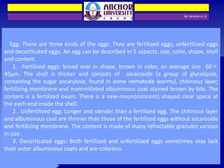
Egg: There are three kinds of the eggs. They are fertilized eggs, unfertilized eggs
and decorticated eggs. An egg can be described in 5 aspects: size, color, shape, shell
and content.
1. Fertilized eggs: broad oval in shape, brown in color, an average size 60×
45µm. The shell is thicker and consists of ascaroside (a group of glycolipids,
containing the sugar ascarylose, found in some nematode worms), chitinous layer,
fertilizing membrane and mammillated albuminous coat stained brown by bile. The
content is a fertilized ovum. There is a new-moon(crescent) shaped clear space at
the each end inside the shell.
2. Unfertilized egg: Longer and slender than a fertilized egg. The chitinous layer
and albuminous coat are thinner than those of the fertilized eggs without ascaroside
and fertilizing membrane. The content is made of many refractable granules various
in size.
3. Decorticated eggs: Both fertilized and unfertilized eggs sometimes may lack
their outer albuminous coats and are colorless.
12
Mr Ibrahim A. G
A fertilized Ascaris egg, still at the unicellular
stage, as they are when passed in stool. Eggs
are normally at this stage when passed in the
stool
The chitinous layer and albuminous coat
of unfertilized egg are thinner than
those of the fertilized eggs without
ascaroside and fertilizing membrane.
The content is made of many
refractable granules various in size.
Decorticated egg:
13
Mr Ibrahim A. G
Life Cycle
1. Site of inhabitation: small intestine
2. Infective stage: embryonated eggs
3. Route of infection: by mouth
4. No intermediate and reservoir hosts
5. Life span of the adult: about 1 year
This worm lives in the lumen of small intestine, feeding on the
intestinal contents, where the fertilized female lays eggs. An adult
female can produce approximately 200,000 eggs per day, which are
passed in feces. When passed, the eggs are unsegmented and
require outside development of about three weeks until a motile
embryo is formed within the egg.
14
Mr Ibrahim A. G
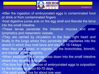
•After the ingestion of embryonated eggs in contaminated food
or drink or from contaminated fingers
•host digestive juices acts on the egg shell and liberate the larva
into the small intestine.
•These larvae penetrate the intestinal mucosa and enter
lymphatics and mesenteric vessels.
•They are carried by circulation to the liver, right heart and
finally to the lungs where they penetrate the capillaries into the
alveoli in which they molt twice and stay for 10-14days
•then they are carried, or migrate, up the bronchioles, bronchi,
and trachea to the epiglottis.
•When swallowed, the larvae pass down into the small intestine
where they develop into adults.
•The time from the ingestion of embryonated eggs to oviposition
by the females is about 60-75 days.
•The adult worms live for about one year.
15
Mr Ibrahim A. G
Adult worms (1) live in the lumen of the small intestine. A female may produce approximately 200,000 eggs per day,
which are passed with the feces (2). Unfertilized eggs may be ingested but are not infective. Fertile eggs embryonate and
become infective after 18 days to several weeks (3), depending on the environmental conditions (optimum: moist, warm,
shaded soil). After infective eggs are swallowed (4), the larvae hatch (5), invade the intestinal mucosa, and are carried via
the portal, then systemic circulation to the lungs (6). The larvae mature further in the lungs (10 to 14 days), penetrate the
alveolar walls, ascend the bronchial tree to the throat, and are swallowed (7). Upon reaching the small intestine, they
develop into adult worms (1). Between 2 and 3 months are required from ingestion of the infective eggs to oviposition by
the adult female. Adult worms can live 1 to 2 years. 16
Mr Ibrahim A. G
Pathogenesis
There are two phase in ascariasis:
1. The blood-lung migration phase of the larvae: During the migration
through the lungs, the larvae may cause a pneumonia. The symptoms of the
pneumonia are low fever, cough, blood-tinged sputum, asthma. Large
numbers of worms may give rise to allergic symptoms. Eosionophilia is
generally present. These clinical manifestation is also called Loeffler’s
syndrome.
2. The intestinal phase of the adults. The presence of a few adult worms in
the lumen of the small intestine usually produces no symptoms, but may
give rise to vague abdominal pains or intermittent colic, especially in
children. A heavy worm burden can result in malnutrition. More serious
manifestations have been observed. Wandering adults may block the
appendical lumen or the common bile duct and even perforate the intestinal
wall. Thus complications of ascariasis, such as intestinal obstruction,
appendicitis, biliary ascariasis, perforation of the intestine, cholecystitis,
pancreatitis and peritonitis, etc., may occur, in which biliary ascariasis is the
most common complication. 17
Mr Ibrahim A. G
Hookworms
The hookworms cause hookworm disease, which
is one of the five major parasitic disease in
China(malaria, shistosomiasis, filariasis, kala-azar
and hookworm disease). At least two species of
hookworms infect man, Necator americanus and
Ancylostoma duodenale. They live in small intestine.
18
Mr Ibrahim A. G
Morphology
1. Adults: They look like an odd piece
thread and are about 1cm. They are
white or light pinkish when living. ♀is
slightly larger than♂.The male’s
posterior end is expanded to form a
copulatory bursa.
2. Eggs: 60×40 µm in size, oval in shape,
shell is thin and colorless. Content is 2-
8cells.
19
Mr Ibrahim A. G
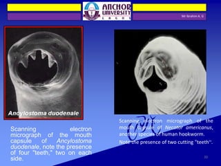
Scanning electron
micrograph of the mouth
capsule of Ancylostoma
duodenale, note the presence
of four "teeth," two on each
side.
Scanning electron micrograph of the
mouth capsule of Necator americanus,
another species of human hookworm.
Note the presence of two cutting "teeth“.
20
Mr Ibrahim A. G
The Morphological Differences between Two
species of Hookworms
_____________________________________________________
A. duodenale N. americanus
______________________________________________________
Size larger smaller
______________________________________________________
Shape single curve, looks like C double curves, looks like S
______________________________________________________
Mouth 2 pairs of ventral teeth 1pair of ventral cutting plates
____________________________________________________________
Copulatory circle in shape oval in shape
Bursa (a top view) (a top view)
____________________________________________________________
Copulatory 1pair with separate 1pair of which unite to form
spicule endings a terminal hooklet
_______________________________________________________
caudal spine present no
_______________________________________________________
vulva position post-equatorial pre-equatorial
_______________________________________________________
21
Mr Ibrahim A. G
Life Cycle
1. Final host: man
2. Infective Stage: Larva 3 or filariform larva
3. Infective Route: by skin
4. Food: blood and tissue fluid
5. Site of inhabitation: small intestine
6. Life span: Ad 15years, Na 3-7years
7. Blood-lung migration: skin- right heart- lungs
22
Mr Ibrahim A. G
Eggs are passed in the stool (1), and under favorable conditions (moisture, warmth, shade), larvae hatch in 1 to 2 days.
The released rhabditiform larvae grow in the feces and/or the soil (2), and after 5 to 10 days (and two molts) they
become filariform (third-stage) larvae that are infective (3) . These infective larvae can survive 3 to 4 weeks in favorable
environmental conditions. On contact with the human host, the larvae penetrate the skin and are carried through the
blood vessels to the heart and then to the lungs. They penetrate into the pulmonary alveoli, ascend the bronchial tree to
the pharynx, and are swallowed (4). The larvae reach the small intestine, where they reside and mature into adults.
Adult worms live in the lumen of the small intestine, where they attach to the intestinal wall with resultant blood loss by
the host (5). Most adult worms are eliminated in 1 to 2 years, but the longevity may reach several years.
Some A. duodenale larvae, following penetration of the host skin, can become dormant (in the intestine or muscle). In
addition, infection by A. duodenale may probably also occur by the oral and transmammary route. N. americanus,
however, requires a transpulmonary migration phase.
23
Mr Ibrahim A. G
Pathogenesis and Clinical Manifestations
1. Larval migration
(1) Dermatitis, known as "ground itch" or "stool poison". The larvae penetrating
the skin cause allergic reaction, petechiae or papule with itching and burning
sensation. Scratching leads to secondary infection.
(2) Pneumonitis (allergic reaction), Loeffler's syndrome: cough, asthma, low fever,
blood-tinged sputum or hemoptysis, chest-pain, inflammation shadows in lungs
under X-ray. These manifestations go on about 2 weeks.
2. Adults in small intestine
(1) Epigastric pain as that of a duodenal ulcer.
(2) A large worm burden results in microcytic hypochromatic anaemia
(character manifestation). The symptoms are lassitude, edema,
palpitation of the heart. In severe case, death may result from cardiac
failure or physical exhaustion.
(3) Allotriophagy (orpica) is due to the lack of trace element iron .
(4) Amenorrhea, sterility, abortion may take place in women.
(5) Gastrointestinal bleeding
(6) Infantile hookworm disease
24
• A common nematode residing in human
colon
• Human infection with whipworm once be
identified in coprolites of prehistoric man
• Worldwide distributed in the same areas
where the Ascaris are found
• Trichuriasis are usually not serious
clinically, but overwhelming infections
leading to death have been reported in
children
Mr Ibrahim A. G
Trichuris trichiura (whipworm)
25
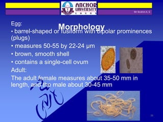
Egg:
• barrel-shaped or fusiform with bipolar prominences
(plugs)
• measures 50-55 by 22-24 µm
• brown, smooth shell
• contains a single-cell ovum
Adult:
The adult female measures about 35-50 mm in
length, and the male about 30-45 mm
Mr Ibrahim A. G
Morphology
26
Mr Ibrahim A. G
Life cycle
• No intermediate host
• Infected through fecal-oral route by the embryonated egg
• Ileocecal portion is the most favorite site of residing
27
Mr Ibrahim A. G
The unembryonated eggs are passed with the stool (1). In the soil, the eggs develop into a 2-cell stage (2), an
advanced cleavage stage(3) , and then they embryonate (4); eggs become infective in 15 to 30 days. After ingestion
(soil-contaminated hands or food), the eggs hatch in the small intestine, and release larvae (5) that mature and
establish themselves as adults in the colon (6). The adult worms (approximately 4 cm in length) live in the cecum and
ascending colon. The adult worms are fixed in that location, with the anterior portions threaded into the mucosa. The
females begin to oviposit 60 to 70 days after infection. Female worms in the cecum shed between 3,000 and 20,000
eggs per day. The life span of the adults is about 1 year.
28
Mr Ibrahim A. G
Pathogenesis
• Symptoms are determined largely by the worm
burden: less than 10 worms are asymptomatic
• Only heavily infected patients develop clinical disease:
Chronic diarrhea, characterized by mucous stools, and associated
with tenesmus
If the diarrhea is protracted, the patient may develop rectal
prolapse, more likely to occur in small children
Many individuals infected with whipworm tend to be
malnourished and anaemic
29
Mr Ibrahim A. G
prolapsed rectum
whipworms are often seen
attached to the rectal tissue
30
Mr Ibrahim A. G
Enterobius vermicularis (Pinworm)
1. A colon residing nematode of smaller size.
2. One of the most widely prevalent parasitic
nematodes of human being.
3. Affecting mainly children below the age of 12
years. No host other than man.
31
Mr Ibrahim A. G
Morphology
Adult :Small nematodes of
8~13mm in length by
0.3~0.6 mm in diameter,
with the spindly shape
Egg : smaller than that of
ascaris, asymmetrical,
with a thin and colorless
shell female
egg
male
anterior end of adult
32
Mr Ibrahim A. G
Life Cycle
1. Adults inhabit in the cecum. Female lay eggs in the perianal region
of the host.
2. No intermediate host, the egg takes around 6 hours to be matured
and becomes the infective stage.
3. Mode of infection may be auto-infection or cross-infection.
33
Mr Ibrahim A. G
Eggs are deposited on perianal folds (1). Self-infection occurs by transferring infective eggs to the mouth with hands
that have scratched the perianal area (2). Person-to-person transmission can also occur through handling of
contaminated clothes or bed linens. Enterobiasis may also be acquired through surfaces in the environment that are
contaminated with pinworm eggs (e.g. , curtains, carpeting). Some small number of eggs may become airborne and
inhaled. These would be swallowed and follow the same development as ingested eggs. Following ingestion of
infective eggs, the larvae hatch in the small intestine (3) and the adults establish themselves in the colon (4). The
time interval from ingestion of infective eggs to oviposition by the adult females is about one month. The life span of
the adults is about two months. Gravid females migrate nocturnally outside the anus and oviposit while crawling on
the skin of the perianal area (5). The larvae contained inside the eggs develop (the eggs become infective) in 4 to 6
hours under optimal conditions (1). Retroinfection, or the migration of newly hatched larvae from the anal skin back
into the rectum, may occur but the frequency with which this happens is unknown. 34
Mr Ibrahim A. G
Pathogenesis
1.The migratory phase of the pinworm is restricted to
the gastrointestinal tract and thus the host does not
experience any systemic reactions.
2. Perianal itching may be complicated. Heavy infections
in children may also produce such symptoms as
sleeplessness, weight loss, hyperactivity, grinding of
teeth, abdominal pain, and vomiting.
3. Ectopic parasitism may happen, which can cause uro-
genital inflammations or even the pelvic cavity can
be involved occasionally.
35
Mr Ibrahim A. G
The Filarial Worms
• The Filariae are long thread-like nematodes. Eight species
inhabit portions of the human subcutaneous tissues and
lymphatic system.
• Adults of all species are parasites of vertebrate hosts.
• Female worms produce eggs. The eggs modify, becoming
elongated and worm-like in appearance and adapting to life
within the vascular system.
• Modified eggs, referred to as microfilariae, are capable of
living a long time in the vertebrate host, but cannot develop
further until ingested by an intermediate host and vector, an
insect.
• Microfilariae transform into infective larvae in the insect and
are deposited in the next host when the insect takes a blood
meal. 36
Mr Ibrahim A. G
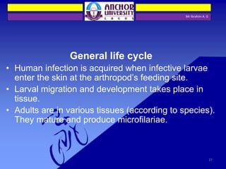
General life cycle
• Human infection is acquired when infective larvae
enter the skin at the arthropod’s feeding site.
• Larval migration and development takes place in
tissue.
• Adults are in various tissues (according to species).
They mature and produce microfilariae.
37
Mr Ibrahim A. G
Wuchereria bancrofti:
“Bancroft's Filariasis.” A blood & lymphatic
dweller. The infection often results in
elephantiasis.
• Vectors - Culex, Aedes, & Anopheles
mosquitoes.
• Diagnosis - Detection and identification of
microfilaria in stained blood smears. Exhibits a
marked circadian migration, best seen at night
after 10 P.M.
• Morphology - Microfilariae are sheathed, and the
nuclear column does not extend to tip of tail.
Wuchereria bancrofti microfilaria in blood smear
38
Mr Ibrahim A. G
Different species of the following genera of mosquitoes are vectors of W. bancrofti filariasis depending on geographical distribution. Among them are:
Culex (C. annulirostris, C. bitaeniorhynchus, C. quinquefasciatus, and C. pipiens); Anopheles (A. arabinensis, A. bancroftii, A. farauti, A. funestus, A.
gambiae, A. koliensis, A. melas, A. merus, A. punctulatus and A. wellcomei); Aedes (A. aegypti, A. aquasalis, A. bellator, A. cooki, A. darlingi, A. kochi, A.
polynesiensis, A. pseudoscutellaris, A. rotumae, A. scapularis, and A. vigilax); Mansonia (M. pseudotitillans, M. uniformis); Coquillettidia (C.
juxtamansonia). During a blood meal, an infected mosquito introduces third-stage filarial larvae onto the skin of the human host, where they penetrate
into the bite wound (1) . They develop in adults that commonly reside in the lymphatics (2) . The female worms measure 80 to 100 mm in length and 0.24
to 0.30 mm in diameter, while the males measure about 40 mm by .1 mm. Adults produce microfilariae measuring 244 to 296 μm by 7.5 to 10 μm, which
are sheathed and have nocturnal periodicity, except the South Pacific microfilariae which have the absence of marked periodicity. The microfilariae
migrate into lymph and blood channels moving actively through lymph and blood (3). A mosquito ingests the microfilariae during a blood meal (4). After
ingestion, the microfilariae lose their sheaths and some of them work their way through the wall of the proventriculus and cardiac portion of the
mosquito's midgut and reach the thoracic muscles (5). There the microfilariae develop into first-stage larvae (6) and subsequently into third-stage
infective larvae (7). The third-stage infective larvae migrate through the hemocoel to the mosquito's prosbocis (8) and can infect another human when
the mosquito takes a blood meal (1)
39
Mr Ibrahim A. G
Wuchereria bancrofti:
Major pathology and symptoms –
• Swelling, due to allergic reaction
occurring around adult worms, produces
obstruction & elephantiasis. Each
individual reacts differently. Very few
develop elephantiasis, but in some this is
extensive.
40
Onchocerca volvulus
Mr Ibrahim A. G
The “blinding filaria.” Infections involve the dermis and subcutaneous
tissues, where adults gather within nodules.
• Vector - Simulium flies (blackfly, or buffalo gnat).
• Diagnosis - microfilariae are found in skin scrapings from around
nodules.
• Morphology - Microfilariae not sheathed; found only in skin, not in the
blood stream.
41
Mr Ibrahim A. G
During a blood meal, an infected blackfly (genus Simulium) introduces third-stage filarial larvae onto the skin of the
human host, where they penetrate into the bite wound (1). In subcutaneous tissues the larvae (2) develop into adult
filariae, which commonly reside in nodules in subcutaneous connective tissues (3). Adults can live in the nodules for
approximately 15 years. Some nodules may contain numerous male and female worms. Females measure 33 to 50 cm in
length and 270 to 400 μm in diameter, while males measure 19 to 42 mm by 130 to 210 μm. In the subcutaneous
nodules, the female worms are capable of producing microfilariae for approximately 9 years. The microfilariae,
measuring 220 to 360 µm by 5 to 9 µm and unsheathed, have a life span that may reach 2 years. They are occasionally
found in peripheral blood, urine, and sputum but are typically found in the skin and in the lymphatics of connective
tissues (4). A blackfly ingests the microfilariae during a blood meal (5). After ingestion, the microfilariae migrate from the
blackfly's midgut through the hemocoel to the thoracic muscles (6). There the microfilariae develop into first-stage
larvae (7) and subsequently into third-stage infective larvae (8). The third-stage infective larvae migrate to the blackfly's
proboscis (9) and can infect another human when the fly takes a blood meal (1). 42
Mr Ibrahim A. G
Onchocerca volvulus:
Major pathology and symptoms -
Characterized by fibrotic nodules which
encapsulate adults, usually on the trunk in
Africa, and on the head in central America. A
progressive, allergic skin rash develops.
Blindness occurs due to the presence of
microfilariae in ocular structures. This parasite
is a major cause of blindness in Africa. Control
is difficult because Simulium flies breed in
running water.
43
Loa loa
Mr Ibrahim A. G
The “eyeworm.” Infections involve the dermis and
subcutaneous tissues (Calabar swellings).
• Vector - Crysops (mango fly), a large fly with biting
mouthparts.
• Diagnosis - Usually made from clinical symptoms,
but if laboratory confirmation is required, blood
should be drawn between 11 am & 1 pm.
• Diagnosis - Microfilariae are sheathed, nuclear
column extends to tip of tail.
44
Mr Ibrahim A. G
The vector for Loa loa filariasis are flies from two species of the genus Chrysops, C. silacea and C. dimidiata. During a
blood meal, an infected fly (genus Chrysops, day-biting flies) introduces third-stage filarial larvae onto the skin of the
human host, where they penetrate into the bite wound (1). The larvae develop into adults that commonly reside in
subcutaneous tissue (2). The female worms measure 40 to 70 mm in length and 0.5 mm in diameter, while the males
measure 30 to 34 mm in length and 0.35 to 0.43 mm in diameter. Adults produce microfilariae measuring 250 to 300 μm
by 6 to 8 μm, which are sheathed and have diurnal periodicity. Microfilariae have been recovered from spinal fluids,
urine, and sputum. During the day they are found in peripheral blood, but during the noncirculation phase, they are
found in the lungs (3). The fly ingests microfilariae during a blood meal (4). After ingestion, the microfilariae lose their
sheaths and migrate from the fly's midgut through the hemocoel to the thoracic muscles of the arthropod (5). There the
microfilariae develop into first-stage larvae (6) and subsequently into third-stage infective larvae (7) . The third-stage
infective larvae migrate to the fly's proboscis (8)and can infect another human when the fly takes a blood meal (1). 45
Mr Ibrahim A. G
Loa loa:
Major pathology and symptoms -
Infections cause a localized
subcutaneous edema, particularly
around the eye, because of larval
migration and death in capillaries. Living
adults cause no inflammation; dying
adults induce granulomatous reactions.
46

More Related Content

What's hot

Entrobius vermicularis
Entrobius vermicularisEntrobius vermicularis
Entrobius vermicularis
MahaGamalAldein
 
Enterobius vermicularis(PINWORM)
Enterobius vermicularis(PINWORM)Enterobius vermicularis(PINWORM)
Enterobius vermicularis(PINWORM)
Syed Ali
 
Cestode
CestodeCestode
Cestode
Aman Ullah
 
Tissue nematodes
Tissue nematodesTissue nematodes
Tissue nematodes
Prasad Gunjal
 
Taeniasis
TaeniasisTaeniasis
Taeniasis
Vanindar Kumar
 
Helminths
HelminthsHelminths
Enterobius vermicularis
Enterobius vermicularisEnterobius vermicularis
Enterobius vermicularis
Ahmadminhas2
 
Hymenolepis nana
Hymenolepis nanaHymenolepis nana
Hymenolepis nana
Raafi Ul Zargar
 
Taenia saginata
Taenia saginataTaenia saginata
Taenia saginata
Usman Ayub Awan
 
Ascaris lumbricoides
Ascaris lumbricoidesAscaris lumbricoides
Ascaris lumbricoides
RaNa MB
 
Hook worm
Hook wormHook worm
Hook worm
Subhash Karna
 
Fleas.pptx
Fleas.pptxFleas.pptx
Fleas.pptx
TehseenKhan34
 
Cestodes (The Taeenid tapeworms)
Cestodes (The Taeenid tapeworms)Cestodes (The Taeenid tapeworms)
Cestodes (The Taeenid tapeworms)
med zar
 
Enterobius vermicularis
Enterobius vermicularisEnterobius vermicularis
Enterobius vermicularis
RaNa MB
 
Helminth introduction
Helminth introductionHelminth introduction
Helminth introduction
Sk. Mizanur Rahman
 
Echinococcus granulosus
Echinococcus granulosusEchinococcus granulosus
Echinococcus granulosus
Santosh Kumar Yadav
 
Coccidian parasites- Cryptosporidiosis
Coccidian parasites- CryptosporidiosisCoccidian parasites- Cryptosporidiosis
Coccidian parasites- Cryptosporidiosis
Suprakash Das
 
Flea lecture
Flea lectureFlea lecture
Flea lecture
Osama Zahid
 
Nematoda
NematodaNematoda
Nematoda
Vivek Dev
 
Hookworms
HookwormsHookworms
Hookworms
MahaGamalAldein
 

What's hot (20)

Entrobius vermicularis
Entrobius vermicularisEntrobius vermicularis
Entrobius vermicularis
 
Enterobius vermicularis(PINWORM)
Enterobius vermicularis(PINWORM)Enterobius vermicularis(PINWORM)
Enterobius vermicularis(PINWORM)
 
Cestode
CestodeCestode
Cestode
 
Tissue nematodes
Tissue nematodesTissue nematodes
Tissue nematodes
 
Taeniasis
TaeniasisTaeniasis
Taeniasis
 
Helminths
HelminthsHelminths
Helminths
 
Enterobius vermicularis
Enterobius vermicularisEnterobius vermicularis
Enterobius vermicularis
 
Hymenolepis nana
Hymenolepis nanaHymenolepis nana
Hymenolepis nana
 
Taenia saginata
Taenia saginataTaenia saginata
Taenia saginata
 
Ascaris lumbricoides
Ascaris lumbricoidesAscaris lumbricoides
Ascaris lumbricoides
 
Hook worm
Hook wormHook worm
Hook worm
 
Fleas.pptx
Fleas.pptxFleas.pptx
Fleas.pptx
 
Cestodes (The Taeenid tapeworms)
Cestodes (The Taeenid tapeworms)Cestodes (The Taeenid tapeworms)
Cestodes (The Taeenid tapeworms)
 
Enterobius vermicularis
Enterobius vermicularisEnterobius vermicularis
Enterobius vermicularis
 
Helminth introduction
Helminth introductionHelminth introduction
Helminth introduction
 
Echinococcus granulosus
Echinococcus granulosusEchinococcus granulosus
Echinococcus granulosus
 
Coccidian parasites- Cryptosporidiosis
Coccidian parasites- CryptosporidiosisCoccidian parasites- Cryptosporidiosis
Coccidian parasites- Cryptosporidiosis
 
Flea lecture
Flea lectureFlea lecture
Flea lecture
 
Nematoda
NematodaNematoda
Nematoda
 
Hookworms
HookwormsHookworms
Hookworms
 

Similar to Nematode

Classes of Aschelminthes Powerpoint Presentation
Classes of Aschelminthes Powerpoint PresentationClasses of Aschelminthes Powerpoint Presentation
Classes of Aschelminthes Powerpoint Presentation
FaizRehman50
 
Ascariasis
Ascariasis Ascariasis
Ascariasis
10gupta
 
General feature of male & female Ascaris, comparison of Ascaris ,Life cycl...
 General feature of male & female  Ascaris, comparison of Ascaris  ,Life cycl... General feature of male & female  Ascaris, comparison of Ascaris  ,Life cycl...
General feature of male & female Ascaris, comparison of Ascaris ,Life cycl...
SoniaBajaj10
 
Ascariasis
AscariasisAscariasis
Ascariasis
raghunathp
 
Pathogenic nemadodes & disease.pptx
Pathogenic nemadodes & disease.pptxPathogenic nemadodes & disease.pptx
Pathogenic nemadodes & disease.pptx
SoniaBajaj10
 
Flatworm.pptx
Flatworm.pptxFlatworm.pptx
Flatworm.pptx
Apzventura1
 
Chapter 8. Medical entomology.pptx
Chapter 8. Medical entomology.pptxChapter 8. Medical entomology.pptx
Chapter 8. Medical entomology.pptx
MesfinMelese4
 
HOOKWORMS.pptx
HOOKWORMS.pptxHOOKWORMS.pptx
HOOKWORMS.pptx
JoshuaKalunda
 
18. phylum platyhelminthes II Full Explanation
18. phylum platyhelminthes II Full Explanation 18. phylum platyhelminthes II Full Explanation
18. phylum platyhelminthes II Full Explanation
Yo yo Nody khan
 
Taenia solium & Taenia saginata
Taenia solium & Taenia saginataTaenia solium & Taenia saginata
Taenia solium & Taenia saginata
Priyanka Patel
 
NEMATYHELMINTHES PDF.pdf
NEMATYHELMINTHES PDF.pdfNEMATYHELMINTHES PDF.pdf
NEMATYHELMINTHES PDF.pdf
MinaBasnet2
 
HELMINTHS lecture-1.pptx
HELMINTHS lecture-1.pptxHELMINTHS lecture-1.pptx
HELMINTHS lecture-1.pptx
RashidUllah7
 
Phylum nematoda 1
Phylum nematoda 1Phylum nematoda 1
Phylum nematoda 1
Hannah Shane Soriano
 
NEMATODA.ppt
NEMATODA.pptNEMATODA.ppt
NEMATODA.ppt
ShadrackKoech7
 
Parasitology Helmin Ascaris & Enterobius.pptx
Parasitology Helmin Ascaris & Enterobius.pptxParasitology Helmin Ascaris & Enterobius.pptx
Parasitology Helmin Ascaris & Enterobius.pptx
mohamederrmali1
 
Earthworm dissection
Earthworm dissectionEarthworm dissection
Earthworm dissection
ewaszolek
 
HOOK WORMS L17 in parasitology (medical lab sciences) .pptx
HOOK WORMS L17 in parasitology (medical lab sciences) .pptxHOOK WORMS L17 in parasitology (medical lab sciences) .pptx
HOOK WORMS L17 in parasitology (medical lab sciences) .pptx
ChaudharyArslanUlHaq
 
Ectoparasites : Ticks , Mites , Fleas , Flies
Ectoparasites : Ticks , Mites , Fleas , FliesEctoparasites : Ticks , Mites , Fleas , Flies
Ectoparasites : Ticks , Mites , Fleas , Flies
Bimochan Poudel
 
Annelida
AnnelidaAnnelida
Annelida
Alaa Elnagar
 
Chagu
ChaguChagu

Similar to Nematode (20)

Classes of Aschelminthes Powerpoint Presentation
Classes of Aschelminthes Powerpoint PresentationClasses of Aschelminthes Powerpoint Presentation
Classes of Aschelminthes Powerpoint Presentation
 
Ascariasis
Ascariasis Ascariasis
Ascariasis
 
General feature of male & female Ascaris, comparison of Ascaris ,Life cycl...
 General feature of male & female  Ascaris, comparison of Ascaris  ,Life cycl... General feature of male & female  Ascaris, comparison of Ascaris  ,Life cycl...
General feature of male & female Ascaris, comparison of Ascaris ,Life cycl...
 
Ascariasis
AscariasisAscariasis
Ascariasis
 
Pathogenic nemadodes & disease.pptx
Pathogenic nemadodes & disease.pptxPathogenic nemadodes & disease.pptx
Pathogenic nemadodes & disease.pptx
 
Flatworm.pptx
Flatworm.pptxFlatworm.pptx
Flatworm.pptx
 
Chapter 8. Medical entomology.pptx
Chapter 8. Medical entomology.pptxChapter 8. Medical entomology.pptx
Chapter 8. Medical entomology.pptx
 
HOOKWORMS.pptx
HOOKWORMS.pptxHOOKWORMS.pptx
HOOKWORMS.pptx
 
18. phylum platyhelminthes II Full Explanation
18. phylum platyhelminthes II Full Explanation 18. phylum platyhelminthes II Full Explanation
18. phylum platyhelminthes II Full Explanation
 
Taenia solium & Taenia saginata
Taenia solium & Taenia saginataTaenia solium & Taenia saginata
Taenia solium & Taenia saginata
 
NEMATYHELMINTHES PDF.pdf
NEMATYHELMINTHES PDF.pdfNEMATYHELMINTHES PDF.pdf
NEMATYHELMINTHES PDF.pdf
 
HELMINTHS lecture-1.pptx
HELMINTHS lecture-1.pptxHELMINTHS lecture-1.pptx
HELMINTHS lecture-1.pptx
 
Phylum nematoda 1
Phylum nematoda 1Phylum nematoda 1
Phylum nematoda 1
 
NEMATODA.ppt
NEMATODA.pptNEMATODA.ppt
NEMATODA.ppt
 
Parasitology Helmin Ascaris & Enterobius.pptx
Parasitology Helmin Ascaris & Enterobius.pptxParasitology Helmin Ascaris & Enterobius.pptx
Parasitology Helmin Ascaris & Enterobius.pptx
 
Earthworm dissection
Earthworm dissectionEarthworm dissection
Earthworm dissection
 
HOOK WORMS L17 in parasitology (medical lab sciences) .pptx
HOOK WORMS L17 in parasitology (medical lab sciences) .pptxHOOK WORMS L17 in parasitology (medical lab sciences) .pptx
HOOK WORMS L17 in parasitology (medical lab sciences) .pptx
 
Ectoparasites : Ticks , Mites , Fleas , Flies
Ectoparasites : Ticks , Mites , Fleas , FliesEctoparasites : Ticks , Mites , Fleas , Flies
Ectoparasites : Ticks , Mites , Fleas , Flies
 
Annelida
AnnelidaAnnelida
Annelida
 
Chagu
ChaguChagu
Chagu
 

Recently uploaded

The use of Nauplii and metanauplii artemia in aquaculture (brine shrimp).pptx
The use of Nauplii and metanauplii artemia in aquaculture (brine shrimp).pptxThe use of Nauplii and metanauplii artemia in aquaculture (brine shrimp).pptx
The use of Nauplii and metanauplii artemia in aquaculture (brine shrimp).pptx
MAGOTI ERNEST
 
Oedema_types_causes_pathophysiology.pptx
Oedema_types_causes_pathophysiology.pptxOedema_types_causes_pathophysiology.pptx
Oedema_types_causes_pathophysiology.pptx
muralinath2
 
Travis Hills' Endeavors in Minnesota: Fostering Environmental and Economic Pr...
Travis Hills' Endeavors in Minnesota: Fostering Environmental and Economic Pr...Travis Hills' Endeavors in Minnesota: Fostering Environmental and Economic Pr...
Travis Hills' Endeavors in Minnesota: Fostering Environmental and Economic Pr...
Travis Hills MN
 
如何办理(uvic毕业证书)维多利亚大学毕业证本科学位证书原版一模一样
如何办理(uvic毕业证书)维多利亚大学毕业证本科学位证书原版一模一样如何办理(uvic毕业证书)维多利亚大学毕业证本科学位证书原版一模一样
如何办理(uvic毕业证书)维多利亚大学毕业证本科学位证书原版一模一样
yqqaatn0
 
What is greenhouse gasses and how many gasses are there to affect the Earth.
What is greenhouse gasses and how many gasses are there to affect the Earth.What is greenhouse gasses and how many gasses are there to affect the Earth.
What is greenhouse gasses and how many gasses are there to affect the Earth.
moosaasad1975
 
Shallowest Oil Discovery of Turkiye.pptx
Shallowest Oil Discovery of Turkiye.pptxShallowest Oil Discovery of Turkiye.pptx
Shallowest Oil Discovery of Turkiye.pptx
Gokturk Mehmet Dilci
 
mô tả các thí nghiệm về đánh giá tác động dòng khí hóa sau đốt
mô tả các thí nghiệm về đánh giá tác động dòng khí hóa sau đốtmô tả các thí nghiệm về đánh giá tác động dòng khí hóa sau đốt
mô tả các thí nghiệm về đánh giá tác động dòng khí hóa sau đốt
HongcNguyn6
 
20240520 Planning a Circuit Simulator in JavaScript.pptx
20240520 Planning a Circuit Simulator in JavaScript.pptx20240520 Planning a Circuit Simulator in JavaScript.pptx
20240520 Planning a Circuit Simulator in JavaScript.pptx
Sharon Liu
 
aziz sancar nobel prize winner: from mardin to nobel
aziz sancar nobel prize winner: from mardin to nobelaziz sancar nobel prize winner: from mardin to nobel
aziz sancar nobel prize winner: from mardin to nobel
İsa Badur
 
Equivariant neural networks and representation theory
Equivariant neural networks and representation theoryEquivariant neural networks and representation theory
Equivariant neural networks and representation theory
Daniel Tubbenhauer
 
ANAMOLOUS SECONDARY GROWTH IN DICOT ROOTS.pptx
ANAMOLOUS SECONDARY GROWTH IN DICOT ROOTS.pptxANAMOLOUS SECONDARY GROWTH IN DICOT ROOTS.pptx
ANAMOLOUS SECONDARY GROWTH IN DICOT ROOTS.pptx
RASHMI M G
 
Cytokines and their role in immune regulation.pptx
Cytokines and their role in immune regulation.pptxCytokines and their role in immune regulation.pptx
Cytokines and their role in immune regulation.pptx
Hitesh Sikarwar
 
Applied Science: Thermodynamics, Laws & Methodology.pdf
Applied Science: Thermodynamics, Laws & Methodology.pdfApplied Science: Thermodynamics, Laws & Methodology.pdf
Applied Science: Thermodynamics, Laws & Methodology.pdf
University of Hertfordshire
 
bordetella pertussis.................................ppt
bordetella pertussis.................................pptbordetella pertussis.................................ppt
bordetella pertussis.................................ppt
kejapriya1
 
THEMATIC APPERCEPTION TEST(TAT) cognitive abilities, creativity, and critic...
THEMATIC  APPERCEPTION  TEST(TAT) cognitive abilities, creativity, and critic...THEMATIC  APPERCEPTION  TEST(TAT) cognitive abilities, creativity, and critic...
THEMATIC APPERCEPTION TEST(TAT) cognitive abilities, creativity, and critic...
Abdul Wali Khan University Mardan,kP,Pakistan
 
Deep Software Variability and Frictionless Reproducibility
Deep Software Variability and Frictionless ReproducibilityDeep Software Variability and Frictionless Reproducibility
Deep Software Variability and Frictionless Reproducibility
University of Rennes, INSA Rennes, Inria/IRISA, CNRS
 
Remote Sensing and Computational, Evolutionary, Supercomputing, and Intellige...
Remote Sensing and Computational, Evolutionary, Supercomputing, and Intellige...Remote Sensing and Computational, Evolutionary, Supercomputing, and Intellige...
Remote Sensing and Computational, Evolutionary, Supercomputing, and Intellige...
University of Maribor
 
Bob Reedy - Nitrate in Texas Groundwater.pdf
Bob Reedy - Nitrate in Texas Groundwater.pdfBob Reedy - Nitrate in Texas Groundwater.pdf
Bob Reedy - Nitrate in Texas Groundwater.pdf
Texas Alliance of Groundwater Districts
 
Sharlene Leurig - Enabling Onsite Water Use with Net Zero Water
Sharlene Leurig - Enabling Onsite Water Use with Net Zero WaterSharlene Leurig - Enabling Onsite Water Use with Net Zero Water
Sharlene Leurig - Enabling Onsite Water Use with Net Zero Water
Texas Alliance of Groundwater Districts
 
Medical Orthopedic PowerPoint Templates.pptx
Medical Orthopedic PowerPoint Templates.pptxMedical Orthopedic PowerPoint Templates.pptx
Medical Orthopedic PowerPoint Templates.pptx
terusbelajar5
 

Recently uploaded (20)

The use of Nauplii and metanauplii artemia in aquaculture (brine shrimp).pptx
The use of Nauplii and metanauplii artemia in aquaculture (brine shrimp).pptxThe use of Nauplii and metanauplii artemia in aquaculture (brine shrimp).pptx
The use of Nauplii and metanauplii artemia in aquaculture (brine shrimp).pptx
 
Oedema_types_causes_pathophysiology.pptx
Oedema_types_causes_pathophysiology.pptxOedema_types_causes_pathophysiology.pptx
Oedema_types_causes_pathophysiology.pptx
 
Travis Hills' Endeavors in Minnesota: Fostering Environmental and Economic Pr...
Travis Hills' Endeavors in Minnesota: Fostering Environmental and Economic Pr...Travis Hills' Endeavors in Minnesota: Fostering Environmental and Economic Pr...
Travis Hills' Endeavors in Minnesota: Fostering Environmental and Economic Pr...
 
如何办理(uvic毕业证书)维多利亚大学毕业证本科学位证书原版一模一样
如何办理(uvic毕业证书)维多利亚大学毕业证本科学位证书原版一模一样如何办理(uvic毕业证书)维多利亚大学毕业证本科学位证书原版一模一样
如何办理(uvic毕业证书)维多利亚大学毕业证本科学位证书原版一模一样
 
What is greenhouse gasses and how many gasses are there to affect the Earth.
What is greenhouse gasses and how many gasses are there to affect the Earth.What is greenhouse gasses and how many gasses are there to affect the Earth.
What is greenhouse gasses and how many gasses are there to affect the Earth.
 
Shallowest Oil Discovery of Turkiye.pptx
Shallowest Oil Discovery of Turkiye.pptxShallowest Oil Discovery of Turkiye.pptx
Shallowest Oil Discovery of Turkiye.pptx
 
mô tả các thí nghiệm về đánh giá tác động dòng khí hóa sau đốt
mô tả các thí nghiệm về đánh giá tác động dòng khí hóa sau đốtmô tả các thí nghiệm về đánh giá tác động dòng khí hóa sau đốt
mô tả các thí nghiệm về đánh giá tác động dòng khí hóa sau đốt
 
20240520 Planning a Circuit Simulator in JavaScript.pptx
20240520 Planning a Circuit Simulator in JavaScript.pptx20240520 Planning a Circuit Simulator in JavaScript.pptx
20240520 Planning a Circuit Simulator in JavaScript.pptx
 
aziz sancar nobel prize winner: from mardin to nobel
aziz sancar nobel prize winner: from mardin to nobelaziz sancar nobel prize winner: from mardin to nobel
aziz sancar nobel prize winner: from mardin to nobel
 
Equivariant neural networks and representation theory
Equivariant neural networks and representation theoryEquivariant neural networks and representation theory
Equivariant neural networks and representation theory
 
ANAMOLOUS SECONDARY GROWTH IN DICOT ROOTS.pptx
ANAMOLOUS SECONDARY GROWTH IN DICOT ROOTS.pptxANAMOLOUS SECONDARY GROWTH IN DICOT ROOTS.pptx
ANAMOLOUS SECONDARY GROWTH IN DICOT ROOTS.pptx
 
Cytokines and their role in immune regulation.pptx
Cytokines and their role in immune regulation.pptxCytokines and their role in immune regulation.pptx
Cytokines and their role in immune regulation.pptx
 
Applied Science: Thermodynamics, Laws & Methodology.pdf
Applied Science: Thermodynamics, Laws & Methodology.pdfApplied Science: Thermodynamics, Laws & Methodology.pdf
Applied Science: Thermodynamics, Laws & Methodology.pdf
 
bordetella pertussis.................................ppt
bordetella pertussis.................................pptbordetella pertussis.................................ppt
bordetella pertussis.................................ppt
 
THEMATIC APPERCEPTION TEST(TAT) cognitive abilities, creativity, and critic...
THEMATIC  APPERCEPTION  TEST(TAT) cognitive abilities, creativity, and critic...THEMATIC  APPERCEPTION  TEST(TAT) cognitive abilities, creativity, and critic...
THEMATIC APPERCEPTION TEST(TAT) cognitive abilities, creativity, and critic...
 
Deep Software Variability and Frictionless Reproducibility
Deep Software Variability and Frictionless ReproducibilityDeep Software Variability and Frictionless Reproducibility
Deep Software Variability and Frictionless Reproducibility
 
Remote Sensing and Computational, Evolutionary, Supercomputing, and Intellige...
Remote Sensing and Computational, Evolutionary, Supercomputing, and Intellige...Remote Sensing and Computational, Evolutionary, Supercomputing, and Intellige...
Remote Sensing and Computational, Evolutionary, Supercomputing, and Intellige...
 
Bob Reedy - Nitrate in Texas Groundwater.pdf
Bob Reedy - Nitrate in Texas Groundwater.pdfBob Reedy - Nitrate in Texas Groundwater.pdf
Bob Reedy - Nitrate in Texas Groundwater.pdf
 
Sharlene Leurig - Enabling Onsite Water Use with Net Zero Water
Sharlene Leurig - Enabling Onsite Water Use with Net Zero WaterSharlene Leurig - Enabling Onsite Water Use with Net Zero Water
Sharlene Leurig - Enabling Onsite Water Use with Net Zero Water
 
Medical Orthopedic PowerPoint Templates.pptx
Medical Orthopedic PowerPoint Templates.pptxMedical Orthopedic PowerPoint Templates.pptx
Medical Orthopedic PowerPoint Templates.pptx
 

Nematode

  • 1. Zoo 211: Lower invertebrate General Characteristics, Biology and Economic Importance of Phylum Nematoda Mr Ibrahim A. G 1
  • 2. Introduction •Nematoda (nematos-thread), about 25,000 species have been named •They live in the sea, freshwater and in good top soils from polar to the tropic region •They parasitized virtually every type of animal and many plants •Virtually every species of vertebrate and invertebrates serve as host for one or more types of parasitic nematodes •Nematodes have numerous pathological impact on human and domestic animals Mr Ibrahim A. G 2
  • 3. Characteristics •Bilaterally symmetrical, unsegmented worms •Triploblastic and pseudocoelomate animals •Body wall with cuticle, cellular or syncytial epidermis and longitudinal muscles in four bands •No cilia, no circulatory and respiratory systems •Excretory system of glandular organ or canal or both •Digestive system complete with muscular pharynx and glands •Sense organs are in the form of pits, papillae, bristles and eye spots •Dioecious; reproductive organs relatively simple and male is smaller than female •Fertilization is internal •Aquatic or terrestrial, free-living or parasitic, enlongated roundworms Mr Ibrahim A. G 3
  • 4. Mr Ibrahim A. G Ascaris Hookworm Wucheraria whipworm Guinea worm Brugia 4
  • 5. Morphology •Nematode body is cylindrical in shape, flexible and made up of nonliving cuticle •Most are less than 5cm long but some parasitic forms are more than 1m in length •Covering is relatively thick, noncellular cuticle, secreted by underlying epidermis (hypodermis) •Cuticle is shed during juvenile growth stages •Several layers of the cuticle are primarily collagen •Fluid-filled pseudocoel constitute a hydrostatic skeleton •The interaction of their muscles, cuticle and fluid- filled pseudocoel aid the movement of nematodes Mr Ibrahim A. G 5
  • 6. Digestive system • The alimentary canal of nematodes consist of: -mouth -muscular pharynx -a long nonmuscular intestine -a short rectum and -a terminal anus •Food is sucked into the pharynx when muscle in its anterior portion contract rapidly and open the lumen •Relaxation of the muscle anterior to the food mass closes the lumen of the pharynx, forcing the food posteriorly towards the intestine (one cell-layer thick) •Food matter moves posteriorly by body movements and by additional food being passed into the intestine from the pharynx •Defecation is accomplished by muscles that simply pull anus open, and expulsive force is provided by the high pseudocoelomic pressure. Mr Ibrahim A. G 6
  • 7. Nervous system •A ring of nerve tissue and ganglia around the pharynx gives rise to small nerves to the anterior end and to two nerve cords, one dorsal and one ventral •Sensory papillae are concentrated around the head and tail •The amphidial opening leads into a deep cuticular pit with sensory endings of modified cilia *The Amphids are a pair of somewhat more complex sensory organs that open on each side of the head at about the same level as the cephalic circle of papillae •Most nematodes bear a bilateral pair of phasmids (similar in structure to amphids) near the posterior end. Mr Ibrahim A. G 7
  • 8. Reproduction •Most nematodes are dioecious •Males are smaller than females and their posterior end usually bears a pair of copulatory spicules •Fertilization is internal, and eggs are usually stored in the uterus until deposition •Development among free-living forms is typically direct •Four juvenile stages are each separated by a molt, or shedding, of the cuticle •Many parasitic nematodes have free-living juvenile stages •Others require intermediate host to complete their life cycles Mr Ibrahim A. G 8
  • 9. Classification of the Phylum Nematoda The Phylum Nematoda is divided into two classes: Class Adenophorea (Aphasmidia) Class Secernentea (Phasmidia) Class Adenophorea (Aphasmidia) • They lack phasmids (caudal sensory organs). • Excretory system is rudimentary or poorly developed. • Males have one spicule. • E.g. Trichuris sp. etc. Class Secernentea (Phasmidia) •They have phasmids • Excretory system is present and well developed. • Males have two spicules. • E.g. Rhabditis, Wuchereria, Onchocerca spp., Ascaris, etc. Mr Ibrahim A. G 9
  • 10. Mr Ibrahim A. G Ascaris Lumbricoides Ascaris lumbricoides , (askaris-intestinal worm) common saying “round worm of man”, is the largest of the intestinal nematodes parasitizing humans. It is the most common worm found in human. It is worldwide in distribution and most prevalent through out the tropics, sub-tropics and more prevalent in the countryside than in the city 10
  • 11. Mr Ibrahim A. G Morphology Adult: •The adults are cylindrical in shape, creamy-white or pinkish in color. •The female averages 20-35cm in length, the largest 49cm. •The male is smaller, averaging 15-31cm in length and distinctly more slender than the female. •The typical curled tail with a pair sickle like copulatory spines. •On the tip of the head there are three lips. •They have a complete digestive tract. •Reproductive organs are tubular. •Male has a single reproductive tubule. •The female has two reproductive tubules and the vulva is ventrally located at the posterior part of the anterior 1/3 of the body. 11
  • 12. Mr Ibrahim A. G Egg: There are three kinds of the eggs. They are fertilized eggs, unfertilized eggs and decorticated eggs. An egg can be described in 5 aspects: size, color, shape, shell and content. 1. Fertilized eggs: broad oval in shape, brown in color, an average size 60× 45µm. The shell is thicker and consists of ascaroside (a group of glycolipids, containing the sugar ascarylose, found in some nematode worms), chitinous layer, fertilizing membrane and mammillated albuminous coat stained brown by bile. The content is a fertilized ovum. There is a new-moon(crescent) shaped clear space at the each end inside the shell. 2. Unfertilized egg: Longer and slender than a fertilized egg. The chitinous layer and albuminous coat are thinner than those of the fertilized eggs without ascaroside and fertilizing membrane. The content is made of many refractable granules various in size. 3. Decorticated eggs: Both fertilized and unfertilized eggs sometimes may lack their outer albuminous coats and are colorless. 12
  • 13. Mr Ibrahim A. G A fertilized Ascaris egg, still at the unicellular stage, as they are when passed in stool. Eggs are normally at this stage when passed in the stool The chitinous layer and albuminous coat of unfertilized egg are thinner than those of the fertilized eggs without ascaroside and fertilizing membrane. The content is made of many refractable granules various in size. Decorticated egg: 13
  • 14. Mr Ibrahim A. G Life Cycle 1. Site of inhabitation: small intestine 2. Infective stage: embryonated eggs 3. Route of infection: by mouth 4. No intermediate and reservoir hosts 5. Life span of the adult: about 1 year This worm lives in the lumen of small intestine, feeding on the intestinal contents, where the fertilized female lays eggs. An adult female can produce approximately 200,000 eggs per day, which are passed in feces. When passed, the eggs are unsegmented and require outside development of about three weeks until a motile embryo is formed within the egg. 14
  • 15. Mr Ibrahim A. G •After the ingestion of embryonated eggs in contaminated food or drink or from contaminated fingers •host digestive juices acts on the egg shell and liberate the larva into the small intestine. •These larvae penetrate the intestinal mucosa and enter lymphatics and mesenteric vessels. •They are carried by circulation to the liver, right heart and finally to the lungs where they penetrate the capillaries into the alveoli in which they molt twice and stay for 10-14days •then they are carried, or migrate, up the bronchioles, bronchi, and trachea to the epiglottis. •When swallowed, the larvae pass down into the small intestine where they develop into adults. •The time from the ingestion of embryonated eggs to oviposition by the females is about 60-75 days. •The adult worms live for about one year. 15
  • 16. Mr Ibrahim A. G Adult worms (1) live in the lumen of the small intestine. A female may produce approximately 200,000 eggs per day, which are passed with the feces (2). Unfertilized eggs may be ingested but are not infective. Fertile eggs embryonate and become infective after 18 days to several weeks (3), depending on the environmental conditions (optimum: moist, warm, shaded soil). After infective eggs are swallowed (4), the larvae hatch (5), invade the intestinal mucosa, and are carried via the portal, then systemic circulation to the lungs (6). The larvae mature further in the lungs (10 to 14 days), penetrate the alveolar walls, ascend the bronchial tree to the throat, and are swallowed (7). Upon reaching the small intestine, they develop into adult worms (1). Between 2 and 3 months are required from ingestion of the infective eggs to oviposition by the adult female. Adult worms can live 1 to 2 years. 16
  • 17. Mr Ibrahim A. G Pathogenesis There are two phase in ascariasis: 1. The blood-lung migration phase of the larvae: During the migration through the lungs, the larvae may cause a pneumonia. The symptoms of the pneumonia are low fever, cough, blood-tinged sputum, asthma. Large numbers of worms may give rise to allergic symptoms. Eosionophilia is generally present. These clinical manifestation is also called Loeffler’s syndrome. 2. The intestinal phase of the adults. The presence of a few adult worms in the lumen of the small intestine usually produces no symptoms, but may give rise to vague abdominal pains or intermittent colic, especially in children. A heavy worm burden can result in malnutrition. More serious manifestations have been observed. Wandering adults may block the appendical lumen or the common bile duct and even perforate the intestinal wall. Thus complications of ascariasis, such as intestinal obstruction, appendicitis, biliary ascariasis, perforation of the intestine, cholecystitis, pancreatitis and peritonitis, etc., may occur, in which biliary ascariasis is the most common complication. 17
  • 18. Mr Ibrahim A. G Hookworms The hookworms cause hookworm disease, which is one of the five major parasitic disease in China(malaria, shistosomiasis, filariasis, kala-azar and hookworm disease). At least two species of hookworms infect man, Necator americanus and Ancylostoma duodenale. They live in small intestine. 18
  • 19. Mr Ibrahim A. G Morphology 1. Adults: They look like an odd piece thread and are about 1cm. They are white or light pinkish when living. ♀is slightly larger than♂.The male’s posterior end is expanded to form a copulatory bursa. 2. Eggs: 60×40 µm in size, oval in shape, shell is thin and colorless. Content is 2- 8cells. 19
  • 20. Mr Ibrahim A. G Scanning electron micrograph of the mouth capsule of Ancylostoma duodenale, note the presence of four "teeth," two on each side. Scanning electron micrograph of the mouth capsule of Necator americanus, another species of human hookworm. Note the presence of two cutting "teeth“. 20
  • 21. Mr Ibrahim A. G The Morphological Differences between Two species of Hookworms _____________________________________________________ A. duodenale N. americanus ______________________________________________________ Size larger smaller ______________________________________________________ Shape single curve, looks like C double curves, looks like S ______________________________________________________ Mouth 2 pairs of ventral teeth 1pair of ventral cutting plates ____________________________________________________________ Copulatory circle in shape oval in shape Bursa (a top view) (a top view) ____________________________________________________________ Copulatory 1pair with separate 1pair of which unite to form spicule endings a terminal hooklet _______________________________________________________ caudal spine present no _______________________________________________________ vulva position post-equatorial pre-equatorial _______________________________________________________ 21
  • 22. Mr Ibrahim A. G Life Cycle 1. Final host: man 2. Infective Stage: Larva 3 or filariform larva 3. Infective Route: by skin 4. Food: blood and tissue fluid 5. Site of inhabitation: small intestine 6. Life span: Ad 15years, Na 3-7years 7. Blood-lung migration: skin- right heart- lungs 22
  • 23. Mr Ibrahim A. G Eggs are passed in the stool (1), and under favorable conditions (moisture, warmth, shade), larvae hatch in 1 to 2 days. The released rhabditiform larvae grow in the feces and/or the soil (2), and after 5 to 10 days (and two molts) they become filariform (third-stage) larvae that are infective (3) . These infective larvae can survive 3 to 4 weeks in favorable environmental conditions. On contact with the human host, the larvae penetrate the skin and are carried through the blood vessels to the heart and then to the lungs. They penetrate into the pulmonary alveoli, ascend the bronchial tree to the pharynx, and are swallowed (4). The larvae reach the small intestine, where they reside and mature into adults. Adult worms live in the lumen of the small intestine, where they attach to the intestinal wall with resultant blood loss by the host (5). Most adult worms are eliminated in 1 to 2 years, but the longevity may reach several years. Some A. duodenale larvae, following penetration of the host skin, can become dormant (in the intestine or muscle). In addition, infection by A. duodenale may probably also occur by the oral and transmammary route. N. americanus, however, requires a transpulmonary migration phase. 23
  • 24. Mr Ibrahim A. G Pathogenesis and Clinical Manifestations 1. Larval migration (1) Dermatitis, known as "ground itch" or "stool poison". The larvae penetrating the skin cause allergic reaction, petechiae or papule with itching and burning sensation. Scratching leads to secondary infection. (2) Pneumonitis (allergic reaction), Loeffler's syndrome: cough, asthma, low fever, blood-tinged sputum or hemoptysis, chest-pain, inflammation shadows in lungs under X-ray. These manifestations go on about 2 weeks. 2. Adults in small intestine (1) Epigastric pain as that of a duodenal ulcer. (2) A large worm burden results in microcytic hypochromatic anaemia (character manifestation). The symptoms are lassitude, edema, palpitation of the heart. In severe case, death may result from cardiac failure or physical exhaustion. (3) Allotriophagy (orpica) is due to the lack of trace element iron . (4) Amenorrhea, sterility, abortion may take place in women. (5) Gastrointestinal bleeding (6) Infantile hookworm disease 24
  • 25. • A common nematode residing in human colon • Human infection with whipworm once be identified in coprolites of prehistoric man • Worldwide distributed in the same areas where the Ascaris are found • Trichuriasis are usually not serious clinically, but overwhelming infections leading to death have been reported in children Mr Ibrahim A. G Trichuris trichiura (whipworm) 25
  • 26. Egg: • barrel-shaped or fusiform with bipolar prominences (plugs) • measures 50-55 by 22-24 µm • brown, smooth shell • contains a single-cell ovum Adult: The adult female measures about 35-50 mm in length, and the male about 30-45 mm Mr Ibrahim A. G Morphology 26
  • 27. Mr Ibrahim A. G Life cycle • No intermediate host • Infected through fecal-oral route by the embryonated egg • Ileocecal portion is the most favorite site of residing 27
  • 28. Mr Ibrahim A. G The unembryonated eggs are passed with the stool (1). In the soil, the eggs develop into a 2-cell stage (2), an advanced cleavage stage(3) , and then they embryonate (4); eggs become infective in 15 to 30 days. After ingestion (soil-contaminated hands or food), the eggs hatch in the small intestine, and release larvae (5) that mature and establish themselves as adults in the colon (6). The adult worms (approximately 4 cm in length) live in the cecum and ascending colon. The adult worms are fixed in that location, with the anterior portions threaded into the mucosa. The females begin to oviposit 60 to 70 days after infection. Female worms in the cecum shed between 3,000 and 20,000 eggs per day. The life span of the adults is about 1 year. 28
  • 29. Mr Ibrahim A. G Pathogenesis • Symptoms are determined largely by the worm burden: less than 10 worms are asymptomatic • Only heavily infected patients develop clinical disease: Chronic diarrhea, characterized by mucous stools, and associated with tenesmus If the diarrhea is protracted, the patient may develop rectal prolapse, more likely to occur in small children Many individuals infected with whipworm tend to be malnourished and anaemic 29
  • 30. Mr Ibrahim A. G prolapsed rectum whipworms are often seen attached to the rectal tissue 30
  • 31. Mr Ibrahim A. G Enterobius vermicularis (Pinworm) 1. A colon residing nematode of smaller size. 2. One of the most widely prevalent parasitic nematodes of human being. 3. Affecting mainly children below the age of 12 years. No host other than man. 31
  • 32. Mr Ibrahim A. G Morphology Adult :Small nematodes of 8~13mm in length by 0.3~0.6 mm in diameter, with the spindly shape Egg : smaller than that of ascaris, asymmetrical, with a thin and colorless shell female egg male anterior end of adult 32
  • 33. Mr Ibrahim A. G Life Cycle 1. Adults inhabit in the cecum. Female lay eggs in the perianal region of the host. 2. No intermediate host, the egg takes around 6 hours to be matured and becomes the infective stage. 3. Mode of infection may be auto-infection or cross-infection. 33
  • 34. Mr Ibrahim A. G Eggs are deposited on perianal folds (1). Self-infection occurs by transferring infective eggs to the mouth with hands that have scratched the perianal area (2). Person-to-person transmission can also occur through handling of contaminated clothes or bed linens. Enterobiasis may also be acquired through surfaces in the environment that are contaminated with pinworm eggs (e.g. , curtains, carpeting). Some small number of eggs may become airborne and inhaled. These would be swallowed and follow the same development as ingested eggs. Following ingestion of infective eggs, the larvae hatch in the small intestine (3) and the adults establish themselves in the colon (4). The time interval from ingestion of infective eggs to oviposition by the adult females is about one month. The life span of the adults is about two months. Gravid females migrate nocturnally outside the anus and oviposit while crawling on the skin of the perianal area (5). The larvae contained inside the eggs develop (the eggs become infective) in 4 to 6 hours under optimal conditions (1). Retroinfection, or the migration of newly hatched larvae from the anal skin back into the rectum, may occur but the frequency with which this happens is unknown. 34
  • 35. Mr Ibrahim A. G Pathogenesis 1.The migratory phase of the pinworm is restricted to the gastrointestinal tract and thus the host does not experience any systemic reactions. 2. Perianal itching may be complicated. Heavy infections in children may also produce such symptoms as sleeplessness, weight loss, hyperactivity, grinding of teeth, abdominal pain, and vomiting. 3. Ectopic parasitism may happen, which can cause uro- genital inflammations or even the pelvic cavity can be involved occasionally. 35
  • 36. Mr Ibrahim A. G The Filarial Worms • The Filariae are long thread-like nematodes. Eight species inhabit portions of the human subcutaneous tissues and lymphatic system. • Adults of all species are parasites of vertebrate hosts. • Female worms produce eggs. The eggs modify, becoming elongated and worm-like in appearance and adapting to life within the vascular system. • Modified eggs, referred to as microfilariae, are capable of living a long time in the vertebrate host, but cannot develop further until ingested by an intermediate host and vector, an insect. • Microfilariae transform into infective larvae in the insect and are deposited in the next host when the insect takes a blood meal. 36
  • 37. Mr Ibrahim A. G General life cycle • Human infection is acquired when infective larvae enter the skin at the arthropod’s feeding site. • Larval migration and development takes place in tissue. • Adults are in various tissues (according to species). They mature and produce microfilariae. 37
  • 38. Mr Ibrahim A. G Wuchereria bancrofti: “Bancroft's Filariasis.” A blood & lymphatic dweller. The infection often results in elephantiasis. • Vectors - Culex, Aedes, & Anopheles mosquitoes. • Diagnosis - Detection and identification of microfilaria in stained blood smears. Exhibits a marked circadian migration, best seen at night after 10 P.M. • Morphology - Microfilariae are sheathed, and the nuclear column does not extend to tip of tail. Wuchereria bancrofti microfilaria in blood smear 38
  • 39. Mr Ibrahim A. G Different species of the following genera of mosquitoes are vectors of W. bancrofti filariasis depending on geographical distribution. Among them are: Culex (C. annulirostris, C. bitaeniorhynchus, C. quinquefasciatus, and C. pipiens); Anopheles (A. arabinensis, A. bancroftii, A. farauti, A. funestus, A. gambiae, A. koliensis, A. melas, A. merus, A. punctulatus and A. wellcomei); Aedes (A. aegypti, A. aquasalis, A. bellator, A. cooki, A. darlingi, A. kochi, A. polynesiensis, A. pseudoscutellaris, A. rotumae, A. scapularis, and A. vigilax); Mansonia (M. pseudotitillans, M. uniformis); Coquillettidia (C. juxtamansonia). During a blood meal, an infected mosquito introduces third-stage filarial larvae onto the skin of the human host, where they penetrate into the bite wound (1) . They develop in adults that commonly reside in the lymphatics (2) . The female worms measure 80 to 100 mm in length and 0.24 to 0.30 mm in diameter, while the males measure about 40 mm by .1 mm. Adults produce microfilariae measuring 244 to 296 μm by 7.5 to 10 μm, which are sheathed and have nocturnal periodicity, except the South Pacific microfilariae which have the absence of marked periodicity. The microfilariae migrate into lymph and blood channels moving actively through lymph and blood (3). A mosquito ingests the microfilariae during a blood meal (4). After ingestion, the microfilariae lose their sheaths and some of them work their way through the wall of the proventriculus and cardiac portion of the mosquito's midgut and reach the thoracic muscles (5). There the microfilariae develop into first-stage larvae (6) and subsequently into third-stage infective larvae (7). The third-stage infective larvae migrate through the hemocoel to the mosquito's prosbocis (8) and can infect another human when the mosquito takes a blood meal (1) 39
  • 40. Mr Ibrahim A. G Wuchereria bancrofti: Major pathology and symptoms – • Swelling, due to allergic reaction occurring around adult worms, produces obstruction & elephantiasis. Each individual reacts differently. Very few develop elephantiasis, but in some this is extensive. 40
  • 41. Onchocerca volvulus Mr Ibrahim A. G The “blinding filaria.” Infections involve the dermis and subcutaneous tissues, where adults gather within nodules. • Vector - Simulium flies (blackfly, or buffalo gnat). • Diagnosis - microfilariae are found in skin scrapings from around nodules. • Morphology - Microfilariae not sheathed; found only in skin, not in the blood stream. 41
  • 42. Mr Ibrahim A. G During a blood meal, an infected blackfly (genus Simulium) introduces third-stage filarial larvae onto the skin of the human host, where they penetrate into the bite wound (1). In subcutaneous tissues the larvae (2) develop into adult filariae, which commonly reside in nodules in subcutaneous connective tissues (3). Adults can live in the nodules for approximately 15 years. Some nodules may contain numerous male and female worms. Females measure 33 to 50 cm in length and 270 to 400 μm in diameter, while males measure 19 to 42 mm by 130 to 210 μm. In the subcutaneous nodules, the female worms are capable of producing microfilariae for approximately 9 years. The microfilariae, measuring 220 to 360 µm by 5 to 9 µm and unsheathed, have a life span that may reach 2 years. They are occasionally found in peripheral blood, urine, and sputum but are typically found in the skin and in the lymphatics of connective tissues (4). A blackfly ingests the microfilariae during a blood meal (5). After ingestion, the microfilariae migrate from the blackfly's midgut through the hemocoel to the thoracic muscles (6). There the microfilariae develop into first-stage larvae (7) and subsequently into third-stage infective larvae (8). The third-stage infective larvae migrate to the blackfly's proboscis (9) and can infect another human when the fly takes a blood meal (1). 42
  • 43. Mr Ibrahim A. G Onchocerca volvulus: Major pathology and symptoms - Characterized by fibrotic nodules which encapsulate adults, usually on the trunk in Africa, and on the head in central America. A progressive, allergic skin rash develops. Blindness occurs due to the presence of microfilariae in ocular structures. This parasite is a major cause of blindness in Africa. Control is difficult because Simulium flies breed in running water. 43
  • 44. Loa loa Mr Ibrahim A. G The “eyeworm.” Infections involve the dermis and subcutaneous tissues (Calabar swellings). • Vector - Crysops (mango fly), a large fly with biting mouthparts. • Diagnosis - Usually made from clinical symptoms, but if laboratory confirmation is required, blood should be drawn between 11 am & 1 pm. • Diagnosis - Microfilariae are sheathed, nuclear column extends to tip of tail. 44
  • 45. Mr Ibrahim A. G The vector for Loa loa filariasis are flies from two species of the genus Chrysops, C. silacea and C. dimidiata. During a blood meal, an infected fly (genus Chrysops, day-biting flies) introduces third-stage filarial larvae onto the skin of the human host, where they penetrate into the bite wound (1). The larvae develop into adults that commonly reside in subcutaneous tissue (2). The female worms measure 40 to 70 mm in length and 0.5 mm in diameter, while the males measure 30 to 34 mm in length and 0.35 to 0.43 mm in diameter. Adults produce microfilariae measuring 250 to 300 μm by 6 to 8 μm, which are sheathed and have diurnal periodicity. Microfilariae have been recovered from spinal fluids, urine, and sputum. During the day they are found in peripheral blood, but during the noncirculation phase, they are found in the lungs (3). The fly ingests microfilariae during a blood meal (4). After ingestion, the microfilariae lose their sheaths and migrate from the fly's midgut through the hemocoel to the thoracic muscles of the arthropod (5). There the microfilariae develop into first-stage larvae (6) and subsequently into third-stage infective larvae (7) . The third-stage infective larvae migrate to the fly's proboscis (8)and can infect another human when the fly takes a blood meal (1). 45
  • 46. Mr Ibrahim A. G Loa loa: Major pathology and symptoms - Infections cause a localized subcutaneous edema, particularly around the eye, because of larval migration and death in capillaries. Living adults cause no inflammation; dying adults induce granulomatous reactions. 46