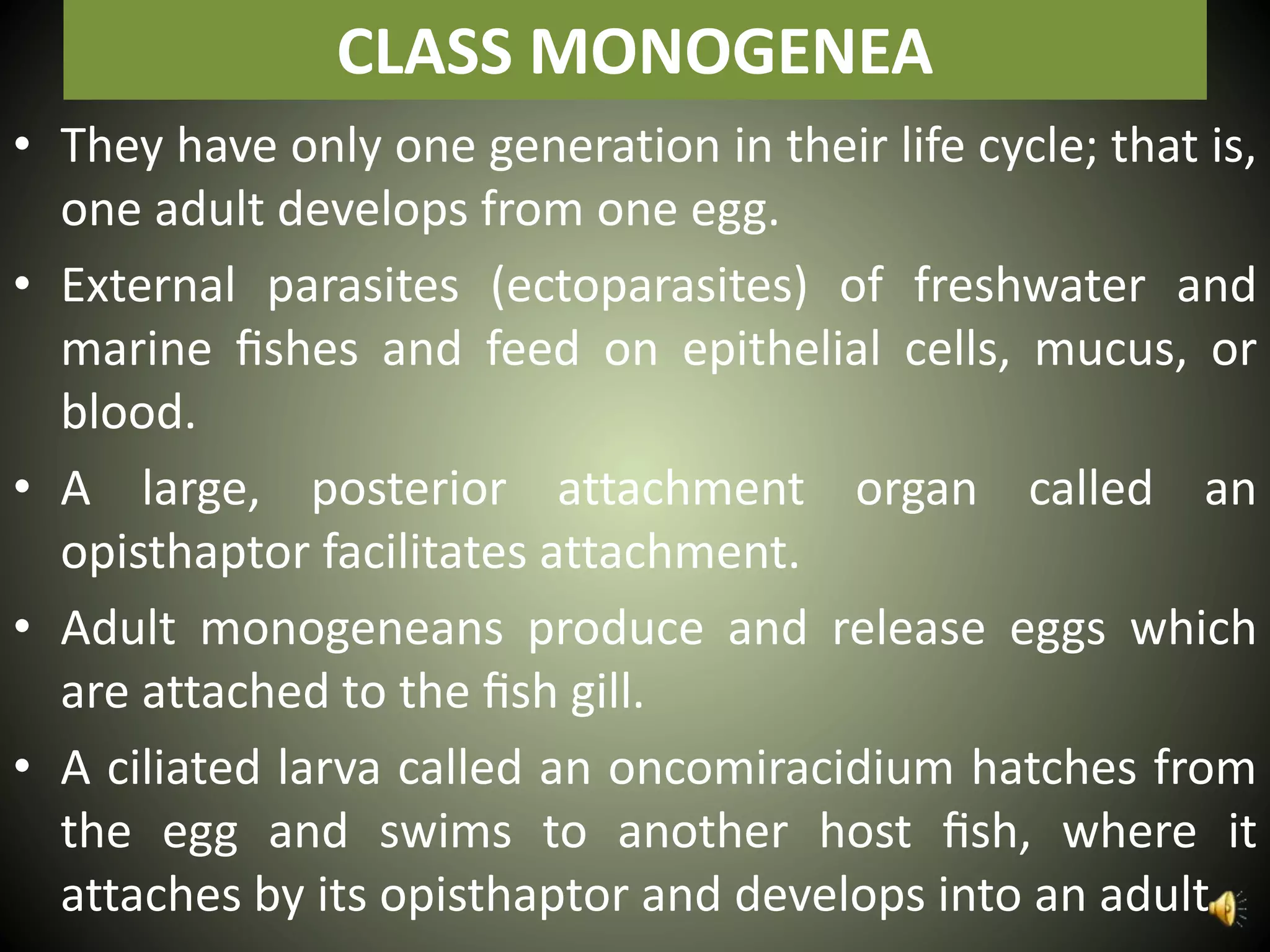
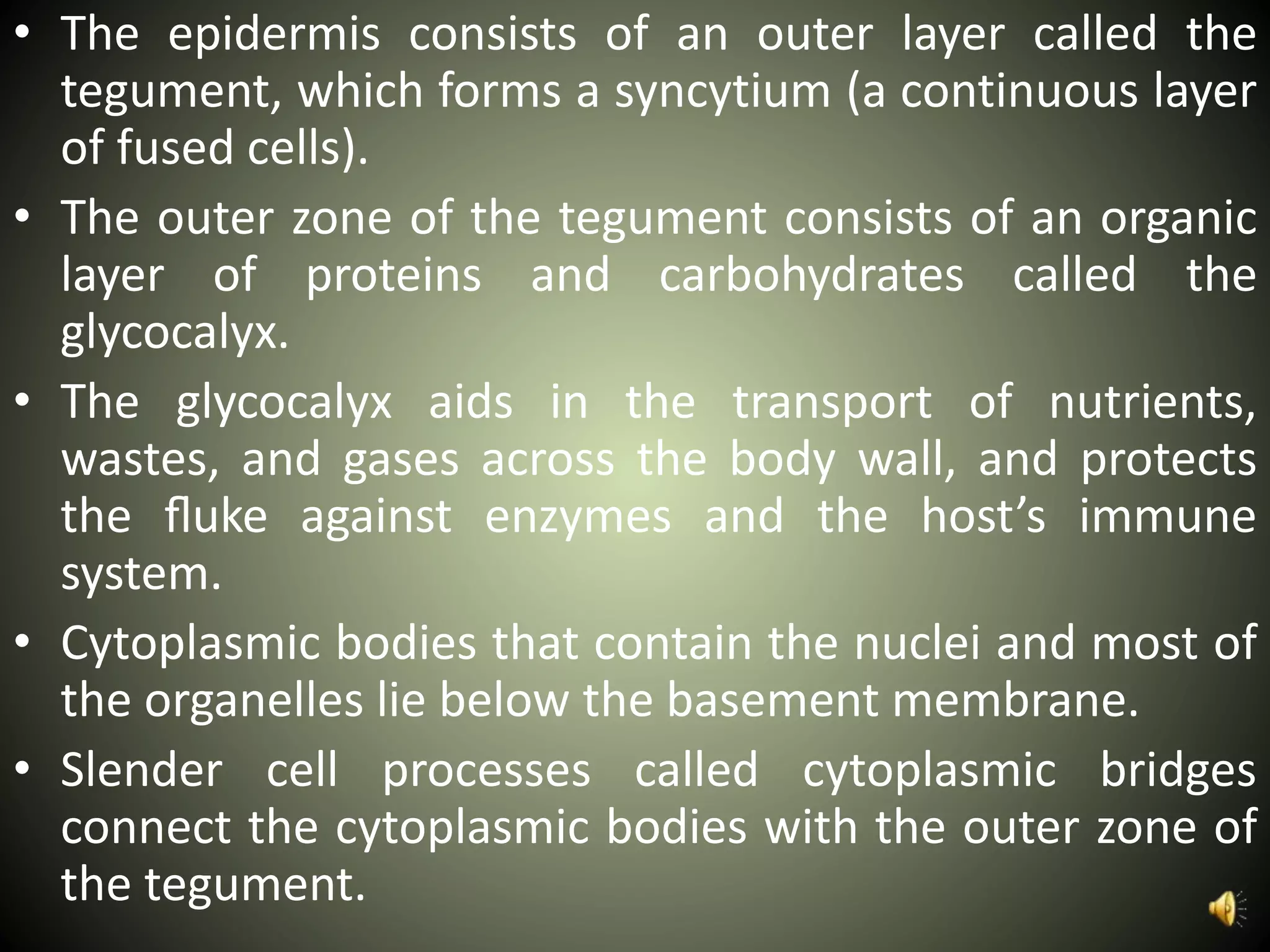
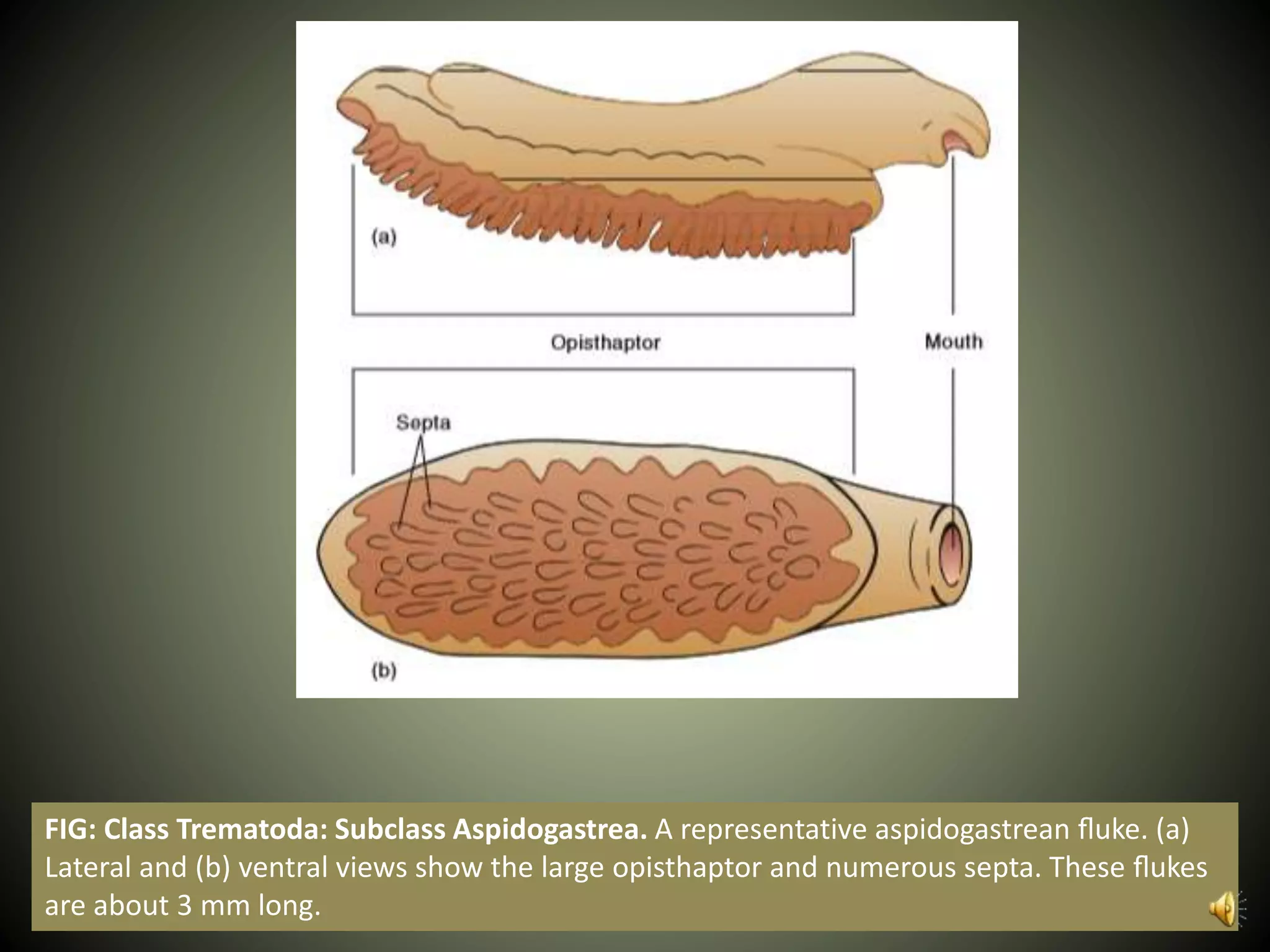
Phylum Platyhelminthes
By
SYED SHAKEEL SHAH
Department of Zoology
University of Narowal
CLASS MONOGENEA
• They have only one generation in their life cycle; that is,
one adult develops from one egg.
• External parasites (ectoparasites) of freshwater and
marine fishes and feed on epithelial cells, mucus, or
blood.
• A large, posterior attachment organ called an
opisthaptor facilitates attachment.
• Adult monogeneans produce and release eggs which
are attached to the fish gill.
• A ciliated larva called an oncomiracidium hatches from
the egg and swims to another host fish, where it
attaches by its opisthaptor and develops into an adult.
FIG: Class Monogenea. Two monogeneid trematodes. (a) Gyrodactylus. (b)
Sphyranura. Note the opisthaptors by which these ectoparasites cling to the gills
of their fish hosts. Both of these monogenea are about 1 cm long.
CLASS TREMATODA
• Class Trematoda are collectively called flukes.
• Almost all adult flukes are parasites of vertebrates,
whereas immature stages may be found in vertebrates
or invertebrates, or encysted on plants.
• Most flukes are flat and oval to elongate, and range
from less than 1 mm to 6 cm in length.
• They feed on host cells and cell fragments.
• The digestive tract includes a mouth and a muscular,
pumping pharynx.
• Posterior to the pharynx, the digestive tract divides into
two blind-ending, variously branched pouches called
cecae.
• Some flukes supplement their feeding by absorbing
nutrients across their body walls.
FIG: Generalized Fluke (Digenetic Trematode). Note the large percentage of the body devoted
to reproduction. The Mehlis’ gland is a conspicuous feature of the female reproductive tract; its
function in trematodes is uncertain.
• The epidermis consists of an outer layer called the
tegument, which forms a syncytium (a continuous layer
of fused cells).
• The outer zone of the tegument consists of an organic
layer of proteins and carbohydrates called the
glycocalyx.
• The glycocalyx aids in the transport of nutrients,
wastes, and gases across the body wall, and protects
the fluke against enzymes and the host’s immune
system.
• Cytoplasmic bodies that contain the nuclei and most of
the organelles lie below the basement membrane.
• Slender cell processes called cytoplasmic bridges
connect the cytoplasmic bodies with the outer zone of
the tegument.
FIG: Trematode Tegument. The fine structure of the tegument of a fluke. The
tegument is an evolutionary adaptation that is highly efficient at absorbing
nutrients and effective for protection.
Subclass Aspidogastrea
• They are primarily internal parasites (endoparasites) of
molluscs.
• A large, oval holdfast organ called the opisthaptor
covers the entire ventral surface of the animal and
characterizes all aspidogastreans.
• The opisthaptor is an extremely strong attachment
organ, and ridges or septa usually subdivide it.
• The oral sucker, characteristic of most other trematode
mouths, is absent.
• The life cycle of aspidogastreans may involve only one
host (a mollusc) or two hosts.
• In the latter case, the final host is usually a vertebrate
(fishes or turtles) that becomes infected by ingesting a
mollusc that contains immature aspidogastreans.
FIG: Class Trematoda: Subclass Aspidogastrea. A representative aspidogastrean fluke. (a)
Lateral and (b) ventral views show the large opisthaptor and numerous septa. These flukes
are about 3 mm long.
Subclass Digenea
• At least two different forms, an adult and one or more larval
stages, develop—a characteristic from which the name of the
subclass was derived.
• Because digenetic flukes require at least two different hosts
to complete their life cycles.
• As adults, they are all endoparasites in the bloodstreams,
digestive tracts, ducts of the digestive organs, or other
visceral organs in a wide variety of vertebrates that serve as
definitive, or final, hosts.
• The one or more intermediate hosts (the hosts that harbor
immature stages) may harbor several different larval stages.
• The adhesive organs are two large suckers.
• The anterior sucker is the oral sucker and surrounds the
mouth.
• The other sucker, the acetabulum, is located ventrally on the
middle portion of the body.
FIG: Generalized Fluke (Digenetic Trematode). Note the large percentage of the body devoted
to reproduction. The Mehlis’ gland is a conspicuous feature of the female reproductive tract; its
function in trematodes is uncertain.
• The eggs of digenetic trematodes are oval and usually
have a lidlike hatch called an operculum.
• When an egg reaches freshwater, the operculum
opens, and a ciliated larva called a miracidium swims
out.
• The miracidium swims until it finds a suitable first
intermediate host (a snail) to which it is chemically
attracted.
• The miracidium penetrates the snail, loses its cilia, and
develops into a sporocyst. (Alternately, the miracidium
may remain in the egg and hatch after a snail eats it.)
• Sporocysts are baglike and contain embryonic cells that
develop into either daughter sporocysts or rediae(sing.,
redia).
• Asexual reproduction first occurs.
• From a single miracidium, hundreds of daughter
sporocysts, and in turn, hundreds of rediae can form by
asexual reproduction.
• Embryonic cells in each daughter sporocyst or redia
produce hundreds of the next larval stage, called
cercariae. (This phenomenon of producing many
cercariae is called polyembryony. It greatly enhances
the chances that one cercaria will further the life cycle.)
• A cercaria has a digestive tract, suckers, and a tail.
• Cercariae leave the snail and swim freely until they
encounter a second intermediate or final host,
which may be a vertebrate, invertebrate, or plant.
• The cercaria penetrates this host and encysts as a
metacercaria (pl., metacercariae).
• When the definitive host eats the second
intermediate host, the metacercaria excysts and
develops into an adult.
FIG: Class Trematoda: Subclass Digenea. The life cycle of the digenetic trematode,
Fasciola hepatica (the common liver fluke). The adult is about 30 mm long and 13 mm
wide. The cercaria is about 0.5 mm long.
Some Important Trematode Parasites of
Humans
Clonorchis sinensis
(The Chinese liver fluke)
• The adult lives in the bile ducts of the liver, where it
feeds on epithelial tissue and blood.
• The adults release embryonated eggs into the
common bile duct.
• The eggs make their way to the intestine and are
eliminated with feces.
FIG: Chinese Liver Fluke, Clonorchis sinensis. (a) Dorsal view. (b) Life cycle. The adult worm
is 10 to 25 mm long and 1 to 5 mm wide.
Fasciola hepatica
(the sheep liver fluke)
• The adults live in the bile duct of the liver.
• Eggs pass via the common bile duct to the intestine,
from which they are eliminated.
• Eggs deposited in freshwater hatch, and the
miracidia must locate the proper species of snail.
• If a snail is found, miracidia penetrate the snails’s
soft tissue and develop into sporocysts that develop
into rediae and give rise to cercariae.
• After the cercariae emerge from the snail, they
encyst on aquatic vegetation.
• Sheep or other animals become infected when they
graze on the aquatic vegetation.
• Humans may become infected with Fasciola
hepatica by eating a freshwater plant called
watercress that contains the encysted metacercaria.
Schistosomes
• Infections are most common in Africa (Schistosoma
haematobium and S. mansoni), South and Central America (S.
mansoni), and Southeast Asia (S. japonicum).
• The adult dioecious worms live in the human bloodstream.
• The male fluke is shorter and thicker than the female, and the
sides of the male body curve under to form a canal along the
ventral surface.
• The female fluke is long and slender, and is carried in the
canal of the male.
• Copulation is continuous, and the female produces thousands
of eggs over her lifetime.
• Each egg contains a spine that mechanically aids it in moving
through host tissue until it is eliminated in either the feces or
urine.
• Unlike other flukes, schistosome eggs lack an operculum.
FIG: Representative Life Cycle of a Schistosome Fluke. The cycle begins in a human (a) when the female fluke lays eggs (b,c)
in the thin-walled, small vessels of the large or small intestine (S. mansoni and S. japonicum) or urinary bladder (S.
haematobium). Secretions from the eggs weaken the walls, and the blood vessels rupture, releasing eggs into the intestinal
lumen or urinary bladder. From there, the eggs leave the body. If they reach freshwater, the eggs hatch into ciliated, free-
swimming larvae called miracidia (d). A miracidium burrows into the tissues of an aquatic snail (e), losing its cilia in the
process, and develops into a sporocyst, then daughter sporocysts. Eventually, forked-tailed larvae (cercariae) are produced
(f). After the cercariae leave the snail, they actively swim about. If they encounter human skin (g), they attach to it and
release tissue-degrading enzymes. The larvae enter the body and migrate to the circulatory system, where they mature.
They end up at the vessels of the intestines or urinary bladder, where sexual reproduction takes place, and the cycle begins
anew. The adult worms are 10 to 20 mm long.
CLASS CESTOIDEA: THE TAPEWORMS
• Commonly called either tapeworms or cestodes.
• They are endoparasites that usually reside in the
vertebrate digestive system.
• Their color is often white with shades of yellow or
gray.
• Adult tapeworms range from 1 mm to 25 m in
length.
• Two unique adaptations to a parasitic lifestyle
characterize tapeworms:
(1) Tapeworms lack a mouth and digestive tract in all of
their life-cycle stages; they absorb nutrients directly
across their body wall.
(2) Most adult tapeworms consist of a long series of
repeating units called proglottids.
• Each proglottid contains a complete set of
reproductive structures.
• The physiology of the intestine of tapeworm’s host
maintains the tapeworm’s homeostasis (internal
constancy).
• In adapting to such a specialized environment,
tapeworms have lost some of the structures
believed to have been present in ancestral
turbellarians.
• Tapeworms are, therefore, a good example of
evolution not always resulting in greater complexity.
Subclass Cestodaria
• They are all endoparasites in the intestine and
coelom of primitive fishes.
• They possess some digenetic trematode features in
that only one set of both reproductive systems is
present in each animal, some bear suckers, and
their bodies are not divided into proglottids like
other cestodes.
• Yet, the absence of a digestive system, the presence
of larval stages similar to those of cestodes, and the
presence of parenchymal muscle cells, which are
not present in any other platyhelminth, all suggest
strong phylogenetic affinities with other cestodes.
Subclass Eucestoda
• They are called true tapeworms.
• The body is divided into three regions.
• At one end is a hold-fast structure called the scolex that
contains circular or leaflike suckers and sometimes a
rostellum of hooks.
• With the scolex, the tapeworm firmly anchors itself to
the intestinal wall of its definitive vertebrate host.
• No mouth is present.
• Posteriorly, the scolex narrows to form the neck.
• Transverse constrictions in the neck give rise to the
third body region, the strobila.
• The strobila consists of a series of linearly arranged
proglottids, which function primarily as reproductive
units.
• As a tapeworm grows, new proglottids are added in the
neck region, and older proglottids are gradually pushed
posteriorly.
• As they move posteriorly, proglottids mature and begin
producing eggs. Thus, anterior proglottids are said to
be immature, those in the midregion of the strobila are
mature, and those at the posterior end that have
accumulated eggs are gravid.
• The outer body wall of tapeworms consists of a
tegument similar in structure to that of trematodes.
• It plays a vital role in nutrient absorption because
tapeworms have no digestive system.
• The nervous system consists of only a pair of lateral
nerve cords that arise from a nerve mass in the scolex
and extend the length of the strobila.
• A protonephridial system also runs the length of the
tapeworm.
• Tapeworms are monoecious.
• Each proglottid contains a complete set of male and
female reproductive organs.
• Numerous testes are scattered throughout the
proglottid and deliver sperm via a duct system to a
copulatory organ called a cirrus.
• The cirrus opens through a genital pore, which is an
opening shared with the female system.
• The male system of a proglottid matures before the
female system, so that copulation usually occurs with
another mature proglottid of the same tapeworm or
with another tapeworm in the same host.
• As previously mentioned, the avoidance of self-
fertilization leads to hybrid vigor.
• A single pair of ovaries in each proglottid produce
eggs.
• Sperm stored in a seminal receptacle fertilize eggs
as the eggs move through the oviduct.
• Vitelline cells from the vitelline gland are then
dumped onto the eggs in the ootype.
• The ootype is an expanded region of the oviduct
that shapes capsules around the eggs.
• The ootype is also surrounded by the Mehlis’ gland,
which aids in the formation of the egg capsule.
• Most tapeworms have a blind ending uterus, where
eggs accumulate.
• As eggs accumulate, the reproductive organs
degenerate; thus, gravid proglottids can be thought
of as “bags of eggs.”
• Eggs are released when gravid proglottids break
free from the end of the tapeworm and pass from
the host with the host’s feces.
• In a few tapeworms, the uterus opens to the
outside of the worm, and eggs are released into the
host’s intestine.
• Because proglottids are not continously lost, adult
tapeworms usually become very long.
FIG: Class Cestoidea: A Tapeworm. (a) The scolex, neck, and proglottids of the pork
tapeworm, Taenia solium. The adult worm attains a length of 2 to 7 m. Included is a
detailed view of a mature proglottid with a complete set of male and female
reproductive structures.
FIG: Class Cestoidea: A Tapeworm. (b) The scolex of the cestode Taenia solium (SEM
x100). Notice the rostellum with two circles of hooks.
Some Important Tapeworm Parasites
of Humans
Taeniarhynchus saginatus
(the beef tapeworm)
• Adults live in the small intestine and may reach
lengths of 25 m.
• About 80,000 eggs per proglottid are released as
proglottids break free of the adult worm.
• As an egg develops, it forms a six-hooked
(hexacanth) larva called the onchosphere.
FIG: Life Cycle of the Beef Tapeworm, Taeniarhynchus saginatus. Adult worms
may attain a length of 25 m.
Taenia solium
(the pork tapeworm)
• It has a life cycle similar to that of Taeniarhynchus
saginatus, except that the intermediate host is the
pig.
• The strobila has been reported as being 10 m long,
but 2 to 3 m is more common.
• The pathology is more serious in the human than in
the pig.
• The disease that results is called cysticercosis and
can be fatal if the cysticerci encyst in the brain.
Diphyllobothrium latum
(The broad fish tapeworm)
• This tapeworm has a scolex with two longitudinal
grooves that act as hold-fast structures.
• The adult worm may attain a length of 10 m and
shed up to a million eggs a day.
• Many proglottids release eggs through uterine
pores.
• When eggs are deposited in freshwater, they hatch,
and ciliated larvae called coracidia emerge.
• These coracidia swim about until small crustaceans
called copepods ingest them.
FIG: Life Cycle of the Broad Fish Tapeworm, Diphyllobothrium latum. Adult
worms may be 3 to 10 m long.
Thank You…!

18. phylum platyhelminthes II Full Explanation

  • 1.
    Phylum Platyhelminthes By SYED SHAKEELSHAH Department of Zoology University of Narowal
  • 2.
    CLASS MONOGENEA • Theyhave only one generation in their life cycle; that is, one adult develops from one egg. • External parasites (ectoparasites) of freshwater and marine fishes and feed on epithelial cells, mucus, or blood. • A large, posterior attachment organ called an opisthaptor facilitates attachment. • Adult monogeneans produce and release eggs which are attached to the fish gill. • A ciliated larva called an oncomiracidium hatches from the egg and swims to another host fish, where it attaches by its opisthaptor and develops into an adult.
  • 3.
    FIG: Class Monogenea.Two monogeneid trematodes. (a) Gyrodactylus. (b) Sphyranura. Note the opisthaptors by which these ectoparasites cling to the gills of their fish hosts. Both of these monogenea are about 1 cm long.
  • 4.
    CLASS TREMATODA • ClassTrematoda are collectively called flukes. • Almost all adult flukes are parasites of vertebrates, whereas immature stages may be found in vertebrates or invertebrates, or encysted on plants. • Most flukes are flat and oval to elongate, and range from less than 1 mm to 6 cm in length. • They feed on host cells and cell fragments. • The digestive tract includes a mouth and a muscular, pumping pharynx. • Posterior to the pharynx, the digestive tract divides into two blind-ending, variously branched pouches called cecae. • Some flukes supplement their feeding by absorbing nutrients across their body walls.
  • 5.
    FIG: Generalized Fluke(Digenetic Trematode). Note the large percentage of the body devoted to reproduction. The Mehlis’ gland is a conspicuous feature of the female reproductive tract; its function in trematodes is uncertain.
  • 6.
    • The epidermisconsists of an outer layer called the tegument, which forms a syncytium (a continuous layer of fused cells). • The outer zone of the tegument consists of an organic layer of proteins and carbohydrates called the glycocalyx. • The glycocalyx aids in the transport of nutrients, wastes, and gases across the body wall, and protects the fluke against enzymes and the host’s immune system. • Cytoplasmic bodies that contain the nuclei and most of the organelles lie below the basement membrane. • Slender cell processes called cytoplasmic bridges connect the cytoplasmic bodies with the outer zone of the tegument.
  • 7.
    FIG: Trematode Tegument.The fine structure of the tegument of a fluke. The tegument is an evolutionary adaptation that is highly efficient at absorbing nutrients and effective for protection.
  • 8.
    Subclass Aspidogastrea • Theyare primarily internal parasites (endoparasites) of molluscs. • A large, oval holdfast organ called the opisthaptor covers the entire ventral surface of the animal and characterizes all aspidogastreans. • The opisthaptor is an extremely strong attachment organ, and ridges or septa usually subdivide it. • The oral sucker, characteristic of most other trematode mouths, is absent. • The life cycle of aspidogastreans may involve only one host (a mollusc) or two hosts. • In the latter case, the final host is usually a vertebrate (fishes or turtles) that becomes infected by ingesting a mollusc that contains immature aspidogastreans.
  • 9.
    FIG: Class Trematoda:Subclass Aspidogastrea. A representative aspidogastrean fluke. (a) Lateral and (b) ventral views show the large opisthaptor and numerous septa. These flukes are about 3 mm long.
  • 10.
    Subclass Digenea • Atleast two different forms, an adult and one or more larval stages, develop—a characteristic from which the name of the subclass was derived. • Because digenetic flukes require at least two different hosts to complete their life cycles. • As adults, they are all endoparasites in the bloodstreams, digestive tracts, ducts of the digestive organs, or other visceral organs in a wide variety of vertebrates that serve as definitive, or final, hosts. • The one or more intermediate hosts (the hosts that harbor immature stages) may harbor several different larval stages. • The adhesive organs are two large suckers. • The anterior sucker is the oral sucker and surrounds the mouth. • The other sucker, the acetabulum, is located ventrally on the middle portion of the body.
  • 11.
    FIG: Generalized Fluke(Digenetic Trematode). Note the large percentage of the body devoted to reproduction. The Mehlis’ gland is a conspicuous feature of the female reproductive tract; its function in trematodes is uncertain.
  • 12.
    • The eggsof digenetic trematodes are oval and usually have a lidlike hatch called an operculum. • When an egg reaches freshwater, the operculum opens, and a ciliated larva called a miracidium swims out. • The miracidium swims until it finds a suitable first intermediate host (a snail) to which it is chemically attracted. • The miracidium penetrates the snail, loses its cilia, and develops into a sporocyst. (Alternately, the miracidium may remain in the egg and hatch after a snail eats it.) • Sporocysts are baglike and contain embryonic cells that develop into either daughter sporocysts or rediae(sing., redia).
  • 13.
    • Asexual reproductionfirst occurs. • From a single miracidium, hundreds of daughter sporocysts, and in turn, hundreds of rediae can form by asexual reproduction. • Embryonic cells in each daughter sporocyst or redia produce hundreds of the next larval stage, called cercariae. (This phenomenon of producing many cercariae is called polyembryony. It greatly enhances the chances that one cercaria will further the life cycle.)
  • 14.
    • A cercariahas a digestive tract, suckers, and a tail. • Cercariae leave the snail and swim freely until they encounter a second intermediate or final host, which may be a vertebrate, invertebrate, or plant. • The cercaria penetrates this host and encysts as a metacercaria (pl., metacercariae). • When the definitive host eats the second intermediate host, the metacercaria excysts and develops into an adult.
  • 15.
    FIG: Class Trematoda:Subclass Digenea. The life cycle of the digenetic trematode, Fasciola hepatica (the common liver fluke). The adult is about 30 mm long and 13 mm wide. The cercaria is about 0.5 mm long.
  • 16.
    Some Important TrematodeParasites of Humans
  • 17.
    Clonorchis sinensis (The Chineseliver fluke) • The adult lives in the bile ducts of the liver, where it feeds on epithelial tissue and blood. • The adults release embryonated eggs into the common bile duct. • The eggs make their way to the intestine and are eliminated with feces.
  • 18.
    FIG: Chinese LiverFluke, Clonorchis sinensis. (a) Dorsal view. (b) Life cycle. The adult worm is 10 to 25 mm long and 1 to 5 mm wide.
  • 19.
    Fasciola hepatica (the sheepliver fluke) • The adults live in the bile duct of the liver. • Eggs pass via the common bile duct to the intestine, from which they are eliminated. • Eggs deposited in freshwater hatch, and the miracidia must locate the proper species of snail. • If a snail is found, miracidia penetrate the snails’s soft tissue and develop into sporocysts that develop into rediae and give rise to cercariae.
  • 20.
    • After thecercariae emerge from the snail, they encyst on aquatic vegetation. • Sheep or other animals become infected when they graze on the aquatic vegetation. • Humans may become infected with Fasciola hepatica by eating a freshwater plant called watercress that contains the encysted metacercaria.
  • 21.
    Schistosomes • Infections aremost common in Africa (Schistosoma haematobium and S. mansoni), South and Central America (S. mansoni), and Southeast Asia (S. japonicum). • The adult dioecious worms live in the human bloodstream. • The male fluke is shorter and thicker than the female, and the sides of the male body curve under to form a canal along the ventral surface. • The female fluke is long and slender, and is carried in the canal of the male. • Copulation is continuous, and the female produces thousands of eggs over her lifetime. • Each egg contains a spine that mechanically aids it in moving through host tissue until it is eliminated in either the feces or urine. • Unlike other flukes, schistosome eggs lack an operculum.
  • 22.
    FIG: Representative LifeCycle of a Schistosome Fluke. The cycle begins in a human (a) when the female fluke lays eggs (b,c) in the thin-walled, small vessels of the large or small intestine (S. mansoni and S. japonicum) or urinary bladder (S. haematobium). Secretions from the eggs weaken the walls, and the blood vessels rupture, releasing eggs into the intestinal lumen or urinary bladder. From there, the eggs leave the body. If they reach freshwater, the eggs hatch into ciliated, free- swimming larvae called miracidia (d). A miracidium burrows into the tissues of an aquatic snail (e), losing its cilia in the process, and develops into a sporocyst, then daughter sporocysts. Eventually, forked-tailed larvae (cercariae) are produced (f). After the cercariae leave the snail, they actively swim about. If they encounter human skin (g), they attach to it and release tissue-degrading enzymes. The larvae enter the body and migrate to the circulatory system, where they mature. They end up at the vessels of the intestines or urinary bladder, where sexual reproduction takes place, and the cycle begins anew. The adult worms are 10 to 20 mm long.
  • 23.
    CLASS CESTOIDEA: THETAPEWORMS • Commonly called either tapeworms or cestodes. • They are endoparasites that usually reside in the vertebrate digestive system. • Their color is often white with shades of yellow or gray. • Adult tapeworms range from 1 mm to 25 m in length.
  • 24.
    • Two uniqueadaptations to a parasitic lifestyle characterize tapeworms: (1) Tapeworms lack a mouth and digestive tract in all of their life-cycle stages; they absorb nutrients directly across their body wall. (2) Most adult tapeworms consist of a long series of repeating units called proglottids. • Each proglottid contains a complete set of reproductive structures.
  • 25.
    • The physiologyof the intestine of tapeworm’s host maintains the tapeworm’s homeostasis (internal constancy). • In adapting to such a specialized environment, tapeworms have lost some of the structures believed to have been present in ancestral turbellarians. • Tapeworms are, therefore, a good example of evolution not always resulting in greater complexity.
  • 26.
    Subclass Cestodaria • Theyare all endoparasites in the intestine and coelom of primitive fishes. • They possess some digenetic trematode features in that only one set of both reproductive systems is present in each animal, some bear suckers, and their bodies are not divided into proglottids like other cestodes. • Yet, the absence of a digestive system, the presence of larval stages similar to those of cestodes, and the presence of parenchymal muscle cells, which are not present in any other platyhelminth, all suggest strong phylogenetic affinities with other cestodes.
  • 27.
    Subclass Eucestoda • Theyare called true tapeworms. • The body is divided into three regions. • At one end is a hold-fast structure called the scolex that contains circular or leaflike suckers and sometimes a rostellum of hooks. • With the scolex, the tapeworm firmly anchors itself to the intestinal wall of its definitive vertebrate host. • No mouth is present. • Posteriorly, the scolex narrows to form the neck. • Transverse constrictions in the neck give rise to the third body region, the strobila. • The strobila consists of a series of linearly arranged proglottids, which function primarily as reproductive units.
  • 28.
    • As atapeworm grows, new proglottids are added in the neck region, and older proglottids are gradually pushed posteriorly. • As they move posteriorly, proglottids mature and begin producing eggs. Thus, anterior proglottids are said to be immature, those in the midregion of the strobila are mature, and those at the posterior end that have accumulated eggs are gravid. • The outer body wall of tapeworms consists of a tegument similar in structure to that of trematodes. • It plays a vital role in nutrient absorption because tapeworms have no digestive system. • The nervous system consists of only a pair of lateral nerve cords that arise from a nerve mass in the scolex and extend the length of the strobila. • A protonephridial system also runs the length of the tapeworm.
  • 29.
    • Tapeworms aremonoecious. • Each proglottid contains a complete set of male and female reproductive organs. • Numerous testes are scattered throughout the proglottid and deliver sperm via a duct system to a copulatory organ called a cirrus. • The cirrus opens through a genital pore, which is an opening shared with the female system. • The male system of a proglottid matures before the female system, so that copulation usually occurs with another mature proglottid of the same tapeworm or with another tapeworm in the same host. • As previously mentioned, the avoidance of self- fertilization leads to hybrid vigor.
  • 30.
    • A singlepair of ovaries in each proglottid produce eggs. • Sperm stored in a seminal receptacle fertilize eggs as the eggs move through the oviduct. • Vitelline cells from the vitelline gland are then dumped onto the eggs in the ootype. • The ootype is an expanded region of the oviduct that shapes capsules around the eggs. • The ootype is also surrounded by the Mehlis’ gland, which aids in the formation of the egg capsule.
  • 31.
    • Most tapewormshave a blind ending uterus, where eggs accumulate. • As eggs accumulate, the reproductive organs degenerate; thus, gravid proglottids can be thought of as “bags of eggs.” • Eggs are released when gravid proglottids break free from the end of the tapeworm and pass from the host with the host’s feces. • In a few tapeworms, the uterus opens to the outside of the worm, and eggs are released into the host’s intestine. • Because proglottids are not continously lost, adult tapeworms usually become very long.
  • 32.
    FIG: Class Cestoidea:A Tapeworm. (a) The scolex, neck, and proglottids of the pork tapeworm, Taenia solium. The adult worm attains a length of 2 to 7 m. Included is a detailed view of a mature proglottid with a complete set of male and female reproductive structures.
  • 33.
    FIG: Class Cestoidea:A Tapeworm. (b) The scolex of the cestode Taenia solium (SEM x100). Notice the rostellum with two circles of hooks.
  • 34.
    Some Important TapewormParasites of Humans
  • 35.
    Taeniarhynchus saginatus (the beeftapeworm) • Adults live in the small intestine and may reach lengths of 25 m. • About 80,000 eggs per proglottid are released as proglottids break free of the adult worm. • As an egg develops, it forms a six-hooked (hexacanth) larva called the onchosphere.
  • 36.
    FIG: Life Cycleof the Beef Tapeworm, Taeniarhynchus saginatus. Adult worms may attain a length of 25 m.
  • 37.
    Taenia solium (the porktapeworm) • It has a life cycle similar to that of Taeniarhynchus saginatus, except that the intermediate host is the pig. • The strobila has been reported as being 10 m long, but 2 to 3 m is more common. • The pathology is more serious in the human than in the pig. • The disease that results is called cysticercosis and can be fatal if the cysticerci encyst in the brain.
  • 38.
    Diphyllobothrium latum (The broadfish tapeworm) • This tapeworm has a scolex with two longitudinal grooves that act as hold-fast structures. • The adult worm may attain a length of 10 m and shed up to a million eggs a day. • Many proglottids release eggs through uterine pores. • When eggs are deposited in freshwater, they hatch, and ciliated larvae called coracidia emerge. • These coracidia swim about until small crustaceans called copepods ingest them.
  • 39.
    FIG: Life Cycleof the Broad Fish Tapeworm, Diphyllobothrium latum. Adult worms may be 3 to 10 m long.
  • 40.