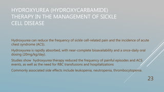
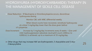
MANAGEMENT OF
SICKLE CELL DISEASE
By
IJEH Cyril E. Pharm D
1
OUTLINE
 Introduction
 Definition
 Sickle Cell Anaemia
 Fibre Formation
 Pathophysiology
 SCD Sequelae
 Hydroxyurea (hydroxycarbamide) Therapy in the Management of Sickle Cell Disease
 Hematopoietic stem cell transplantation (HSCT)
 Role of pharmacist
 Conclusion
 References 2
INTRODUCTION
Blood is a connective tissue. One of its primary roles include circulation, immune modulation
etc.
Its chiefly made up of Blood cells (of which red blood cells constitute more than 99%) and
Plasma.
Red Blood Cells (RBC) or Erythrocytes are tiny biconcave disc-shaped cells. Their cytoplasm
is rich in hemoglobin. They are non-motile, but withstand deformation with remarkable
elasticity. RBC have an average life span of 4 months. Old RBCs are sequestered in the
spleen and bone marrow by phagocytosis.
3
INTRODUCTION
 Anemia is present when there is a decrease in hemoglobin in the blood below the
reference level for the age and sex of the individual. It occurs due to varying reasons such
as blood loss, iron deficiency, hemolysis etc.
 In hemolytic anemia, the rate of hemolysis is at an excess to the compensatory mechanism
of the bone marrow. Hemolytic anemia may be Inherited or Acquired in nature.
Age Description Hemoglobin level (g/dL)
Newborns 17-22
Children 11-13
Adult Males 14-18
Adult Women 12-16
4
DEFINITIONS
 Hemolytic anemia causes include
Red Cell Membrane defect elliptocytosis
Metabolic defects G6PD deficiency
Hemoglobinopathies Sickle cell disease
 Sickle cell disease (SCD) or Depranocytosis: a recessively inherited chronic hemolytic
anemia.
 Caused by a single nucleotide substitution (adenine to thymine) in the β globin
gene on chromosome 11
 Hemoglobin S (most common): GTG  GAG results in substitution of valine
(hydrophobic) for glutamate (hydrophilic) at the sixth codon of the β globin
chain(α2β2
6gluval)7
5
SICKLE CELL ANAEMIA
 Sickle cell anemia (SCA) refers to the clinically
similar disorders HbSS or HbSβ0-thalassemia.
 Sickle cell disease (SCD) refers to all disease
genotypes, including SCA and compound
heterozygous disorders, such as HbSC, HbSD,
and HbS β+-thalassemia. The carrier state for
hemoglobin S (HbAS or sickle cell trait) is not
a form of SCD1.
6
SICKLE CELL ANAEMIA CONT’D
Disease Severity is Genotype –Dependent
Genotype
Hgb SS
Hgb S / β0 thalassemia
Hgb SC
Hgb S / α thalassemia
Hgb S / D
Hgb S / A
Hgb S / E
Hgb S / β+ thalassemia
Hgb S / HPFH
WorseningDisease
Severity
Asymptomatic
+ / - Mild Anemia
Genotype Hb
(g/dL)
HbS% HbA% HbA2% HbF% HbC%
SS 6-9 >90 0 <3.5 <10 0
Sβ0 7-9 >80 0 >3.5 <20 0
Sβ+ 9-12 >60 10-30 >3.5 <20 0
SC 9-14 50 0 <3.5 ≤1.0 45
7
EPIDEMIOLOGY
 Sickle cell disease (SCD) is the most prevalent genetic disease in the World Health
Organization’s (WHO) African Region. It is estimated more than 300,000 babies are
born with severe forms of hemoglobinopathies worldwide each year. 75% of all
patients with SCD live in Sub-Saharan Africa.3
 In countries such as Cameroon, Republic of Congo, Gabon, Ghana and Nigeria, the
trait prevalence is 20%-30%. In countries where the trait prevalence is above 20% the
disease affects about 2% of the population. The geographic distribution of the sickle-
cell trait is very similar to that of malaria.4
 Other affected parts of the World include Sicily, Greece, southern Turkey, and India, all
of which have areas in which malaria is endemic.
8
FIBRE FORMATION
 The amino acid change causes the hemoglobin to associate
with other hemoglobin molecules rather than with the
cellular environment. The amino acid residues that interact
with each other, β6 Valine, β85 Phenylalanine and β88
Leucine. This leads to Fibre formation.
9
FIBRE FORMATION
 Fibre formation only occurs in the deoxy or T-
state due to the structural change to form the
T-state, a different region of the protein
exposes a hydrophobic surface area. The area
containing the mutated amino acid residue and
the area exposed by forming the T-state
associate together to form the
fibres. Hemoglobin molecules associate into
double strands and then, seven of these strands
associate together to form a fibre.
10
PATHOPHYSIOLOGY
 These fibres polymerizes under low oxygen conditions
(40mmHg O2 for 2-4minutes)2 and form bundles that
distort red cells into the classic sickle shape
 The sickling process is initially reversible, but recurrence
leads to red cells losing membrane flexibility. This is caused
by dehydration, partly caused by potassium leaving the red
cells via calcium activated potassium channels called the
Gados channel.
11
PATHOPHYSIOLOGY
Pathophysiology of Sickle
cell Disease
Robbins and Cotran: Pathologic Basis of Disease, 8th Ed. 2010, pp 646
12
SCD SEQUELAE
AND MANAGEMENT
13
SCD SEQUELAE
 Sickled cells have a life span of 10-20 days5. The lifespan of SCD patients is
shortened by 2-3 decades compared to the general population6,8 Life
expectancy in SCA patients in our environment is still low although many now
survive beyond the 4th decade with optimal management9
 Clinical features set in after the first six months of life, when the level of HbF
begin to diminish.
 Types of Crises associated with SCD broadly include
Vaso-occlusive crisis Aplastic Crisis
Sequestration crisis Hemolytic crisis
14
SCD SEQUELAE
Vaso-Occlusive Crisis (VOC) is defined as pain resulting from tissue ischemia caused by vaso-
occlusion most commonly in the bone(s) and bone marrow.
The first VOC may occur as early as 6 months of age, often presenting as dactylitis, but
thereafter VOCs occur with variable frequency.
VOCs and their accompanying pain most commonly occur in the extremities, chest, and back.
When they occur in other sites, they can be confused with, or can be the prodromal stage of,
other acute complications (e.g., head (stroke), flank (papillary necrosis), and abdomen (hepatic
or splenic sequestration, constipation from opioid toxicity, or another hepatobiliary
complication))1.
15
SCD SEQUELAE
Stroke 10% of children; Cause of 20% death1
Strongly advised children undergo Transcranial Doppler Ultrasonagraphy
Blood Transfusion
Acute chest syndrome 40% of patients. Child mortality 1.1%; Adult mortality 4.8%1
(ACS) Other etiology include infection, Pulmonary edema.
Following VOC admisision, ACS may be prevented by Incentive
spirometry every 2-4hours while awake.
Patients are given an intravenous cephalosporin, an oral macrolide
antibiotic, supplemental oxygen (O2 saturation>95%), and close
monitoring for bronchospasm, acute anemia, and hypoxemia.
In rapid progression of ACS (O2 saturation<90% despite supplemental
oxygen, increasing respiratory distress, progressive pulmonary infiltrates,
and/or decline in hemoglobin concentration despite simple transfusion) urgent
exchange transfusion.
16
SCD SEQUELAE
Renal tubular necrosis 15-30% of SS Adults1
Do not give blood transfusions to treat ARF unless there are other
indications for transfusion
Use renal replacement therapy (e.g., hemodialysis) when needed for
acute renal failure
Priapism 35% of boys and men
Its usually of the low flow ischaemic type. Characterized by pain and soft glans.
Prompt recognition and initial conservative medical management with analgesics,
intravenous fluids, oxygen, and sedation if needed
Less conservative therapy include penile aspiration, corporal irrigation using α-adrenergic agents
(e.g., pseudoephedrine, epinephrine, etilefrine), and the use of oral agents (PDE-5 inhibitors, pseudo-
ephedrine). Surgical intervention which involves a shunt. 17
SCD SEQUELAE
Aplastic Crisis: Leads to Acute anemia (decline of 2g/dL or more).
The reticulocyte count is reduced or even zero.
Parvovirus B19 has been implicated in this event. This virus destroys erythroid
precursors in the bone marrow.
Recovery is marked by reticulocytosis and rising haemoglobin levels,
increased immunoglobulin G (IgG). The resulting humoral immunity is
lifelong, preventing recurrent events.
Red blood cell transfusion.
Isolation of hospitalized patients (droplet precautions) to prevent spread of the
parvovirus B19 to pregnant women and others with SCD or compromised
immunity.
18
SCA SEQUELAE
Splenic Sequestration 7-30% incidence in SS individuals. Most typical in children
between 1-4years1.
Sudden spleen enlargement amidst acute anemia. Reticulocyte count is elevated, and platelet
count is generally decreased
Increased risk of infection by encapsulated organisms eg S. pneumonia, H. influenzae
(meningitis or septicaemia) especially in younger children.
Blood transfusion; however, excessive transfusion (>8 g/dL) should be avoided, as the sequestered
erythrocytes in the enlarged spleen typically re-enter the circulation several days later.
Intravenous hydration for severe anaemic cases. Splenectomy in cases recurrent splenic sequestration or
hypersplenism.
All infants with SCD should receive the complete series of the 13-valent conjugate pneumococcal vaccine
series beginning shortly after birth.
23-valent pneumococcal polysaccharide vaccine at 2 years, with a second dose at 5 years.
Children aged 6 to 18 years with functional or anatomic asplenia should receive one dose of PCV1319
SCA SEQUELAE
 Avascular Necrosis (AVN): 10% SS Adults. Increases to about 50% by age 331.
HbSS-α-thalassemia and HbS β0-thalassemia, are at a particularly high risk
to develop AVN at a younger age.
Its bone death due to compromised blood supply. Occurs mostly at
the Hip joint (bilateral cases 40-80%).
The therapeutic approach to AVN depends on the stage of the disease. Ficat
proposed a four-stage radiographic classification of AVN of the hip based on plain
radiography. MRI was not available at the time. Steinberg et al. expanded the Ficat
staging system into six stages using MRI data.
EARLY STAGES Analgesics. Biphosphonates (Alendronate). Cholesterol-lowering drugs.
Blood thinners if indicated.
LATE STAGES Surgical intervention, which include core decompression, osteotomy.
However clinical results gotten are poor, with about 40% of patients progressing to total
hip arthroplasty.
20
SCA SEQUELAE
Stage Radiographic signs
EARLY: Stage 0. Preclinical
EARLY: Stage I. Preradiographic
None; marrow necrosis may be present histologically.
None; abnormal MRI with marrow and bone necrosis
EARLY: Stage II. Before flattening of head or
sequestrum formation
Diffuse porosis, sclerosis, or cysts
TRANSITION Femoral head flattening
Crescent sign
LATE: Stage III. Collapse Broken contour of head
Sequestrum
Joint space normal
LATE: Stage IV. Osteoarthritis Flattened contour
Decreased joint space
Collapse of head
Stages of Avascular Necrosis1 21
HYDROXYUREA (HYDROXYCARBAMIDE) THERAPY IN
THE MANAGEMENT OF SICKLE CELL DISEASE
Hydroxyurea inhibits ribonucleotide reductase. Prior to its use in SCD, this medication has been
in use for several decades to treat people with myeloproliferative disorders.
Effects include: Increases HbF (15-20%)
Lowering amount of circulating leukocytes, and reticulocytes. Alters
the expression of adhesion molecules (all these contribute to VOC)
Nitric oxide released directly from its metabolism. This may contribute
to local vasodilation1.
Fetal haemoglobin consist of α2γ2 the Gamma globin chain binds HbS, and prevents
polymerisation process.
γ subunit production  α2 γS  does not polymerize
22
HYDROXYUREA (HYDROXYCARBAMIDE)
THERAPY IN THE MANAGEMENT OF SICKLE
CELL DISEASE
Hydroxyurea can reduce the frequency of sickle cell-related pain and the incidence of acute
chest syndrome (ACS).
Hydroxyurea is rapidly absorbed, with near-complete bioavailability and a once-daily oral
dosing (20mg/kg/day).
Studies show hydroxyurea therapy reduced the frequency of painful episodes and ACS
events, as well as the need for RBC transfusions and hospitalizations
Commonly associated side effects include leukopenia, neutropenia, thrombocytopenia.
23
HYDROXYUREA (HYDROXYCARBAMIDE) THERAPY IN
THE MANAGEMENT OF SICKLE CELL DISEASE
Hydroxyurea Treament Protocol1
 Initiation: CBC with WBC differential, reticulocyte count, platelet count, and RBC MCV
Quantitative measurement of HbF
Comprehensive metabolic profile, including renal and liver function tests
Starting dosage for adults (500 mg capsules): 15 mg/kg/day (round up to the nearest
500 mg); 5–10 mg/kg/day if patient has chronic kidney disease
Starting dosage for infants and children: 20 mg/kg/day
Monitoring: Monitor CBC with WBC differential and reticulocyte count at least every 4 weeks when
adjusting dosage.
neutrophil count ≥2,000/uL
platelet count ≥80,000/uL
Once a stable dose is established, CBC with WBC differential, reticulocyte count, and
platelet count every 2–3 months
24
HYDROXYUREA (HYDROXYCARBAMIDE) THERAPY IN
THE MANAGEMENT OF SICKLE CELL DISEASE
 Dose Reduction: If Neutropnia or thrombocytopenia occurs, discontinue
hydroxyurea dosing
Monitor CBC with WBC differential weekly
When blood counts have recovered, reinstitute hydroxyurea
at a dose 5 mg/kg/day lower than the dose given before onset
of cytopenias
Dose Increment: Increase by 5 mg/kg/day increments every 8 weeks – Give until
mild myelosuppression (absolute neutrophil count 2,000/uL to
4,000/uL) is achieved, up to a maximum of 35 mg/kg/day.
 Other drugs that may increase HbF are Erythropoietin, 5-Azacytidine and 5-Aza-
2’deoxycytidine 25
HYDROXYUREA (HYDROXYCARBAMIDE) THERAPY IN
THE MANAGEMENT OF SICKLE CELL DISEASE
Hydroxyurea is recommended10 in
 In adults with SCA who have ≥3 moderate to severe pain episodes in a 12-month period
 In adults with SCA who have a history of ACS or symptomatic anemia.
 In children with SCA who have ≥3 moderate to severe pain episodes in a 12-month period.
 In children with SCA who have a history of ACS or symptomatic anemia.
 For infants and children age 9 months or older with SCA who are asymptomatic or have infrequent
pain episodes, we recommend hydroxyurea therapy.
 In patients with SCA who have a history of stroke and a contraindication to chronic transfusions,
suggest hydroxyurea therapy as compared with no therapy.
 In adults with HbSβ+-thalassemia with ≥3 moderate to severe pain episodes in a 12-month period
or a history of ACS, suggest hydroxyurea therapy.
 In patients with HbSC and children with HbSβ+-thalassemia, there is insufficient evidence to
provide recommendations on hydroxyurea therapy.
26
HEMATOPOIETIC STEM CELL TRANSPLANTATION
(HSCT)
 The first successful allogeneic stem cell transplantation (SCT) for sickle cell disease (SCD) was
reported in 1984, establishing the procedure as the only curative option for the disease12
 The procedure involves “conditioning” therapy, utilizing myelosuppressive and/or immune-
modifying drugs, followed by infusion of histocompatible stem cells (derived from bone
marrow, peripheral blood, or umbilical cord blood).
 Recipients undergo myeloablative conditioning, prior to replacement with donor stem cells
(human leukocyte antigen (HLA) matched sibling donor (MSD) transplants offer the best
outcomes for SCD) 11.
 Disease-free survival was over 80% and graft rejection rates were low at 10% or less. As disease
parameters were followed long-term (2 or more years after transplant), most patients were
relieved of pain, had no further strokes or ACS, had stabilized pulmonary function, and had
stable neurologic and cognitive evaluations z
 Stem cell recipients typically need to take immunosuppressants for months to a few years. 27
HEMATOPOIETIC STEM CELL TRANSPLANTATION
(HSCT)
 This procedure is less risky in children (<16years) than adults patients. Likely due to the
absence of comorbid conditions and organ decompensation11.
 To reduce the risk of transplant, a non-myeloblative approach will be necessary. This
however increases the possibility of graft rejection, recurrence of disease symptoms, though
they were tolerated well. Mixed chimerism (presence of both donor and recipient
hematopoiesis) is a frequently observed outcome of non-myeloablative SCT11.
 In mixed chimerism, there is an observed immunologic tolerance which is associated with a
reduced frequency of severe graft-versus-host disease.
28
HEMATOPOIETIC STEM CELL TRANSPLANTATION
(HSCT)
 However, widespread adoption of this treatment modality has been slow to evolve.
Reasons bordering on the complex risk-versus-benefit considerations (e.g., failure of
engraftment and chronic graft-versus-host disease).
 SCD patients should be risk stratified.
 Patients with SCD are characterized as high-risk if they have central nervous system
pathology (clinical or subclinical stroke, seizures), recurrent severe acute chest syndrome,
chronic unremitting pain, or early evidence of end organ damage such as pulmonary
hypertension. The appropriateness of SCT can be more firmly established in the presence
of these high-risk features.
 Increasing age at transplant as well as suboptimal HLA matching increase risks of organ
toxicity, graft-versus-host-disease, morbidity and transplant-related mortality
exponentially
29
OTHER ASSOCIATED DRUGS
 Folic Acid: rapid hemolysis leads to high cell turnover, which leads to depletion
of folate stores in the body.
Deficiency of folate or vitamin B12 inhibits purine and thymidylate syntheses,
impairs DNA synthesis, and causes erythroblast apoptosis, resulting in anemia
from ineffective erythropoiesis.
Folic acid replenishes the depleted folate stores necessary for erythropoiesis.
Recommended dose of 1-5mg/day for SCD patients.
 Thiocyanate: found abundantly in staple African foods, such as the African yam
(Dioscorea sp) and cassava (Manihot utilissima). It exerts anti-sickling
activities on the erythrocytes.
It reduced potassium loss with no significant change in sodium gain, and
significantly inhibited Ca2+ -Mg2+ and Na+ -K + ATPases activities.
Effects include increased mean corpuscular volume (MCV)
Reduces blood pH, serum bicarbonate and bilirubin.
Lowered haemoglobin concentration, RBC and platelets count.
significantly elevated serum alkaline phosphatase (ALP), alanine
aminotransferase (ALT) and aspartate aminotransferase (AST)
30
ROLE OF PHARMACIST
 To be active in counselling SCD patients and their relatives
 To help patients identify preluding symptoms to SCD crisis and/or related
complications
 To educate patients on how best to manage the disease condition. To identify and
prevent precipitating factors that may lead to crisis.
 To foster compliance in hydroxyurea therapy, stating the importance for patients
not missing their daily dosing. Informing them it may take up to 6months before
observable chages.
31
CONCLUSION
SCD is a hereditary non-infective blood disorder. Adequate genotyping
and/or emergency hospital practice helps forestall against crisis in life.
32
REFERENCE
 1National Heart, Lung, and Blood Institute: Evidence-Based Management of Sickle Cell Disease. Expert Panel Report, 2014.
 2Marchant WA, Walker I: Anaesthetic management of the child with sickle cell disease. Paediatr Anaesth 2003; 13:473–89.
 3http://www.cdc.gov/globalhealth/countries/nigeria/what/scd.htm - Assessed 14/06/2015
 4http://www.afro.who.int/en/nigeria/nigeria-publications/1775-sickle%20cell%20disease.html – Assessed 14/06/2015
 5Quinn CT, et al.: Minor elective surgical procedures using general anesthesia in children with sickle cell anemia without pre-
operative blood transfusion. Pediatr Blood Cancer 2005; 45:43–7
 6 Platt OS, Brambilla DJ, Rosse WF, Milner PF, Castro O, Steinberg MH, et al. Mortality in sickle cell disease. Life expectancy and risk
factors for early death. N Engl J Med. 1994;330(23):1639-44.
 7Ingram VM. A specific chemical difference between the globins of normal human and sickle cell anaemia haemoglobin. Nature.
1956;178(4537):792-4.
 8 Powars DR, Chan LS, Hiti A, Ramicone E, Johnson C. Outcome of sickle cell anemia: a 4decade observational study of 1056 patients.
Medicine (Baltimore). 2005;84(6):363-76.
 9Chijioke A., Kolo P. M.: The Longevity and Clinical Pattern of Adult Sickle Cell Anaemia in Ilorin. European Journal of Scientific
Research Vol.32 No.4 (2009); pp.528-532.
 10American Society of Hematology. Update on the use of hydroxyurea therapy in sickle cell disease. Blood: 124 (26). December 18,
2014.
 11Shalini Shenoy. Hematopoietic stem-cell transplantation for sickle cell disease: current evidence and opinions. Ther Adv Hematol .
2013 Oct; 4(5): 335-344
 12Johnson FL, Look AT, Gockerman J, et al. Bone-marrow transplantation in a patient with sickle-cell anemia. N Engl J Med.
1984;311:780-783. 33

Sickle cell disease

  • 1.
    MANAGEMENT OF SICKLE CELLDISEASE By IJEH Cyril E. Pharm D 1
  • 2.
    OUTLINE  Introduction  Definition Sickle Cell Anaemia  Fibre Formation  Pathophysiology  SCD Sequelae  Hydroxyurea (hydroxycarbamide) Therapy in the Management of Sickle Cell Disease  Hematopoietic stem cell transplantation (HSCT)  Role of pharmacist  Conclusion  References 2
  • 3.
    INTRODUCTION Blood is aconnective tissue. One of its primary roles include circulation, immune modulation etc. Its chiefly made up of Blood cells (of which red blood cells constitute more than 99%) and Plasma. Red Blood Cells (RBC) or Erythrocytes are tiny biconcave disc-shaped cells. Their cytoplasm is rich in hemoglobin. They are non-motile, but withstand deformation with remarkable elasticity. RBC have an average life span of 4 months. Old RBCs are sequestered in the spleen and bone marrow by phagocytosis. 3
  • 4.
    INTRODUCTION  Anemia ispresent when there is a decrease in hemoglobin in the blood below the reference level for the age and sex of the individual. It occurs due to varying reasons such as blood loss, iron deficiency, hemolysis etc.  In hemolytic anemia, the rate of hemolysis is at an excess to the compensatory mechanism of the bone marrow. Hemolytic anemia may be Inherited or Acquired in nature. Age Description Hemoglobin level (g/dL) Newborns 17-22 Children 11-13 Adult Males 14-18 Adult Women 12-16 4
  • 5.
    DEFINITIONS  Hemolytic anemiacauses include Red Cell Membrane defect elliptocytosis Metabolic defects G6PD deficiency Hemoglobinopathies Sickle cell disease  Sickle cell disease (SCD) or Depranocytosis: a recessively inherited chronic hemolytic anemia.  Caused by a single nucleotide substitution (adenine to thymine) in the β globin gene on chromosome 11  Hemoglobin S (most common): GTG  GAG results in substitution of valine (hydrophobic) for glutamate (hydrophilic) at the sixth codon of the β globin chain(α2β2 6gluval)7 5
  • 6.
    SICKLE CELL ANAEMIA Sickle cell anemia (SCA) refers to the clinically similar disorders HbSS or HbSβ0-thalassemia.  Sickle cell disease (SCD) refers to all disease genotypes, including SCA and compound heterozygous disorders, such as HbSC, HbSD, and HbS β+-thalassemia. The carrier state for hemoglobin S (HbAS or sickle cell trait) is not a form of SCD1. 6
  • 7.
    SICKLE CELL ANAEMIACONT’D Disease Severity is Genotype –Dependent Genotype Hgb SS Hgb S / β0 thalassemia Hgb SC Hgb S / α thalassemia Hgb S / D Hgb S / A Hgb S / E Hgb S / β+ thalassemia Hgb S / HPFH WorseningDisease Severity Asymptomatic + / - Mild Anemia Genotype Hb (g/dL) HbS% HbA% HbA2% HbF% HbC% SS 6-9 >90 0 <3.5 <10 0 Sβ0 7-9 >80 0 >3.5 <20 0 Sβ+ 9-12 >60 10-30 >3.5 <20 0 SC 9-14 50 0 <3.5 ≤1.0 45 7
  • 8.
    EPIDEMIOLOGY  Sickle celldisease (SCD) is the most prevalent genetic disease in the World Health Organization’s (WHO) African Region. It is estimated more than 300,000 babies are born with severe forms of hemoglobinopathies worldwide each year. 75% of all patients with SCD live in Sub-Saharan Africa.3  In countries such as Cameroon, Republic of Congo, Gabon, Ghana and Nigeria, the trait prevalence is 20%-30%. In countries where the trait prevalence is above 20% the disease affects about 2% of the population. The geographic distribution of the sickle- cell trait is very similar to that of malaria.4  Other affected parts of the World include Sicily, Greece, southern Turkey, and India, all of which have areas in which malaria is endemic. 8
  • 9.
    FIBRE FORMATION  Theamino acid change causes the hemoglobin to associate with other hemoglobin molecules rather than with the cellular environment. The amino acid residues that interact with each other, β6 Valine, β85 Phenylalanine and β88 Leucine. This leads to Fibre formation. 9
  • 10.
    FIBRE FORMATION  Fibreformation only occurs in the deoxy or T- state due to the structural change to form the T-state, a different region of the protein exposes a hydrophobic surface area. The area containing the mutated amino acid residue and the area exposed by forming the T-state associate together to form the fibres. Hemoglobin molecules associate into double strands and then, seven of these strands associate together to form a fibre. 10
  • 11.
    PATHOPHYSIOLOGY  These fibrespolymerizes under low oxygen conditions (40mmHg O2 for 2-4minutes)2 and form bundles that distort red cells into the classic sickle shape  The sickling process is initially reversible, but recurrence leads to red cells losing membrane flexibility. This is caused by dehydration, partly caused by potassium leaving the red cells via calcium activated potassium channels called the Gados channel. 11
  • 12.
    PATHOPHYSIOLOGY Pathophysiology of Sickle cellDisease Robbins and Cotran: Pathologic Basis of Disease, 8th Ed. 2010, pp 646 12
  • 13.
  • 14.
    SCD SEQUELAE  Sickledcells have a life span of 10-20 days5. The lifespan of SCD patients is shortened by 2-3 decades compared to the general population6,8 Life expectancy in SCA patients in our environment is still low although many now survive beyond the 4th decade with optimal management9  Clinical features set in after the first six months of life, when the level of HbF begin to diminish.  Types of Crises associated with SCD broadly include Vaso-occlusive crisis Aplastic Crisis Sequestration crisis Hemolytic crisis 14
  • 15.
    SCD SEQUELAE Vaso-Occlusive Crisis(VOC) is defined as pain resulting from tissue ischemia caused by vaso- occlusion most commonly in the bone(s) and bone marrow. The first VOC may occur as early as 6 months of age, often presenting as dactylitis, but thereafter VOCs occur with variable frequency. VOCs and their accompanying pain most commonly occur in the extremities, chest, and back. When they occur in other sites, they can be confused with, or can be the prodromal stage of, other acute complications (e.g., head (stroke), flank (papillary necrosis), and abdomen (hepatic or splenic sequestration, constipation from opioid toxicity, or another hepatobiliary complication))1. 15
  • 16.
    SCD SEQUELAE Stroke 10%of children; Cause of 20% death1 Strongly advised children undergo Transcranial Doppler Ultrasonagraphy Blood Transfusion Acute chest syndrome 40% of patients. Child mortality 1.1%; Adult mortality 4.8%1 (ACS) Other etiology include infection, Pulmonary edema. Following VOC admisision, ACS may be prevented by Incentive spirometry every 2-4hours while awake. Patients are given an intravenous cephalosporin, an oral macrolide antibiotic, supplemental oxygen (O2 saturation>95%), and close monitoring for bronchospasm, acute anemia, and hypoxemia. In rapid progression of ACS (O2 saturation<90% despite supplemental oxygen, increasing respiratory distress, progressive pulmonary infiltrates, and/or decline in hemoglobin concentration despite simple transfusion) urgent exchange transfusion. 16
  • 17.
    SCD SEQUELAE Renal tubularnecrosis 15-30% of SS Adults1 Do not give blood transfusions to treat ARF unless there are other indications for transfusion Use renal replacement therapy (e.g., hemodialysis) when needed for acute renal failure Priapism 35% of boys and men Its usually of the low flow ischaemic type. Characterized by pain and soft glans. Prompt recognition and initial conservative medical management with analgesics, intravenous fluids, oxygen, and sedation if needed Less conservative therapy include penile aspiration, corporal irrigation using α-adrenergic agents (e.g., pseudoephedrine, epinephrine, etilefrine), and the use of oral agents (PDE-5 inhibitors, pseudo- ephedrine). Surgical intervention which involves a shunt. 17
  • 18.
    SCD SEQUELAE Aplastic Crisis:Leads to Acute anemia (decline of 2g/dL or more). The reticulocyte count is reduced or even zero. Parvovirus B19 has been implicated in this event. This virus destroys erythroid precursors in the bone marrow. Recovery is marked by reticulocytosis and rising haemoglobin levels, increased immunoglobulin G (IgG). The resulting humoral immunity is lifelong, preventing recurrent events. Red blood cell transfusion. Isolation of hospitalized patients (droplet precautions) to prevent spread of the parvovirus B19 to pregnant women and others with SCD or compromised immunity. 18
  • 19.
    SCA SEQUELAE Splenic Sequestration7-30% incidence in SS individuals. Most typical in children between 1-4years1. Sudden spleen enlargement amidst acute anemia. Reticulocyte count is elevated, and platelet count is generally decreased Increased risk of infection by encapsulated organisms eg S. pneumonia, H. influenzae (meningitis or septicaemia) especially in younger children. Blood transfusion; however, excessive transfusion (>8 g/dL) should be avoided, as the sequestered erythrocytes in the enlarged spleen typically re-enter the circulation several days later. Intravenous hydration for severe anaemic cases. Splenectomy in cases recurrent splenic sequestration or hypersplenism. All infants with SCD should receive the complete series of the 13-valent conjugate pneumococcal vaccine series beginning shortly after birth. 23-valent pneumococcal polysaccharide vaccine at 2 years, with a second dose at 5 years. Children aged 6 to 18 years with functional or anatomic asplenia should receive one dose of PCV1319
  • 20.
    SCA SEQUELAE  AvascularNecrosis (AVN): 10% SS Adults. Increases to about 50% by age 331. HbSS-α-thalassemia and HbS β0-thalassemia, are at a particularly high risk to develop AVN at a younger age. Its bone death due to compromised blood supply. Occurs mostly at the Hip joint (bilateral cases 40-80%). The therapeutic approach to AVN depends on the stage of the disease. Ficat proposed a four-stage radiographic classification of AVN of the hip based on plain radiography. MRI was not available at the time. Steinberg et al. expanded the Ficat staging system into six stages using MRI data. EARLY STAGES Analgesics. Biphosphonates (Alendronate). Cholesterol-lowering drugs. Blood thinners if indicated. LATE STAGES Surgical intervention, which include core decompression, osteotomy. However clinical results gotten are poor, with about 40% of patients progressing to total hip arthroplasty. 20
  • 21.
    SCA SEQUELAE Stage Radiographicsigns EARLY: Stage 0. Preclinical EARLY: Stage I. Preradiographic None; marrow necrosis may be present histologically. None; abnormal MRI with marrow and bone necrosis EARLY: Stage II. Before flattening of head or sequestrum formation Diffuse porosis, sclerosis, or cysts TRANSITION Femoral head flattening Crescent sign LATE: Stage III. Collapse Broken contour of head Sequestrum Joint space normal LATE: Stage IV. Osteoarthritis Flattened contour Decreased joint space Collapse of head Stages of Avascular Necrosis1 21
  • 22.
    HYDROXYUREA (HYDROXYCARBAMIDE) THERAPYIN THE MANAGEMENT OF SICKLE CELL DISEASE Hydroxyurea inhibits ribonucleotide reductase. Prior to its use in SCD, this medication has been in use for several decades to treat people with myeloproliferative disorders. Effects include: Increases HbF (15-20%) Lowering amount of circulating leukocytes, and reticulocytes. Alters the expression of adhesion molecules (all these contribute to VOC) Nitric oxide released directly from its metabolism. This may contribute to local vasodilation1. Fetal haemoglobin consist of α2γ2 the Gamma globin chain binds HbS, and prevents polymerisation process. γ subunit production  α2 γS  does not polymerize 22
  • 23.
    HYDROXYUREA (HYDROXYCARBAMIDE) THERAPY INTHE MANAGEMENT OF SICKLE CELL DISEASE Hydroxyurea can reduce the frequency of sickle cell-related pain and the incidence of acute chest syndrome (ACS). Hydroxyurea is rapidly absorbed, with near-complete bioavailability and a once-daily oral dosing (20mg/kg/day). Studies show hydroxyurea therapy reduced the frequency of painful episodes and ACS events, as well as the need for RBC transfusions and hospitalizations Commonly associated side effects include leukopenia, neutropenia, thrombocytopenia. 23
  • 24.
    HYDROXYUREA (HYDROXYCARBAMIDE) THERAPYIN THE MANAGEMENT OF SICKLE CELL DISEASE Hydroxyurea Treament Protocol1  Initiation: CBC with WBC differential, reticulocyte count, platelet count, and RBC MCV Quantitative measurement of HbF Comprehensive metabolic profile, including renal and liver function tests Starting dosage for adults (500 mg capsules): 15 mg/kg/day (round up to the nearest 500 mg); 5–10 mg/kg/day if patient has chronic kidney disease Starting dosage for infants and children: 20 mg/kg/day Monitoring: Monitor CBC with WBC differential and reticulocyte count at least every 4 weeks when adjusting dosage. neutrophil count ≥2,000/uL platelet count ≥80,000/uL Once a stable dose is established, CBC with WBC differential, reticulocyte count, and platelet count every 2–3 months 24
  • 25.
    HYDROXYUREA (HYDROXYCARBAMIDE) THERAPYIN THE MANAGEMENT OF SICKLE CELL DISEASE  Dose Reduction: If Neutropnia or thrombocytopenia occurs, discontinue hydroxyurea dosing Monitor CBC with WBC differential weekly When blood counts have recovered, reinstitute hydroxyurea at a dose 5 mg/kg/day lower than the dose given before onset of cytopenias Dose Increment: Increase by 5 mg/kg/day increments every 8 weeks – Give until mild myelosuppression (absolute neutrophil count 2,000/uL to 4,000/uL) is achieved, up to a maximum of 35 mg/kg/day.  Other drugs that may increase HbF are Erythropoietin, 5-Azacytidine and 5-Aza- 2’deoxycytidine 25
  • 26.
    HYDROXYUREA (HYDROXYCARBAMIDE) THERAPYIN THE MANAGEMENT OF SICKLE CELL DISEASE Hydroxyurea is recommended10 in  In adults with SCA who have ≥3 moderate to severe pain episodes in a 12-month period  In adults with SCA who have a history of ACS or symptomatic anemia.  In children with SCA who have ≥3 moderate to severe pain episodes in a 12-month period.  In children with SCA who have a history of ACS or symptomatic anemia.  For infants and children age 9 months or older with SCA who are asymptomatic or have infrequent pain episodes, we recommend hydroxyurea therapy.  In patients with SCA who have a history of stroke and a contraindication to chronic transfusions, suggest hydroxyurea therapy as compared with no therapy.  In adults with HbSβ+-thalassemia with ≥3 moderate to severe pain episodes in a 12-month period or a history of ACS, suggest hydroxyurea therapy.  In patients with HbSC and children with HbSβ+-thalassemia, there is insufficient evidence to provide recommendations on hydroxyurea therapy. 26
  • 27.
    HEMATOPOIETIC STEM CELLTRANSPLANTATION (HSCT)  The first successful allogeneic stem cell transplantation (SCT) for sickle cell disease (SCD) was reported in 1984, establishing the procedure as the only curative option for the disease12  The procedure involves “conditioning” therapy, utilizing myelosuppressive and/or immune- modifying drugs, followed by infusion of histocompatible stem cells (derived from bone marrow, peripheral blood, or umbilical cord blood).  Recipients undergo myeloablative conditioning, prior to replacement with donor stem cells (human leukocyte antigen (HLA) matched sibling donor (MSD) transplants offer the best outcomes for SCD) 11.  Disease-free survival was over 80% and graft rejection rates were low at 10% or less. As disease parameters were followed long-term (2 or more years after transplant), most patients were relieved of pain, had no further strokes or ACS, had stabilized pulmonary function, and had stable neurologic and cognitive evaluations z  Stem cell recipients typically need to take immunosuppressants for months to a few years. 27
  • 28.
    HEMATOPOIETIC STEM CELLTRANSPLANTATION (HSCT)  This procedure is less risky in children (<16years) than adults patients. Likely due to the absence of comorbid conditions and organ decompensation11.  To reduce the risk of transplant, a non-myeloblative approach will be necessary. This however increases the possibility of graft rejection, recurrence of disease symptoms, though they were tolerated well. Mixed chimerism (presence of both donor and recipient hematopoiesis) is a frequently observed outcome of non-myeloablative SCT11.  In mixed chimerism, there is an observed immunologic tolerance which is associated with a reduced frequency of severe graft-versus-host disease. 28
  • 29.
    HEMATOPOIETIC STEM CELLTRANSPLANTATION (HSCT)  However, widespread adoption of this treatment modality has been slow to evolve. Reasons bordering on the complex risk-versus-benefit considerations (e.g., failure of engraftment and chronic graft-versus-host disease).  SCD patients should be risk stratified.  Patients with SCD are characterized as high-risk if they have central nervous system pathology (clinical or subclinical stroke, seizures), recurrent severe acute chest syndrome, chronic unremitting pain, or early evidence of end organ damage such as pulmonary hypertension. The appropriateness of SCT can be more firmly established in the presence of these high-risk features.  Increasing age at transplant as well as suboptimal HLA matching increase risks of organ toxicity, graft-versus-host-disease, morbidity and transplant-related mortality exponentially 29
  • 30.
    OTHER ASSOCIATED DRUGS Folic Acid: rapid hemolysis leads to high cell turnover, which leads to depletion of folate stores in the body. Deficiency of folate or vitamin B12 inhibits purine and thymidylate syntheses, impairs DNA synthesis, and causes erythroblast apoptosis, resulting in anemia from ineffective erythropoiesis. Folic acid replenishes the depleted folate stores necessary for erythropoiesis. Recommended dose of 1-5mg/day for SCD patients.  Thiocyanate: found abundantly in staple African foods, such as the African yam (Dioscorea sp) and cassava (Manihot utilissima). It exerts anti-sickling activities on the erythrocytes. It reduced potassium loss with no significant change in sodium gain, and significantly inhibited Ca2+ -Mg2+ and Na+ -K + ATPases activities. Effects include increased mean corpuscular volume (MCV) Reduces blood pH, serum bicarbonate and bilirubin. Lowered haemoglobin concentration, RBC and platelets count. significantly elevated serum alkaline phosphatase (ALP), alanine aminotransferase (ALT) and aspartate aminotransferase (AST) 30
  • 31.
    ROLE OF PHARMACIST To be active in counselling SCD patients and their relatives  To help patients identify preluding symptoms to SCD crisis and/or related complications  To educate patients on how best to manage the disease condition. To identify and prevent precipitating factors that may lead to crisis.  To foster compliance in hydroxyurea therapy, stating the importance for patients not missing their daily dosing. Informing them it may take up to 6months before observable chages. 31
  • 32.
    CONCLUSION SCD is ahereditary non-infective blood disorder. Adequate genotyping and/or emergency hospital practice helps forestall against crisis in life. 32
  • 33.
    REFERENCE  1National Heart,Lung, and Blood Institute: Evidence-Based Management of Sickle Cell Disease. Expert Panel Report, 2014.  2Marchant WA, Walker I: Anaesthetic management of the child with sickle cell disease. Paediatr Anaesth 2003; 13:473–89.  3http://www.cdc.gov/globalhealth/countries/nigeria/what/scd.htm - Assessed 14/06/2015  4http://www.afro.who.int/en/nigeria/nigeria-publications/1775-sickle%20cell%20disease.html – Assessed 14/06/2015  5Quinn CT, et al.: Minor elective surgical procedures using general anesthesia in children with sickle cell anemia without pre- operative blood transfusion. Pediatr Blood Cancer 2005; 45:43–7  6 Platt OS, Brambilla DJ, Rosse WF, Milner PF, Castro O, Steinberg MH, et al. Mortality in sickle cell disease. Life expectancy and risk factors for early death. N Engl J Med. 1994;330(23):1639-44.  7Ingram VM. A specific chemical difference between the globins of normal human and sickle cell anaemia haemoglobin. Nature. 1956;178(4537):792-4.  8 Powars DR, Chan LS, Hiti A, Ramicone E, Johnson C. Outcome of sickle cell anemia: a 4decade observational study of 1056 patients. Medicine (Baltimore). 2005;84(6):363-76.  9Chijioke A., Kolo P. M.: The Longevity and Clinical Pattern of Adult Sickle Cell Anaemia in Ilorin. European Journal of Scientific Research Vol.32 No.4 (2009); pp.528-532.  10American Society of Hematology. Update on the use of hydroxyurea therapy in sickle cell disease. Blood: 124 (26). December 18, 2014.  11Shalini Shenoy. Hematopoietic stem-cell transplantation for sickle cell disease: current evidence and opinions. Ther Adv Hematol . 2013 Oct; 4(5): 335-344  12Johnson FL, Look AT, Gockerman J, et al. Bone-marrow transplantation in a patient with sickle-cell anemia. N Engl J Med. 1984;311:780-783. 33

Editor's Notes

  • #6 7Ingram VM. A specific chemical difference between the globins of normal human and sicklecell anaemia haemoglobin. Nature. 1956;178(4537):792-4.
  • #7 1National Heart, Lung, and Blood Institute: Evidence-Based Management of Sickle Cell Disease. Expert Panel Report, 2014.
  • #9 3http://www.cdc.gov/globalhealth/countries/nigeria/what/scd.htm - Assessed 14/06/2015 4http://www.afro.who.int/en/nigeria/nigeria-publications/1775-sickle%20cell%20disease.html – Assessed 14/06/2015
  • #12 2Marchant WA, Walker I: Anaesthetic management of the child with sickle cell disease. Paediatr Anaesth 2003; 13:473–89. Anesthesiology Problem-oriented Patient Management, 6th ed. 2008, pp 980–92.
  • #15 5Quinn CT, et al.: Minor elective surgical procedures using general anesthesia in children with sickle cell anemia without pre-operative blood transfusion. Pediatr Blood Cancer 2005; 45:43–7 6 Platt OS, Brambilla DJ, Rosse WF, Milner PF, Castro O, Steinberg MH, et al. Mortality in sickle cell disease. Life expectancy and risk factors for early death. N Engl J Med. 1994;330(23):1639-44. 8 Powars DR, Chan LS, Hiti A, Ramicone E, Johnson C. Outcome of sickle cell anemia: a 4decade observational study of 1056 patients. Medicine (Baltimore). 2005;84(6):363-76. 9Chijioke A., Kolo P. M.: The Longevity and Clinical Pattern of Adult Sickle Cell Anaemia in Ilorin. Europpean Journal of Scientific Research Vol.32 No.4 (2009); pp.528-532
  • #16 59National Heart, Lung, and Blood Institute: Evidence-Based Management of Sickle Cell Disease. Expert Panel Report, 2014.
  • #22 59National Heart, Lung, and Blood Institute: Evidence-Based Management of Sickle Cell Disease. Expert Panel Report, 2014.
  • #27 10American Society of Hematology. Update on the use of hydroxyurea therapy in sickle cell disease. Blood: 124 (26). December 18, 2014.
  • #28 12Johnson FL, Look AT, Gockerman J, et al. Bone-marrow transplantation in a patient with sickle-cell anemia. N Engl J Med. 1984;311:780-783. 11Shalini Shenoy. Hematopoietic stem-cell transplantation for sickle cell disease: current evidence and opinions. Ther Adv Hematol . 2013 Oct; 4(5): 335-344
  • #29 11Ther Adv Hematol. Hematopoietic stem-cell transplantation for sickle cell disease: current evidence and opinions. Shalini Shenoy. 2013 Oct; 4(5): 335-344