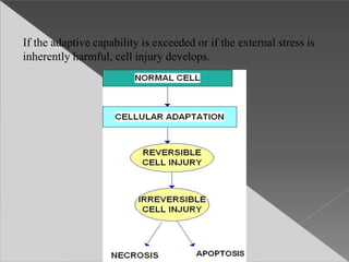
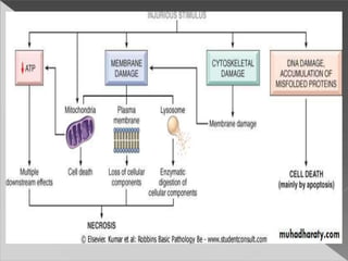
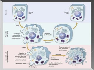
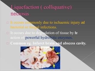
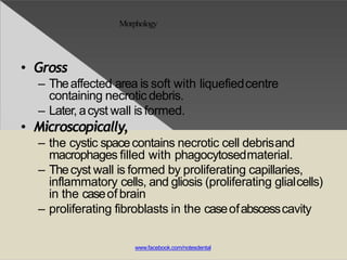
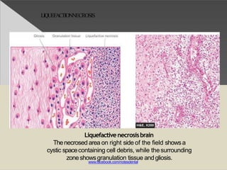
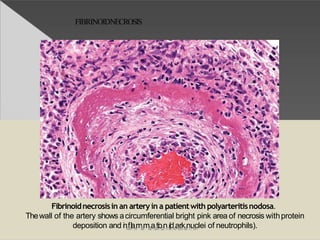
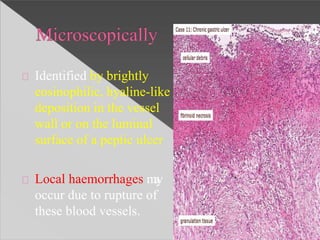
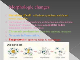
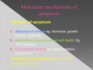
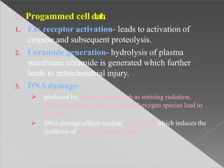
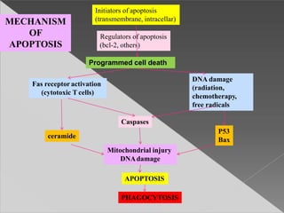
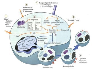
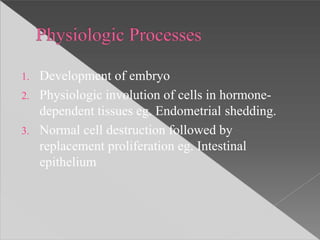
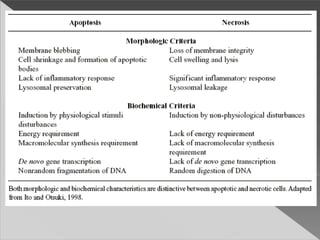
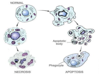
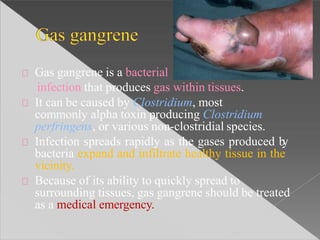
CELL DEATH AND
NECROSIS
Presented by-
Dr Ekta Dwivedi
INTRODUCTION
Most forms of disease state begin with cell injury
and consequently loss of cellular function.
Cell injury is defined as- a variety of stresses a cell
encounters as a result of changes in its internal and
external environment.
The cellular response to injury varies and
depends upon
 Type of cell and tissue invovled
 Extent of injury
 Type of injury
CELLULAR RESPONSES TO STRESS AND NOXIOUS
STIMULI
Cells are active participants in their environment, constantly
adjusting their structure and function to accommodate changing
demands and extracellular stresses.
Cells tends to maintain their intracellular milieu within a fairy
narrow range of physiologic parameters; that is, they maintain
normal homeostasis.
As cells encounter physiologic stresses or pathologic stimuli,
they can undergo adaptation.
The principle adaptative responses are:-
-Hypertrophy,
- Hyperplasia,
-Atrophy,
-Metaplasia
If the adaptive capability is exceeded or if the external stress is
inherently harmful, cell injury develops.
OVERVIEW OF CELL INJURY OR CELL DEATH
REVERSIBLE CELL INJURY-
In early stages or mild forms of injury the functional or morphologic changes
are reversible if the damaging stimulus is removed.
IRRIVERSIBLE CELL INJURY (CELL DEATH)-
With continuing damage, the injury becomes irreversible, at which time the
cell cannot recover and it dies. There are two types of cell death-
Necrosis
Apoptosis
Genetic cause
Acquired causes
Based on underlying agent:
Hypoxia and ischaemia
Physical agents
Chemical agents and drugs
Microbial agents
Immunologic agents
Nutritional Imbalences
Psychological factors
Deficiency of oxygen or hypoxia results in
failure to carry out cellular activities.
Most common, causes of hypoxia are:
Reduced supply of blood to cells- ischaemia
Oxygen deprivation can result from other causes like-
anaemia, carbon monoxide poisoning,
cardiorespiration insufficiency and increase in
demand of tissues
Mechanical trauma
Thermal trauma
Electricity
Radiation rapid changes in atmospheric pressure
Chemical poisons: cyanide, arsenic,mercury.
Strong acid and alkalis
Insecticides and pesticides
High oxygen concentration
Hypertonic glucose and salt
Alcohol and narcotic drugs
Theraputic drugs
Infection caused by
Bacteria
Rickettsiae
Viruses
Fungi
Protozoa
Metazoa
Parasites
Immunity is a double edged word :
Hypersensitivity reactions
Anaphylatic reactions
Autoimmune diseases
Nutritional deficiency diseases :
deficiency of nutrients ( eg. Starvation), of protein
calorie ( eg. Marasmus, kwashiorkor)
Of minerals ( eg. Anaemia)
Nutritional excess
Obesity
Atterosclerosis
Heart disease
Hypertension
Mental stress
Strain
Anxiety
Overwork
Frustration
Reversible cell injury
Irreversible cell injury
HYPOXIA/ISCHAEMIA
↓ ATP
↓ Intercellular pH
(cytosol)
Damaged sodium
pump
(membrane)
↓ Protein
synthesis (RER)
Ultrastructural/Functional Changes
REVERSIBLE CELLINJURY
2 essential features:
Inability of the cell to reverse mitochondrial
dysfunction
Disturbance in cell membrane function
In addition: depletion of proteins, leakage of
lysosomal enzymes into cytoplasm, reduced
intracellular pH and further reduction inATP.
Continued hypoxia leads to influx of large amount of
Calcium ions.
Normal Ca ions – in ECF 10-3 M (millimoles)
Cytosole 10-7 M
Leads to mitochondrial dysfunction
Morphological mitochondrial changes:
 Vacuoles in mitochondria
 Deposition of amorphous Ca in mitochondrial matrix.
Mechanism:
Accelerated degradation of membrane phospholipids.
Cytoskeletal damage
Toxic oxygen radicals
Hydrolytic enzymes
Serum estimation of liberated intracellular enzymes
MEMBRANE DAMAGE
Nuclear changes
(Pyknosis, Karyolysis,
Karyorrhexis)
Cell Death
(myelin figures)
Serum enzyme
estimation ( SGOT,
LDH)
Libration of intacellular enzymes
See Ch. 1, p. 9,
Fig. 1-9
MECHANISMS OF CELL INJURY
ATP DEPLETION : failure of energy dependent functions-
reversible injury-necrosis
MITOCHONRIAL DAMAGE: ATP depletion-failure of energy
dependent functions- ultimately, necrosis; under some conditions,
leakage of proteins that cause apoptosis
INFLUX OF CALCIUM: activation of enzymes that damage
cellular components and may also trigger apoptosis
ACCUMULATIN OF REACTIVE OXYGEN SPECIES: covalent
modification of cellular proteins, lipids, nucleic acids
INCREASED PERMEABILITY OF CELLULAR
MEMBRANES: may affect plasma membrane, lysosomal
membranes, mitochondrial membranes; typically culminates in
necrosis
ACCUMULATION OF DAMAGED DNA AND MISFOLDED
PROTEINS: triggers apoptosis
MORPHOLOGY OF CELL INJURY
REVERSIBLE CELL INJURY-
The two main morphologic correlates of reversible cell injury are cellular
swelling and fatty change.
-Cellular swelling is the result of energy-dependent ion pumps in the plasma
membrane, leading to an inability to maintain ionic and fluid homeostasis.
-Fatty change occur in hypoxic injury and various forms of toxic or metabolic
injury.
It is manifested by the appearance of small or large lipid vacuoles in the
cytoplasm.
OTHER MORPHOLOGIC FEATURES:-
-Membrane blebbing and loss of microvilli, mitochondrial swelling, dilation of
ER, and eosinophilia ( due to decreased cytoplasmic RNA)
Cell death is a state of irreversible
injury
In living body it may occur as:
Local or focal change (autolysis, necrosis and
apoptosis)
Changes that follow( gangrene and pathologic
calcification)
End of life ( somatic death)
-Necrotic cells are unable to maintain membrane integrity, and
their contents often leak out.
-The enzymes responsible for digestion of the cell are derived
either from the lysosomes of the dying cells themselves or from
the lysosomes of leukocytes that are recruited as part of the
inflammatory reaction to the dead cells.
Disintegration of the cell by its own
hydrolytic enzymes liberated from lysosomes
It is rapid in some tissues rich in hydrolytic
enzymes such as pancreas and gastric mucosa.
Morphologically , autolysis is identified by :
 Homogeneous and eosinophilic cytoplasm
 Loss of cellular details
 Remains of cell as debris
Defined as- focal death along
with degradation of tissue by
hydrolytic enzymes librated by
cells, accompanied by
inflammation.
2 essential features:
Cell digestion by lytic
enzymes
Denaturation of proteins
Morphologic changes in
necrosis:
Cytoplasmic
 Homogenous and intensely eosinophilic
 Occasionally: vacuolation or dystrophic calcification
Nuclear
 Pyknosis- condensation of nuclear chromatin
 Karyolysis – undergo dissolution
 Karyorrhexis- fragmentation into many clumps
5 types :
Coagulative necrosis
Liquefaction (colliquative) necrosis
Caseous necrosis
Fat necrosis
Fibrinoid necrosis
Most common type
Caused by irreversible focal injury, mostly from sudden
cessation of blood flow (ischaemia)and less often from
bacterial and chemical agents
Organs affected are: heart, kidney and spleen.
Foci of coagulative necrosis:
In early stages: pale, firm,
and slightly swollen.
With progression: become more
yellowish, softer, and shrunken.
Hallmark of coagulative
necrosis: conversion of normal
cells into their ‘tombstones’ i.e.
outlines of the cells are retained
so that the cell type can still be
recognised but their cytoplasm
and nuclear details are lost.
Necrosed cells are swollen and
appear more eosinophillic than
normal
InfarctKidney
Theaffected area on right shows cells with intensely eosinophilic cytoplasmof
tubular cells but the outlines of tubules are still maintained.
Thenuclei show granular debris.
Theinterface between viable and non-viable area shows nonspecific chronic
inflammation and proliferating vessels
www.facebook.com/notesdenta
Microscopic changes are the result of:
 Denaturation of proteins
 Enzymatic digestion of the cell.
Eventually, the necrosed focus is infiltrated by
inflammatory cells and
The dead cells are phagocytosed leaving
granular debris and fragments of cells.
It occurs commonly due to ischaemic injury and
bacterial or fungal infections.
It occurs due to degradation of tissue by the
action of powerful hydrolytic enzymes.
Common eg. Infarct brain and abscess cavity.
Morphology
• Gross
– Theaffected area is soft with liquefiedcentre
containing necrotic debris.
– Later, acyst wall isformed.
• Microscopically,
– the cystic spacecontains necrotic cell debrisand
macrophages filled with phagocytosedmaterial.
– Thecyst wall is formed by proliferating capillaries,
inflammatory cells, and gliosis (proliferating glialcells)
in the caseof brain
– proliferating fibroblasts in the caseofabscesscavity
www.facebook.com/notesdental
LIQUEFACTIONNECROSIS
Liquefactivenecrosisbrain
Thenecrosed area on right side of the field showsa
cystic spacecontaining cell debris, while thesurrounding
zoneshowsgranulation tissue andgliosis.
www.facebook.com/notesdental
Found in the centre of foci of tuberculous
infection.
It is a combined feature of coagulative and
liquefactive necrosis.
Foci of caseous necrosis, as the
name implies, resemble dry
cheese and are soft, granular and
yellowish.
This appearence is partly
attributed to the histotoxic
effects of lipopolysaccharides
present in the capsule of the
tubercle bacilli, Mycobacterium
tuberculosis.
The necrosed foci are
structureless, eosinophilic and
contain granular debris.
The surrounding tissue shows
characteristic granulomatous
iflammatory reaction
consisting of epitheloid cells
with interspersed giant cells of
langhan’s or foreign body type
and peripherally lymphocytes.
CASEOUSNECROSIS
Caseous necrosis lymphnode
There is eosinophilic, amorphous, granular material, while the peripheryshows
granulomatous inflammation.
www.facebook.com/notesdental
It is a special form of cell death occurring at two
anatomically different locations but
morphologically similar lesions.
These are:
Following pancreatic necrosis
Traumatic fat necrosis commonly in breast
Fat necrosis in either of the 2 instances results in
hydrolylsis of neutral fat present in adipose
cells into glycerol and free fatty acids.
The damaged adipose cells assume cloudy
appearance when only free fatty remain
behind , after glycerol leaks out.
The leaked out free fatty acids, complex with Ca
to form Ca soaps (sponification)
FATNECROSIS
Fatnecrosis in acutepancreatitis.
Theareas of white chalky deposits represent foci of fat necrosis with
calcium soapformation (saponification) at sites oflipid breakdown in the
www.facmebeooske.cnomte/nroyt.esdenta
Morphology
• Grossly
– fat necrosis appears as
yellowish-white andfirm
deposits.
– Formation of calcium
soapsimparts the
necrosed foci firmer and
chalky white appearance.
• Microscopically
– the necrosed fat cells
havecloudy appearance
– surrounded by an
inflammatory reaction.
Formation of calciumsoaps
is identified in the tissue
sections asamorphous,
granular andbasophilic
terialwww.facebook.com/notesde
mnta
al
It is characterized by the deposition of fibrin-
like material which has the staining properties of
fibrin.
It is encountered in various examples of
immunologic tissue injury (eg. Autoimmune
diseases, arthus reaction), artioles in
hypertension, peptic ulcer etc.
FIBRINOIDNECROSIS
Fibrinoidnecrosisin an artery in a patientwith polyarteritisnodosa.
Thewall of the artery shows acircumferential bright pink area of necrosis withprotein
deposition and inwfwlawm.facmebaootkio.conm(/dnoaterskdennutacllei of neutrophils).
Identified by brightly
eosinophilic, hyaline-like
deposition in the vessel
wall or on the luminal
surface of a peptic ulcer
Local haemorrhages may
occur due to rupture of
these blood vessels.
Apoptosis is a form of ‘coordinated and
internally programmed cell death’ which is of
significance in variety of physiologic pathologic
conditions.
Apoptosis in Greek meaning ‘falling off’ or
‘dropping off’.
Shrinking of cell : with dense cytoplasm and almost
normal organelles.
Convolution of cell membrane with formation of membrane-
bound near-spherical bodies called apoptotic bodies
containing compacted organelles.
Chromatin condensation around the periphery of nucleus
No acute inflammation.
Phagocytosis of apoptotic bodies by macrophages
Initiators of apoptosis
1. Absence of stimuli eg. Hormone, growth
factors, cytokines.
2. Activators of programmed cell death. Eg.
TNF receptors.
3. Intracellular stimuli eg. Heat, radiation,
hypoxia etc.
Regulators of apoptosis.eg. bcl-2, p53,
caspases, bax etc.
Progammed cell death.
1. Fas receptor activation- leads to activation of
caspase and subsequent proteolysis.
2. Ceramide generation- hydrolysis of plasma
membrane ceramide is generated which further
leads to mitochondrial injury.
3. DNAdamage-
 produced by various agents such as ionising radiation,
chemotherapeutic agents, activated oxygen species lead to
apoptosis
 DNA damage affects nuclear protein p53 which induces the
synthesis of cell death protein bax.
Phagocytosis
The dead apoptotic cells and their fragments
possess cell surfacereceptors which facilitate
their identification by adjucent phagocytes.
Initiators of apoptosis
(transmembrane, intracellar)
Regulators of apoptosis
(bcl-2, others)
Programmed cell death
Fas receptoractivation
(cytotoxic T cells)
DNA damage
(radiation,
chemotherapy,
free radicals
Caspases
ceramide
P53
Bax
Mitochondrial injury
DNAdamage
APOPTOSIS
PHAGOCYTOSIS
MECHANISM
OF
APOPTOSIS
1. Development of embryo
2. Physiologic involution of cells in hormone-
dependent tissues eg. Endometrial shedding.
3. Normal cell destruction followed by
replacement proliferation eg. Intestinal
epithelium
1. Cell death in tumor
2. Cell death by cytotoxic T cells.
3. Cell death in viral infections
4. Pathologic atrophy of organ and tissues on
withdrawal of stimuli eg. Atrophy of kidney or
salivary glandon obstruction of ureter or ducts
respectively.
5. Cell death in response to injurious agents involved
in causation of necrosis eg. radiation., hypoxia and
mild thermal injury
6. Pgrogressive depletion of CD4+ T cells inAIDS
Gangrene is a potentially life-threatening condition
caused by a critically insufficient blood supply
(necrosis).
This may occur after an injury or infection, or in
people suffering from any chronic health problem
affecting blood circulation.
The primary cause of gangrene is reduced blood
supply to the affected tissues, which results
in cell death.
Diabetes and long-term smoking increase the risk of
suffering from gangrene.
Dry
Wet
Gas
In either type of gangrene, coagulation necrosis
undergo liquefaction by the action of putrefactive
bacteria.
Dry gangrene is a form of coagulative
necrosis that develops in ischemic tissue, where the blood
supply is inadequate to keep tissue viable.
Dry gangrene is often due to peripheral artery disease, but can
be due to acute limb ischemia.
The limited oxygen in the ischemic limb
limits putrefaction and bacteria fail to survive.
The affected part is dry, shrunken and dark reddish-black.
The line of separation usually brings about complete
separation, with eventual falling off of the gangrenous tissue if
it is not removed surgically, a process called autoamputation.
Wet, or infected, gangrene is characterized by thriving
bacteria and has a poor prognosis (compared to dry gangrene)
due to septicemia resulting from the free communication
between infected fluid and circulatory fluid.
The tissue is infected by saprogenic microorganisms
eg.Clostridium perfringens or Bacillus fusiformis , which
cause tissue to swell and emit a fetid smell.
Wet gangrene usually develops rapidly due to blockage of
venous (mainly) and/or arterial blood flow.
The affected part is saturated
with stagnant blood, which
promotes the rapid growth of
bacteria.
The toxic products formed by
bacteria are absorbed, causing
systemic manifestation
of septicemia and finally death.
The affected part is edematous,
soft, putrid, rotten and dark
Gas gangrene is a bacterial
infection that produces gas within tissues.
It can be caused by Clostridium, most
commonly alpha toxin producing Clostridium
perfringens, or various non-clostridial species.
Infection spreads rapidly as the gases produced by
bacteria expand and infiltrate healthy tissue in the
vicinity.
Because of its ability to quickly spread to
surrounding tissues, gas gangrene should be treated
as a medical emergency.
Gas gangrene is caused by bacterial exotoxin-
producing clostridial species, which are mostly
found in soil.
These environmental bacteria may enter the muscle
through a wound and subsequently proliferate in
necrotic tissue and secrete powerful toxins.
These toxins destroy nearby tissue, generating gas at
the same time.
A gas composition of 5.9% hydrogen, 3.4% carbon
dioxide, 74.5% nitrogen, and 16.1% oxygen was
reported in one clinical case.
Progression to toxemia and shock is often very rapid.
Metastatic calcification is
deposition of calcium salts in
otherwise normal tissue,
because of elevated serum
levels of calcium.
Occur because of deranged
metabolism as well as increased
absorption or decreased
excretion of calcium and related
minerals, as seen in
hyperparathyroidism.
Dystrophic calcification is caused
by abnormalities or degeneration
of tissues resulting in mineral
deposition, though blood levels of
calcium remain normal.
Metastatic calcification is often
found in many tissues throughout
a person or animal, whereas
dystrophic calcification is
localized.
References:-
• Robbinson's basic pathology 8 ed
• HarshMohan - Textbook of
Pathology 6thEd.
• Color atlas of pathology
Cell death and necrosis

Cell death and necrosis

  • 2.
  • 3.
    INTRODUCTION Most forms ofdisease state begin with cell injury and consequently loss of cellular function. Cell injury is defined as- a variety of stresses a cell encounters as a result of changes in its internal and external environment. The cellular response to injury varies and depends upon  Type of cell and tissue invovled  Extent of injury  Type of injury
  • 4.
    CELLULAR RESPONSES TOSTRESS AND NOXIOUS STIMULI Cells are active participants in their environment, constantly adjusting their structure and function to accommodate changing demands and extracellular stresses. Cells tends to maintain their intracellular milieu within a fairy narrow range of physiologic parameters; that is, they maintain normal homeostasis. As cells encounter physiologic stresses or pathologic stimuli, they can undergo adaptation. The principle adaptative responses are:- -Hypertrophy, - Hyperplasia, -Atrophy, -Metaplasia
  • 5.
    If the adaptivecapability is exceeded or if the external stress is inherently harmful, cell injury develops.
  • 6.
    OVERVIEW OF CELLINJURY OR CELL DEATH REVERSIBLE CELL INJURY- In early stages or mild forms of injury the functional or morphologic changes are reversible if the damaging stimulus is removed. IRRIVERSIBLE CELL INJURY (CELL DEATH)- With continuing damage, the injury becomes irreversible, at which time the cell cannot recover and it dies. There are two types of cell death- Necrosis Apoptosis
  • 7.
  • 8.
    Based on underlyingagent: Hypoxia and ischaemia Physical agents Chemical agents and drugs Microbial agents Immunologic agents Nutritional Imbalences Psychological factors
  • 9.
    Deficiency of oxygenor hypoxia results in failure to carry out cellular activities. Most common, causes of hypoxia are: Reduced supply of blood to cells- ischaemia Oxygen deprivation can result from other causes like- anaemia, carbon monoxide poisoning, cardiorespiration insufficiency and increase in demand of tissues
  • 10.
    Mechanical trauma Thermal trauma Electricity Radiationrapid changes in atmospheric pressure
  • 11.
    Chemical poisons: cyanide,arsenic,mercury. Strong acid and alkalis Insecticides and pesticides High oxygen concentration Hypertonic glucose and salt Alcohol and narcotic drugs Theraputic drugs
  • 12.
  • 13.
    Immunity is adouble edged word : Hypersensitivity reactions Anaphylatic reactions Autoimmune diseases
  • 14.
    Nutritional deficiency diseases: deficiency of nutrients ( eg. Starvation), of protein calorie ( eg. Marasmus, kwashiorkor) Of minerals ( eg. Anaemia) Nutritional excess Obesity Atterosclerosis Heart disease Hypertension
  • 15.
  • 16.
  • 17.
    HYPOXIA/ISCHAEMIA ↓ ATP ↓ IntercellularpH (cytosol) Damaged sodium pump (membrane) ↓ Protein synthesis (RER) Ultrastructural/Functional Changes REVERSIBLE CELLINJURY
  • 18.
    2 essential features: Inabilityof the cell to reverse mitochondrial dysfunction Disturbance in cell membrane function In addition: depletion of proteins, leakage of lysosomal enzymes into cytoplasm, reduced intracellular pH and further reduction inATP.
  • 19.
    Continued hypoxia leadsto influx of large amount of Calcium ions. Normal Ca ions – in ECF 10-3 M (millimoles) Cytosole 10-7 M Leads to mitochondrial dysfunction Morphological mitochondrial changes:  Vacuoles in mitochondria  Deposition of amorphous Ca in mitochondrial matrix.
  • 21.
    Mechanism: Accelerated degradation ofmembrane phospholipids. Cytoskeletal damage Toxic oxygen radicals Hydrolytic enzymes Serum estimation of liberated intracellular enzymes
  • 23.
    MEMBRANE DAMAGE Nuclear changes (Pyknosis,Karyolysis, Karyorrhexis) Cell Death (myelin figures) Serum enzyme estimation ( SGOT, LDH) Libration of intacellular enzymes
  • 26.
    See Ch. 1,p. 9, Fig. 1-9
  • 27.
    MECHANISMS OF CELLINJURY ATP DEPLETION : failure of energy dependent functions- reversible injury-necrosis MITOCHONRIAL DAMAGE: ATP depletion-failure of energy dependent functions- ultimately, necrosis; under some conditions, leakage of proteins that cause apoptosis INFLUX OF CALCIUM: activation of enzymes that damage cellular components and may also trigger apoptosis ACCUMULATIN OF REACTIVE OXYGEN SPECIES: covalent modification of cellular proteins, lipids, nucleic acids INCREASED PERMEABILITY OF CELLULAR MEMBRANES: may affect plasma membrane, lysosomal membranes, mitochondrial membranes; typically culminates in necrosis ACCUMULATION OF DAMAGED DNA AND MISFOLDED PROTEINS: triggers apoptosis
  • 29.
    MORPHOLOGY OF CELLINJURY REVERSIBLE CELL INJURY- The two main morphologic correlates of reversible cell injury are cellular swelling and fatty change. -Cellular swelling is the result of energy-dependent ion pumps in the plasma membrane, leading to an inability to maintain ionic and fluid homeostasis. -Fatty change occur in hypoxic injury and various forms of toxic or metabolic injury. It is manifested by the appearance of small or large lipid vacuoles in the cytoplasm. OTHER MORPHOLOGIC FEATURES:- -Membrane blebbing and loss of microvilli, mitochondrial swelling, dilation of ER, and eosinophilia ( due to decreased cytoplasmic RNA)
  • 30.
    Cell death isa state of irreversible injury In living body it may occur as: Local or focal change (autolysis, necrosis and apoptosis) Changes that follow( gangrene and pathologic calcification) End of life ( somatic death)
  • 31.
    -Necrotic cells areunable to maintain membrane integrity, and their contents often leak out. -The enzymes responsible for digestion of the cell are derived either from the lysosomes of the dying cells themselves or from the lysosomes of leukocytes that are recruited as part of the inflammatory reaction to the dead cells.
  • 33.
    Disintegration of thecell by its own hydrolytic enzymes liberated from lysosomes It is rapid in some tissues rich in hydrolytic enzymes such as pancreas and gastric mucosa. Morphologically , autolysis is identified by :  Homogeneous and eosinophilic cytoplasm  Loss of cellular details  Remains of cell as debris
  • 34.
    Defined as- focaldeath along with degradation of tissue by hydrolytic enzymes librated by cells, accompanied by inflammation. 2 essential features: Cell digestion by lytic enzymes Denaturation of proteins
  • 35.
    Morphologic changes in necrosis: Cytoplasmic Homogenous and intensely eosinophilic  Occasionally: vacuolation or dystrophic calcification Nuclear  Pyknosis- condensation of nuclear chromatin  Karyolysis – undergo dissolution  Karyorrhexis- fragmentation into many clumps
  • 36.
    5 types : Coagulativenecrosis Liquefaction (colliquative) necrosis Caseous necrosis Fat necrosis Fibrinoid necrosis
  • 37.
    Most common type Causedby irreversible focal injury, mostly from sudden cessation of blood flow (ischaemia)and less often from bacterial and chemical agents Organs affected are: heart, kidney and spleen.
  • 38.
    Foci of coagulativenecrosis: In early stages: pale, firm, and slightly swollen. With progression: become more yellowish, softer, and shrunken.
  • 39.
    Hallmark of coagulative necrosis:conversion of normal cells into their ‘tombstones’ i.e. outlines of the cells are retained so that the cell type can still be recognised but their cytoplasm and nuclear details are lost. Necrosed cells are swollen and appear more eosinophillic than normal
  • 40.
    InfarctKidney Theaffected area onright shows cells with intensely eosinophilic cytoplasmof tubular cells but the outlines of tubules are still maintained. Thenuclei show granular debris. Theinterface between viable and non-viable area shows nonspecific chronic inflammation and proliferating vessels www.facebook.com/notesdenta
  • 41.
    Microscopic changes arethe result of:  Denaturation of proteins  Enzymatic digestion of the cell. Eventually, the necrosed focus is infiltrated by inflammatory cells and The dead cells are phagocytosed leaving granular debris and fragments of cells.
  • 42.
    It occurs commonlydue to ischaemic injury and bacterial or fungal infections. It occurs due to degradation of tissue by the action of powerful hydrolytic enzymes. Common eg. Infarct brain and abscess cavity.
  • 43.
    Morphology • Gross – Theaffectedarea is soft with liquefiedcentre containing necrotic debris. – Later, acyst wall isformed. • Microscopically, – the cystic spacecontains necrotic cell debrisand macrophages filled with phagocytosedmaterial. – Thecyst wall is formed by proliferating capillaries, inflammatory cells, and gliosis (proliferating glialcells) in the caseof brain – proliferating fibroblasts in the caseofabscesscavity www.facebook.com/notesdental
  • 44.
    LIQUEFACTIONNECROSIS Liquefactivenecrosisbrain Thenecrosed area onright side of the field showsa cystic spacecontaining cell debris, while thesurrounding zoneshowsgranulation tissue andgliosis. www.facebook.com/notesdental
  • 45.
    Found in thecentre of foci of tuberculous infection. It is a combined feature of coagulative and liquefactive necrosis.
  • 46.
    Foci of caseousnecrosis, as the name implies, resemble dry cheese and are soft, granular and yellowish. This appearence is partly attributed to the histotoxic effects of lipopolysaccharides present in the capsule of the tubercle bacilli, Mycobacterium tuberculosis.
  • 47.
    The necrosed fociare structureless, eosinophilic and contain granular debris. The surrounding tissue shows characteristic granulomatous iflammatory reaction consisting of epitheloid cells with interspersed giant cells of langhan’s or foreign body type and peripherally lymphocytes.
  • 48.
    CASEOUSNECROSIS Caseous necrosis lymphnode Thereis eosinophilic, amorphous, granular material, while the peripheryshows granulomatous inflammation. www.facebook.com/notesdental
  • 49.
    It is aspecial form of cell death occurring at two anatomically different locations but morphologically similar lesions. These are: Following pancreatic necrosis Traumatic fat necrosis commonly in breast
  • 50.
    Fat necrosis ineither of the 2 instances results in hydrolylsis of neutral fat present in adipose cells into glycerol and free fatty acids. The damaged adipose cells assume cloudy appearance when only free fatty remain behind , after glycerol leaks out. The leaked out free fatty acids, complex with Ca to form Ca soaps (sponification)
  • 51.
    FATNECROSIS Fatnecrosis in acutepancreatitis. Theareasof white chalky deposits represent foci of fat necrosis with calcium soapformation (saponification) at sites oflipid breakdown in the www.facmebeooske.cnomte/nroyt.esdenta
  • 52.
    Morphology • Grossly – fatnecrosis appears as yellowish-white andfirm deposits. – Formation of calcium soapsimparts the necrosed foci firmer and chalky white appearance. • Microscopically – the necrosed fat cells havecloudy appearance – surrounded by an inflammatory reaction. Formation of calciumsoaps is identified in the tissue sections asamorphous, granular andbasophilic terialwww.facebook.com/notesde mnta al
  • 53.
    It is characterizedby the deposition of fibrin- like material which has the staining properties of fibrin. It is encountered in various examples of immunologic tissue injury (eg. Autoimmune diseases, arthus reaction), artioles in hypertension, peptic ulcer etc.
  • 54.
    FIBRINOIDNECROSIS Fibrinoidnecrosisin an arteryin a patientwith polyarteritisnodosa. Thewall of the artery shows acircumferential bright pink area of necrosis withprotein deposition and inwfwlawm.facmebaootkio.conm(/dnoaterskdennutacllei of neutrophils).
  • 55.
    Identified by brightly eosinophilic,hyaline-like deposition in the vessel wall or on the luminal surface of a peptic ulcer Local haemorrhages may occur due to rupture of these blood vessels.
  • 56.
    Apoptosis is aform of ‘coordinated and internally programmed cell death’ which is of significance in variety of physiologic pathologic conditions. Apoptosis in Greek meaning ‘falling off’ or ‘dropping off’.
  • 57.
    Shrinking of cell: with dense cytoplasm and almost normal organelles. Convolution of cell membrane with formation of membrane- bound near-spherical bodies called apoptotic bodies containing compacted organelles. Chromatin condensation around the periphery of nucleus No acute inflammation. Phagocytosis of apoptotic bodies by macrophages
  • 58.
    Initiators of apoptosis 1.Absence of stimuli eg. Hormone, growth factors, cytokines. 2. Activators of programmed cell death. Eg. TNF receptors. 3. Intracellular stimuli eg. Heat, radiation, hypoxia etc. Regulators of apoptosis.eg. bcl-2, p53, caspases, bax etc.
  • 59.
    Progammed cell death. 1.Fas receptor activation- leads to activation of caspase and subsequent proteolysis. 2. Ceramide generation- hydrolysis of plasma membrane ceramide is generated which further leads to mitochondrial injury. 3. DNAdamage-  produced by various agents such as ionising radiation, chemotherapeutic agents, activated oxygen species lead to apoptosis  DNA damage affects nuclear protein p53 which induces the synthesis of cell death protein bax.
  • 60.
    Phagocytosis The dead apoptoticcells and their fragments possess cell surfacereceptors which facilitate their identification by adjucent phagocytes.
  • 61.
    Initiators of apoptosis (transmembrane,intracellar) Regulators of apoptosis (bcl-2, others) Programmed cell death Fas receptoractivation (cytotoxic T cells) DNA damage (radiation, chemotherapy, free radicals Caspases ceramide P53 Bax Mitochondrial injury DNAdamage APOPTOSIS PHAGOCYTOSIS MECHANISM OF APOPTOSIS
  • 63.
    1. Development ofembryo 2. Physiologic involution of cells in hormone- dependent tissues eg. Endometrial shedding. 3. Normal cell destruction followed by replacement proliferation eg. Intestinal epithelium
  • 64.
    1. Cell deathin tumor 2. Cell death by cytotoxic T cells. 3. Cell death in viral infections 4. Pathologic atrophy of organ and tissues on withdrawal of stimuli eg. Atrophy of kidney or salivary glandon obstruction of ureter or ducts respectively. 5. Cell death in response to injurious agents involved in causation of necrosis eg. radiation., hypoxia and mild thermal injury 6. Pgrogressive depletion of CD4+ T cells inAIDS
  • 67.
    Gangrene is apotentially life-threatening condition caused by a critically insufficient blood supply (necrosis). This may occur after an injury or infection, or in people suffering from any chronic health problem affecting blood circulation. The primary cause of gangrene is reduced blood supply to the affected tissues, which results in cell death. Diabetes and long-term smoking increase the risk of suffering from gangrene.
  • 68.
    Dry Wet Gas In either typeof gangrene, coagulation necrosis undergo liquefaction by the action of putrefactive bacteria.
  • 69.
    Dry gangrene isa form of coagulative necrosis that develops in ischemic tissue, where the blood supply is inadequate to keep tissue viable. Dry gangrene is often due to peripheral artery disease, but can be due to acute limb ischemia. The limited oxygen in the ischemic limb limits putrefaction and bacteria fail to survive. The affected part is dry, shrunken and dark reddish-black. The line of separation usually brings about complete separation, with eventual falling off of the gangrenous tissue if it is not removed surgically, a process called autoamputation.
  • 70.
    Wet, or infected,gangrene is characterized by thriving bacteria and has a poor prognosis (compared to dry gangrene) due to septicemia resulting from the free communication between infected fluid and circulatory fluid. The tissue is infected by saprogenic microorganisms eg.Clostridium perfringens or Bacillus fusiformis , which cause tissue to swell and emit a fetid smell. Wet gangrene usually develops rapidly due to blockage of venous (mainly) and/or arterial blood flow.
  • 71.
    The affected partis saturated with stagnant blood, which promotes the rapid growth of bacteria. The toxic products formed by bacteria are absorbed, causing systemic manifestation of septicemia and finally death. The affected part is edematous, soft, putrid, rotten and dark
  • 72.
    Gas gangrene isa bacterial infection that produces gas within tissues. It can be caused by Clostridium, most commonly alpha toxin producing Clostridium perfringens, or various non-clostridial species. Infection spreads rapidly as the gases produced by bacteria expand and infiltrate healthy tissue in the vicinity. Because of its ability to quickly spread to surrounding tissues, gas gangrene should be treated as a medical emergency.
  • 73.
    Gas gangrene iscaused by bacterial exotoxin- producing clostridial species, which are mostly found in soil. These environmental bacteria may enter the muscle through a wound and subsequently proliferate in necrotic tissue and secrete powerful toxins. These toxins destroy nearby tissue, generating gas at the same time. A gas composition of 5.9% hydrogen, 3.4% carbon dioxide, 74.5% nitrogen, and 16.1% oxygen was reported in one clinical case. Progression to toxemia and shock is often very rapid.
  • 74.
    Metastatic calcification is depositionof calcium salts in otherwise normal tissue, because of elevated serum levels of calcium. Occur because of deranged metabolism as well as increased absorption or decreased excretion of calcium and related minerals, as seen in hyperparathyroidism.
  • 75.
    Dystrophic calcification iscaused by abnormalities or degeneration of tissues resulting in mineral deposition, though blood levels of calcium remain normal. Metastatic calcification is often found in many tissues throughout a person or animal, whereas dystrophic calcification is localized.
  • 76.
    References:- • Robbinson's basicpathology 8 ed • HarshMohan - Textbook of Pathology 6thEd. • Color atlas of pathology