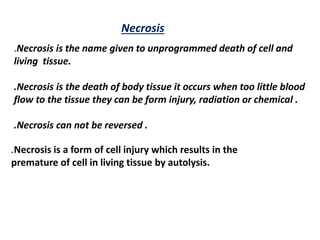
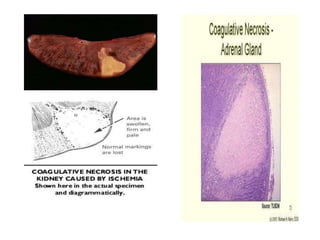
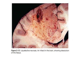
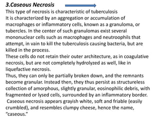
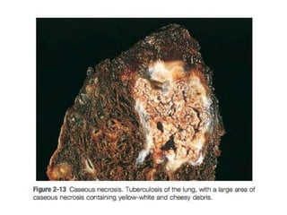
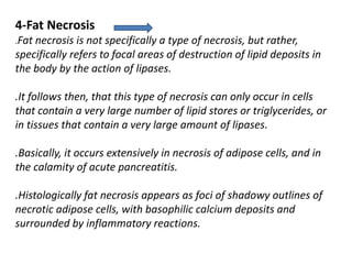
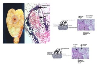
ADVANCE COLLEGE OF PHARMACY
Kanpur
DEPARTMENT OF PHARMACOLOGY
Mr. ANUBHAV DUBEY
M.Pharma
pharmacology
NECROSIS
Presented- By
Necrosis
.Necrosis is the name given to unprogrammed death of cell and
living tissue.
.Necrosis is the death of body tissue it occurs when too little blood
flow to the tissue they can be form injury, radiation or chemical .
.Necrosis can not be reversed .
.Necrosis is a form of cell injury which results in the
premature of cell in living tissue by autolysis.
Types of necrosis
1-cogulative necrosis.
2-liquefative necrosis
.3- caseous necrosis.
4-fat necrosis.
5-fibrnoid necrosis
1-cogulative necrosis
this is a type of necrosis in which the external architecture of the
cell is preserved for a span of at least a couple days.
It is most commonly seen in necrosis associated with conditions
of ischemia. Because the external architecture persists, it is
highly unlikely this type of necrosis is associated with extremely
damaging traumas, destructive acute or chronic immune
responses or toxins.
This is because the mechanism that would cause coagulative
necrosis would have to be one that damages all parts of the cell,
including both structural proteins and enzymes.
2-Liquefactive Necrosis
. In contrast to coagulative necrosis, liquefactive necrosis is
characterized by literal liquefaction.
.these cells are completely digested and broken down by the
excessive and extreme action of lysosomes and enzymes.
.Consequently, it is most commonly associated with infection, by
focal bacteria and focal fungi.
This is because these microbes stimulate the accumulation of a very
large number of leukocytes via an immune reaction, and the
lysosomal enzymes from these leukocytes destroy a wide range of
microbe infected cells.
3.Caseous Necrosis
This type of necrosis is characteristic of tuberculosis
It is characterized by an aggregation or accumulation of
macrophages or inflammatory cells, known as a granuloma, or
tubercles. In the center of such granulomas exist several
mononuclear cells such as macrophages and neutroophils that
attempt, in vain to kill the tuberculosis causing bacteria, but are
killed in the process.
These cells do not retain their outer architecture, as in coagulative
necrosis, but are not completely hydrolyzed as well, like in
liquefactive necrosis.
Thus, they can only be partially broken down, and the remnants
become granular. Instead then, they thus persist as structureless
collection of amorphous, slightly granular, eosinophilic debris, with
fragmented or lysed cells, surrounded by an inflammatory border.
Caseous necrosis appears grayish white, soft and friable (easily
crumbled), and resembles clumpy cheese, hence the name,
“caseous.”
4-Fat Necrosis
.Fat necrosis is not specifically a type of necrosis, but rather,
specifically refers to focal areas of destruction of lipid deposits in
the body by the action of lipases.
.It follows then, that this type of necrosis can only occur in cells
that contain a very large number of lipid stores or triglycerides, or
in tissues that contain a very large amount of lipases.
.Basically, it occurs extensively in necrosis of adipose cells, and in
the calamity of acute pancreatitis.
.Histologically fat necrosis appears as foci of shadowy outlines of
necrotic adipose cells, with basophilic calcium deposits and
surrounded by inflammatory reactions.
5-Fibrinoid Necrosis
.This is a special type of necrosis seen specifically in immune reactions
involving the blood vessels.
.It occurs when complexes of antigens and antibodies are deposited
at the walls of arteries.
.Deposits of these “immune complexes” coupled with a substance
known as fibrin that would have leaked out of arteries forms a bright
pink, amorphous (shapeless) appearance in hematoxylin and eosin
stains.
.It is associated with immune vasculitis, where the immune system
attacks blood vessels.
.If these immune complexes and fibrin aggregations are not promptly
removed from the walls of the arteries, then they attract Calcium
ions and induce calcification, which will have serious consequences.
Causes of necrosis
1- ischemia
2-physical agent
3- chemical agent
4-immunological agent
Difference between apoptosis and necrosis
Apoptosis Necrosis
Affects single cell Affects group of neighboring
cell
No inflamation response Inflamation response
Cell shrinkage Cell swelling
Chromatin can dentations and
non random DNA fragmentation
Random degradation of DNA
Apoptotic bodies integrate
ingested by neighboring cell
Lysed cell ingested by
macrophage
Presentation necrosis

Presentation necrosis

  • 1.
    ADVANCE COLLEGE OFPHARMACY Kanpur DEPARTMENT OF PHARMACOLOGY Mr. ANUBHAV DUBEY M.Pharma pharmacology NECROSIS Presented- By
  • 2.
    Necrosis .Necrosis is thename given to unprogrammed death of cell and living tissue. .Necrosis is the death of body tissue it occurs when too little blood flow to the tissue they can be form injury, radiation or chemical . .Necrosis can not be reversed . .Necrosis is a form of cell injury which results in the premature of cell in living tissue by autolysis.
  • 4.
    Types of necrosis 1-cogulativenecrosis. 2-liquefative necrosis .3- caseous necrosis. 4-fat necrosis. 5-fibrnoid necrosis
  • 5.
    1-cogulative necrosis this isa type of necrosis in which the external architecture of the cell is preserved for a span of at least a couple days. It is most commonly seen in necrosis associated with conditions of ischemia. Because the external architecture persists, it is highly unlikely this type of necrosis is associated with extremely damaging traumas, destructive acute or chronic immune responses or toxins. This is because the mechanism that would cause coagulative necrosis would have to be one that damages all parts of the cell, including both structural proteins and enzymes.
  • 7.
    2-Liquefactive Necrosis . Incontrast to coagulative necrosis, liquefactive necrosis is characterized by literal liquefaction. .these cells are completely digested and broken down by the excessive and extreme action of lysosomes and enzymes. .Consequently, it is most commonly associated with infection, by focal bacteria and focal fungi. This is because these microbes stimulate the accumulation of a very large number of leukocytes via an immune reaction, and the lysosomal enzymes from these leukocytes destroy a wide range of microbe infected cells.
  • 9.
    3.Caseous Necrosis This typeof necrosis is characteristic of tuberculosis It is characterized by an aggregation or accumulation of macrophages or inflammatory cells, known as a granuloma, or tubercles. In the center of such granulomas exist several mononuclear cells such as macrophages and neutroophils that attempt, in vain to kill the tuberculosis causing bacteria, but are killed in the process. These cells do not retain their outer architecture, as in coagulative necrosis, but are not completely hydrolyzed as well, like in liquefactive necrosis. Thus, they can only be partially broken down, and the remnants become granular. Instead then, they thus persist as structureless collection of amorphous, slightly granular, eosinophilic debris, with fragmented or lysed cells, surrounded by an inflammatory border. Caseous necrosis appears grayish white, soft and friable (easily crumbled), and resembles clumpy cheese, hence the name, “caseous.”
  • 11.
    4-Fat Necrosis .Fat necrosisis not specifically a type of necrosis, but rather, specifically refers to focal areas of destruction of lipid deposits in the body by the action of lipases. .It follows then, that this type of necrosis can only occur in cells that contain a very large number of lipid stores or triglycerides, or in tissues that contain a very large amount of lipases. .Basically, it occurs extensively in necrosis of adipose cells, and in the calamity of acute pancreatitis. .Histologically fat necrosis appears as foci of shadowy outlines of necrotic adipose cells, with basophilic calcium deposits and surrounded by inflammatory reactions.
  • 13.
    5-Fibrinoid Necrosis .This isa special type of necrosis seen specifically in immune reactions involving the blood vessels. .It occurs when complexes of antigens and antibodies are deposited at the walls of arteries. .Deposits of these “immune complexes” coupled with a substance known as fibrin that would have leaked out of arteries forms a bright pink, amorphous (shapeless) appearance in hematoxylin and eosin stains. .It is associated with immune vasculitis, where the immune system attacks blood vessels. .If these immune complexes and fibrin aggregations are not promptly removed from the walls of the arteries, then they attract Calcium ions and induce calcification, which will have serious consequences.
  • 15.
    Causes of necrosis 1-ischemia 2-physical agent 3- chemical agent 4-immunological agent Difference between apoptosis and necrosis
  • 16.
    Apoptosis Necrosis Affects singlecell Affects group of neighboring cell No inflamation response Inflamation response Cell shrinkage Cell swelling Chromatin can dentations and non random DNA fragmentation Random degradation of DNA Apoptotic bodies integrate ingested by neighboring cell Lysed cell ingested by macrophage