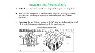
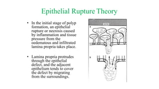
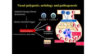
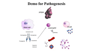
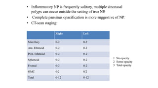
Nasal polyposis is a condition characterized by benign growths called polyps that develop in the nasal cavity and sinuses. The document discusses the epidemiology, etiology, pathogenesis, clinical presentation, diagnosis, and histopathology of nasal polyposis. Key points include:
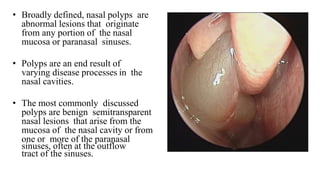
- Nasal polyps originate from the nasal mucosa and paranasal sinuses. Common causes include chronic sinus inflammation and allergy.
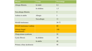
- Prevalence is 1-4% generally but higher in patients with asthma, cystic fibrosis, or aspirin sensitivity.
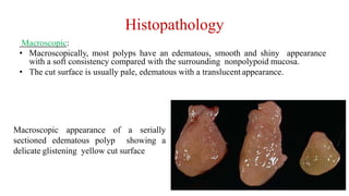
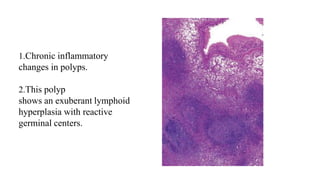
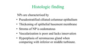
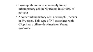
- Histopathology shows edematous polyps with eosinophils or chronic inflammatory polyps with fibrosis.
- Symptoms include nasal
Case report - discussion about presentation and managements of laryngoceles.
Published in Journal of Evolution of Medical and Dental Sciences 2015; Vol. 4, Issue 32, April 20; Page: 5586-5591
Abstract: Laryngoceles are rare, cystic dilatation of saccule of ventricle of larynx. Three types are recognized –internal, external and mixed types. Many of the laryngoceles are asymptomatic; few require surgical excision via internal/endoscopic or external approach. Contrast CT is the investigation of choice. A 40year old male presented to our OPD with a neck Scar, later diagnosed as laryngocele. Here is the case report about presentation, diagnosis and management of a large mixed layngocele.
KEYWORDS: Layngocele, Neck swelling, Saccule, Ventricle of larynx.
Slides prepared and compiled by highly experienced ENT teacher, Dr. Krishna Koirala from Nepal , for teaching undergraduate and postgraduate ENT students in the field of otorhinolaryngology.
A clear and concise explanation of the basic concepts in the subject matter concerned.
He is the Head of department with a sound knowledge in the field of ENT to teach both undergraduate and postgraduate ENT students
Skull base osteomyelitis is a rare complication of otitis externa in which soft tissue pathogens have spread to the periosteum and temporal bone of the skull causing necrosis.
Complications of rhinosinusitis(Dr ravindra daggupati)Ravindra Daggupati
orbital complications of rhino sinusitis,intra cranial complications of rhino sinusitis,classification of complications,diagnosis and treatment of complications
Nasal polyps are soft, painless, noncancerous growths on the lining of your nasal passages or sinuses. They hang down like teardrops or grapes. They result from chronic inflammation and are associated with asthma, recurring infection, allergies, drug sensitivity or certain immune disorders.
to differentiate b/w wheezing and stridor....lead to know to make clinical dx for asthma, croup, laryngomalacia, epiglottis...there many noisy breathing....our focus wheezing n stridor....
Updated version of molecular basis, with implied clinical aspect of the molecular basis.
(contents are taken from standard textbook and i dont own the copyright for the content details.)
Thyroid swelling and management. In detail case discussion of thyroid swelling and its management. Details of examination as well included in the slide.
- Video recording of this lecture in English language: https://youtu.be/lK81BzxMqdo
- Video recording of this lecture in Arabic language: https://youtu.be/Ve4P0COk9OI
- Link to download the book free: https://nephrotube.blogspot.com/p/nephrotube-nephrology-books.html
- Link to NephroTube website: www.NephroTube.com
- Link to NephroTube social media accounts: https://nephrotube.blogspot.com/p/join-nephrotube-on-social-media.html
Pulmonary Thromboembolism - etilogy, types, medical- Surgical and nursing man...VarunMahajani
Disruption of blood supply to lung alveoli due to blockage of one or more pulmonary blood vessels is called as Pulmonary thromboembolism. In this presentation we will discuss its causes, types and its management in depth.
NVBDCP.pptx Nation vector borne disease control programSapna Thakur
NVBDCP was launched in 2003-2004 . Vector-Borne Disease: Disease that results from an infection transmitted to humans and other animals by blood-feeding arthropods, such as mosquitoes, ticks, and fleas. Examples of vector-borne diseases include Dengue fever, West Nile Virus, Lyme disease, and malaria.
263778731218 Abortion Clinic /Pills In Harare ,sisternakatoto
263778731218 Abortion Clinic /Pills In Harare ,ABORTION WOMEN’S CLINIC +27730423979 IN women clinic we believe that every woman should be able to make choices in her pregnancy. Our job is to provide compassionate care, safety,affordable and confidential services. That’s why we have won the trust from all generations of women all over the world. we use non surgical method(Abortion pills) to terminate…Dr.LISA +27730423979women Clinic is committed to providing the highest quality of obstetrical and gynecological care to women of all ages. Our dedicated staff aim to treat each patient and her health concerns with compassion and respect.Our dedicated group ABORTION WOMEN’S CLINIC +27730423979 IN women clinic we believe that every woman should be able to make choices in her pregnancy. Our job is to provide compassionate care, safety,affordable and confidential services. That’s why we have won the trust from all generations of women all over the world. we use non surgical method(Abortion pills) to terminate…Dr.LISA +27730423979women Clinic is committed to providing the highest quality of obstetrical and gynecological care to women of all ages. Our dedicated staff aim to treat each patient and her health concerns with compassion and respect.Our dedicated group of receptionists, nurses, and physicians have worked together as a teamof receptionists, nurses, and physicians have worked together as a team wwww.lisywomensclinic.co.za/
Prix Galien International 2024 Forum ProgramLevi Shapiro
June 20, 2024, Prix Galien International and Jerusalem Ethics Forum in ROME. Detailed agenda including panels:
- ADVANCES IN CARDIOLOGY: A NEW PARADIGM IS COMING
- WOMEN’S HEALTH: FERTILITY PRESERVATION
- WHAT’S NEW IN THE TREATMENT OF INFECTIOUS,
ONCOLOGICAL AND INFLAMMATORY SKIN DISEASES?
- ARTIFICIAL INTELLIGENCE AND ETHICS
- GENE THERAPY
- BEYOND BORDERS: GLOBAL INITIATIVES FOR DEMOCRATIZING LIFE SCIENCE TECHNOLOGIES AND PROMOTING ACCESS TO HEALTHCARE
- ETHICAL CHALLENGES IN LIFE SCIENCES
- Prix Galien International Awards Ceremony
Title: Sense of Smell
Presenter: Dr. Faiza, Assistant Professor of Physiology
Qualifications:
MBBS (Best Graduate, AIMC Lahore)
FCPS Physiology
ICMT, CHPE, DHPE (STMU)
MPH (GC University, Faisalabad)
MBA (Virtual University of Pakistan)
Learning Objectives:
Describe the primary categories of smells and the concept of odor blindness.
Explain the structure and location of the olfactory membrane and mucosa, including the types and roles of cells involved in olfaction.
Describe the pathway and mechanisms of olfactory signal transmission from the olfactory receptors to the brain.
Illustrate the biochemical cascade triggered by odorant binding to olfactory receptors, including the role of G-proteins and second messengers in generating an action potential.
Identify different types of olfactory disorders such as anosmia, hyposmia, hyperosmia, and dysosmia, including their potential causes.
Key Topics:
Olfactory Genes:
3% of the human genome accounts for olfactory genes.
400 genes for odorant receptors.
Olfactory Membrane:
Located in the superior part of the nasal cavity.
Medially: Folds downward along the superior septum.
Laterally: Folds over the superior turbinate and upper surface of the middle turbinate.
Total surface area: 5-10 square centimeters.
Olfactory Mucosa:
Olfactory Cells: Bipolar nerve cells derived from the CNS (100 million), with 4-25 olfactory cilia per cell.
Sustentacular Cells: Produce mucus and maintain ionic and molecular environment.
Basal Cells: Replace worn-out olfactory cells with an average lifespan of 1-2 months.
Bowman’s Gland: Secretes mucus.
Stimulation of Olfactory Cells:
Odorant dissolves in mucus and attaches to receptors on olfactory cilia.
Involves a cascade effect through G-proteins and second messengers, leading to depolarization and action potential generation in the olfactory nerve.
Quality of a Good Odorant:
Small (3-20 Carbon atoms), volatile, water-soluble, and lipid-soluble.
Facilitated by odorant-binding proteins in mucus.
Membrane Potential and Action Potential:
Resting membrane potential: -55mV.
Action potential frequency in the olfactory nerve increases with odorant strength.
Adaptation Towards the Sense of Smell:
Rapid adaptation within the first second, with further slow adaptation.
Psychological adaptation greater than receptor adaptation, involving feedback inhibition from the central nervous system.
Primary Sensations of Smell:
Camphoraceous, Musky, Floral, Pepperminty, Ethereal, Pungent, Putrid.
Odor Detection Threshold:
Examples: Hydrogen sulfide (0.0005 ppm), Methyl-mercaptan (0.002 ppm).
Some toxic substances are odorless at lethal concentrations.
Characteristics of Smell:
Odor blindness for single substances due to lack of appropriate receptor protein.
Behavioral and emotional influences of smell.
Transmission of Olfactory Signals:
From olfactory cells to glomeruli in the olfactory bulb, involving lateral inhibition.
Primitive, less old, and new olfactory systems with different path
Couples presenting to the infertility clinic- Do they really have infertility...Sujoy Dasgupta
Dr Sujoy Dasgupta presented the study on "Couples presenting to the infertility clinic- Do they really have infertility? – The unexplored stories of non-consummation" in the 13th Congress of the Asia Pacific Initiative on Reproduction (ASPIRE 2024) at Manila on 24 May, 2024.
Knee anatomy and clinical tests 2024.pdfvimalpl1234
This includes all relevant anatomy and clinical tests compiled from standard textbooks, Campbell,netter etc..It is comprehensive and best suited for orthopaedicians and orthopaedic residents.
micro teaching on communication m.sc nursing.pdfAnurag Sharma
Microteaching is a unique model of practice teaching. It is a viable instrument for the. desired change in the teaching behavior or the behavior potential which, in specified types of real. classroom situations, tends to facilitate the achievement of specified types of objectives.
Ozempic: Preoperative Management of Patients on GLP-1 Receptor Agonists Saeid Safari
Preoperative Management of Patients on GLP-1 Receptor Agonists like Ozempic and Semiglutide
ASA GUIDELINE
NYSORA Guideline
2 Case Reports of Gastric Ultrasound
Lung Cancer: Artificial Intelligence, Synergetics, Complex System Analysis, S...Oleg Kshivets
RESULTS: Overall life span (LS) was 2252.1±1742.5 days and cumulative 5-year survival (5YS) reached 73.2%, 10 years – 64.8%, 20 years – 42.5%. 513 LCP lived more than 5 years (LS=3124.6±1525.6 days), 148 LCP – more than 10 years (LS=5054.4±1504.1 days).199 LCP died because of LC (LS=562.7±374.5 days). 5YS of LCP after bi/lobectomies was significantly superior in comparison with LCP after pneumonectomies (78.1% vs.63.7%, P=0.00001 by log-rank test). AT significantly improved 5YS (66.3% vs. 34.8%) (P=0.00000 by log-rank test) only for LCP with N1-2. Cox modeling displayed that 5YS of LCP significantly depended on: phase transition (PT) early-invasive LC in terms of synergetics, PT N0—N12, cell ratio factors (ratio between cancer cells- CC and blood cells subpopulations), G1-3, histology, glucose, AT, blood cell circuit, prothrombin index, heparin tolerance, recalcification time (P=0.000-0.038). Neural networks, genetic algorithm selection and bootstrap simulation revealed relationships between 5YS and PT early-invasive LC (rank=1), PT N0—N12 (rank=2), thrombocytes/CC (3), erythrocytes/CC (4), eosinophils/CC (5), healthy cells/CC (6), lymphocytes/CC (7), segmented neutrophils/CC (8), stick neutrophils/CC (9), monocytes/CC (10); leucocytes/CC (11). Correct prediction of 5YS was 100% by neural networks computing (area under ROC curve=1.0; error=0.0).
CONCLUSIONS: 5YS of LCP after radical procedures significantly depended on: 1) PT early-invasive cancer; 2) PT N0--N12; 3) cell ratio factors; 4) blood cell circuit; 5) biochemical factors; 6) hemostasis system; 7) AT; 8) LC characteristics; 9) LC cell dynamics; 10) surgery type: lobectomy/pneumonectomy; 11) anthropometric data. Optimal diagnosis and treatment strategies for LC are: 1) screening and early detection of LC; 2) availability of experienced thoracic surgeons because of complexity of radical procedures; 3) aggressive en block surgery and adequate lymph node dissection for completeness; 4) precise prediction; 5) adjuvant chemoimmunoradiotherapy for LCP with unfavorable prognosis.
3. Introduction
• A polyp presents in the nasal cavity with a grape-like
appearance, having a 'body' and a 'stalk'. The surface is
smooth and the color is more yellow than the pink mucous
membrane.
• Nasal polyps originate in the upper part of the nose around
the openings to the ethmoidal sinuses.
4. • Broadly defined, nasal polyps are
abnormal lesions that originate
from any portion of the nasal
mucosa or paranasal sinuses.
• Polyps are an end result of
varying disease processes in the
nasal cavities.
• The most commonly discussed
polyps are benign semitransparent
nasal lesions that arise from the
mucosa of the nasal cavity or from
one or more of the paranasal
sinuses, often at the outflow
tract of the sinuses.
5. Earliest records- 2000 BC
500 BC
“Nasal Polyposus”
1744- Elongation of
lining of sinus
membrane
1940-
Immunohistochemistr
y
19th Century
6. EPIDEMIOLOGY
• The prevalence of nasal polyps (NP) in the population has been grossly estimated as
1–4%.
• It increases with age, reaching a peak in those aged 50 years and older.
• Male: Female = 2:1
• An association between NP and allergic rhinitis (AR) is weak, with NP prevalence in patients
with AR estimated between 1.5-1.7% and this incidence is similar to that of the general
population.
• Nasal polyposis occurs with a high frequency in groups of patients
having specific airway diseases
7. Disease Group Percentage
Allergic Rhinitis InAdult 0.1
In children 1.5
Non-allergic Rhinitis 5
Asthma in adults Allergic 5
Non-allergic 13
NSAID intolerance 36-72
NSAID intolerance+asthma 80
Allergic fungal
rhinosinusitis
>80
Churg-strauss syndrome 50
Cystic fibrosis In children 10
In adults 40
Primary ciliary dyskinesia 40
8. • Genetic inheritance has been proposed as a possible etiology
of NP. Studies have suggested that up to 14% of patients
with NP have a family history of NP.
• In a Caucasian population, NP have been shown to have a
strong eosinophilic component while NP in Asian countries
showa neutrophilic pattern.
• Up to 80% of NSAID insensitive patients have NP.
9. ETIOLOGY andASSOCIATED DISEASES
• The aspirin triad: A triad ofnasal
polyposis, aspirin and asthma was described (Samtard’s triad)
and it represents the most aggressive forms of disease.
• It is a nonallergic entity and the intolerance is not confined to
acetylsalicylic acid, as the patients react to other NSAIDs.
10. ALLERGY?
• Previously believed to have strong association with NP
• Studies have failed to prove this association
• According to Keith et al there is no deterioration of nasal
symptoms or eosinophilia during the pollen season in polyp
patients having a positive skin test to pollen. Thus, it appears that
allergy is not a well documented cause or aggravating factor in
nasal polyposis.
11. Cystic fibrosis:
• Cystic fibrosis (CF), also known as mucoviscidosis, is an
autosomal recessive genetic disorder that affects most critically
the lungs, and also the pancreas, liver, and intestine.
• It is characterized by abnormal transport of chloride and
sodium across an epithelium, leading to thick, viscous
secretions.
• 45% of adults with cystic fibrosis.
12. Kartagener’s syndrome:
• Primary ciliary dyskinesia (PCD), also known as
immotile ciliary syndrome.
• Rare, ciliopathic, autosomal recessive genetic disorder.
• It causes a defect in the action of the cilia lining the
respiratory tract (lower and upper, sinuses, Eustachian tube,
middle ear) and fallopian tube, and also of the flagella of
sperm in males.
• 40%
13. Young syndrome
• It is a rare condition which consists of recurrent respiratory
disease with
• Chronic rhinosinusitis,
• Nasal polyps,
• Bronchiectasis and
• Azoospermia.
14. Pathogenesis & Pathophysiology
• The pathogenesis of nasal polyps explains how the polyps start and
grow.
• The pathophysiology of nasal polyps explains the events and processes
taking place in the outgrowth of nasal polyps.
• Several pathogenetic theories on the formation of nasal polyps have been
published during the last 150 years
15. Adenoma and fibroma theory
• Billroth found increased number of long tubulous glands in the polyps.
• The NP were interpreted as adenomas that began by growing underthe
nasal mucosa, pushing the epithelium and the original nasal glands
outwards.
• Hopmann did not find any glands in the NP from his study andinterpreted
NP as soft fibromas, protruding towards the nasalmucosa.
16. Necrotizing Ethmoiditis Theory
• This theory supposes that ethmoiditis leads to periostitis and
osteitis of the ethmoid bone and causes bone necrosis.
• Hayek argued strongly against this theory, based on the fact
that he could not found bone necrosis in the ethmoid sinus.
17. Glandular-Cyst Theory
• This theory is based upon the presence of cystic glands and mucus-
filled cysts in NP.
• It is hypothesised that oedema of the nasal mucosa causes
obstruction of the ducts of basal glands, leading to the formation
of cysts in the nasal mucosa.
• The cysts expand and push the nasal mucosa downwards, forming a
polyp.
18. Mucosal Exudate Theory
• Hayek believed that the formation of NP started via an
exudate localised deep in the nasal mucosa, which pressed
outwards caudally.
19. Theory on Cystic Dilatation of the Excretory Duct of Nasal Glands
and Vessel Obstruction
• According to this theory in chronic inflammation of the nasal mucosa,
excretory ducts of nasal tubulo-alveolar glands are obstructed, distended
and dilated into cystic structures.
• The capillaries and veins (which are arranged around the excretory
ducts and the gland mass) become stretched and obstructed, resulting in
increased permeability, transudation and oedema.
• This theory has been used to explain polyp formation in cystic fibrosis.
20. Blockade Theory
• The theory of Jenkins
• The polyp itself is an accumulation of intercellular fluid
dammed up in a localised tissue.
• The dam is usually caused by an infiltration of round cells,
producing blockade of intercellular spaces and local lymph
oedema.
21. Peri-Phlebitis andPeri- Lymphangitis Theory
• The theory of Eggston and Wolff is based upon the recurrent
infections that lead to the blocking of intercellular fluid
transport in the mucosa and oedema of the lamina propria.
• If the oedema involves major areas, the result is the prolapse
of the mucosa and formation of polyps.
22. Glandular Hyperplasia Theory
• Krajina found in cases of chronic infection or allergy
localised infiltrates in the nasal mucosa and localised
hyperplasia of nasal glands.
• The glands will increase in size and cause bulging of
the mucosa.
23. Epithelial Rupture Theory
• In the initial stage of polyp
formation, an epithelial
rupture or necrosis caused
by inflammation and tissue
pressure from the
oedematous and infiltrated
lamina propria takes place.
• Lamina propria protrudes
through the epithelial
defect, and the adjacent
epithelium tends to cover
the defect by migrating
from the surroundings.
24. • If the epithelial defect
is not covered soon
enough or if it is
insufficiently covered,
the prolapsed lamina
propria continues to
grow and the polyp,
with its vascular stalk,
is established.
• After epithelialization
of the polyp, the
characteristic new, long
tubulous glands are
formed
25. • In the ASA intolerant patients, a lowered prostaglandin E2
(PGE2) production has been observed.
• PGE2 has a significant anti-inflammatory activity, including
inhibition of eosinophils.
• A possible intrinsic defect in PGE2 production might, therefore,
be responsible for a further increase of eosinophilic accumulation
in ASA intolerant patients.
26. Role of Staphylococcus aureus
enterotoxins (SAE)
• Multiclonal IgE antibody formation to SAE
can be seen in nasal polyp tissue, but rarely in
CRS.
• It is positive in about 30-50% of the patients
with NP and in about 60-80% of nasal polyp
subjects with asthma
27. Nasal polyposis: aetiology and pathogenesis
Chemokines
T
Cytokines
B
Hyper IgE
Eosinophils
( apoptosis)
Superantigens
IL-5
ECP
Albumin
Eotaxin
Polyclonal IgE
Epithelial damage (barrier
dysfunction)
chronic microbial trigger
S. Aureusenterotoxins: diseasemodifiers
30. Histopathology
Macroscopic:
• Macroscopically, most polyps have an edematous, smooth and shiny appearance
with a soft consistency compared with the surrounding nonpolypoid mucosa.
• The cut surface is usually pale, edematous with a translucent appearance.
Macroscopic appearance of a serially
sectioned edematous polyp showing a
delicate glistening yellow cut surface
31. • Microscopic:
Polyps have historically been classified based on their histological
structural appearance and the nature of predominant inflammatory cell
population into 3 types.
1. Allergic polyp
2. Chronic inflammatory polyp
32. (1) Edematous, Eosinophilic, or “ Allergic” polyps:
• . Most common type.
• These polyps are lined with respiratory epithelium
• The basement membrane is often thickened, and there is abundant submucosal
edema.
• Mucus retention cysts are common and varying amounts of mixed inflammatory cell
infiltrates contain mostly eosinophils, plasma cells, and scattered lymphocytes.
• The edematous and eosinophilic polyps are seen in the whole spectrum of associated
disorders including, EMCRS, allergic fungal sinusitis, Sampter’s triad, cystic fibrosis, and
Churg–Strauss syndrome.
33. 1.Edematous polyp
2.Thickened basement membranes
(arrows)
3.Marked submucosal edema resulting in
extensive clear space between
submucosal connective tissue fibers.
4.Scattered throughout are mixed
inflammatory infiltrates including
eosinophils and plasma cells
34. • 2. Chronic inflammatory polyp:
• This type is less common, forming less than 10% of inflammatory nasal
polyps.
• The main histological features are the presence of submucosal fibrosis
and an often prominent mixed inflammatory infiltrate with a lymphoid
predominance often with germinal centers.
• The surface epithelium is likely to show squamous metaplasia as a
marker of chronicity.
36. 3) Seromucinous, glandular polyps:
• Polyps with hyperplasia of seromucinous glands are less
common.
• Lesions in this category are relatively new and somewhat
controversial as to their relationship with true epithelial
neoplasms, and include respiratory epithelial adenomatoid
hamartoma and seromucinous hamartoma.
37. Mucus Histopathology
• Secretions from CRS with polyps generally contain more eosinophils than those
without polyps, regardless of consistency of the mucus.
• Secretions are typically thick, almost solid.
• Stained sections of this mucus typically show clusters of eosinophils,
eosinophil breakdown products (Charcot– Leyden crystals), and other
inflammatory and epithelial cells.
• This mucus, termed eosinophilic mucus, is the diagnostic criteria for EMCRS
and allergic fungal sinusitis.
38. Antrochoanal polyp
• Antrochoanal polyps are the most common type of choanal polyp.
• Other sites of origin may be sphenoid, ethmoid, rarely septum, and
inferior turbinate.
• Antrochoanal polyps represent 4–6% of all polyps, and in the pediatric
population, up to 33%.
• They have an antral and choanal component. These typically arise
from the posterior wall of maxillary antrum and often have a thin
“neck” that passes through the maxillary sinus ostium (or accessory ostium).
• They are often unilateral, but may be bilateral on rare occasions.
39.
40. • Bernoullis phenomenon
• Jacksons abnormal mucopolysacharide deposition
• Infections cause
• Vasomotor imbalance theory
41. • Macroscopically, these range from erythematous to cystic with the latter
often seen in the antral portion.
• Microscopically, these polyps are lined by ciliated pseudostratified
epithelium that is usually intact, with a thin basement membrane.
• Stroma may exhibit myxoid change and stromal giant cells, but usually
lacks a significant inflammatory component.
• Rarely, degenerative changes including cholesterol granulomas and
angiomatous change may be found.
42. Clinical presentation
• Small polyps may not produce symptoms and may be identified
only during routine examination.
• Small polyps in middle meatus may produce symptoms and
block the outflow tract of the sinuses causing chronic or
recurrent sinusitis symptoms.
43. Symptom-producing polyps can cause:
• 1) Nasal airway obstruction
• 2) Post nasal drainage
• 3) Dull headache
• 4) Snoring
• 5) Rhinorrhoea
• 6) Hyposmia/anosmia
• 7) Epistaxis if preset may give suspicion of
more serious nasal cavity lesions.
44. • Massive polyposis or a single large polyp (eg:
antrochoanal polyp) can cause:
• 1) Obstructive sleep symptoms
• 2) Chronic mouth breathing
•3) Altered craniofacial structure.
(Frog face)
• 4) Proptosis
• 5) Hypertelorism
• 6) Diplopia
45. Diagnosis and scoring
• Rhinoscopy: Large polyp can be identified by
anterior rhinoscopy.
• In contrast to hyperplastic turbinate, a polyp can
be made to move by touching with a probe.
• Seen as multiple glistening, grayish white, smooth
pedunculated mass in both side of nasal cavity.
• On probing insensitive to touch, do not bleed and
probe can be passed all around the masses.
46. • Direct nasal endoscopy:
- Preferred method of examination.
- Can diagnose small polyp in middle meatus.
- It gives superior assessment of the extent of
the disease and anatomical abnormalities
47. • Polyp grading system. ( Meltzer et al)
Endoscopic appearance
Score
no visible NP 0
small amount of polypoid
disease confined within the
middle
meatus
1
multiple polyps
occupying the middle
meatus
2
polyps
extending beyond the
middle meatus
3
polyps completely
obstructing the nasal
cavity
4
48. Imaging modalities
• CT scan is the primary modality used to evaluate patients with NP, but MRI does have an
occasional role.
• Conventional radiographs are no longer within the standard of care for the evaluation of the
paranasal sinuses.
• Modern helical multislice CT scanners can produce reconstructed images of diagnostic quality
in any plane of imaging.
• Cystic fibrosis patients have preferential opacification of the paranasal sinuses, whereas
non-CF patients with NP have preferential opacification of the nasal cavity.
• On MRI, fungal infection may have very low signal on T2- weighted images, and may
thus mimic aerated sinuses.
49. • Polyps appear radiographically as rounded nodules of soft tissue along the mucosal
surfaces of the paranasal sinuses and nasal cavity.
• More radiodense than the surrounding mucosal thickening or secretions, which make
them appear slightly brighter on CT.
• This pattern may be reversed if the secretions become inspissated.
50. • Polyps do not erode into the surrounding bone, but pressure from a polyp may produce a
benign local remodeling pattern that scallops the underlying bone. (Fig 1)
• This bone remodeling will occasionally thin bony septations beyond the resolution of CT,
giving the appearance of bone erosion, particularly in the ethmoid septations. (Fig 2)
Fig 1 Fig 2
51. • In antronasal polyps sometimes, the polyp will be narrowed as it passes through the
maxillary os, resulting in a dumbbell configuration on coronal CT. The medial wall of
the maxillary sinus is usually bowed into the nasal cavity. (Fig 1)
• In antrochoanal polyp the most helpful radiologic feature is the
mass itself extending into the nasopharynx. (Fig 2)
Fig 1 Fig 2
52. • Inflammatory NP is frequently solitary, multiple sinonasal
polyps can occur outside the setting of true NP.
• Complete pansinus opacification is more suggestive of NP.
• CT-scan staging:
Right Left
Maxillary 0-2 0-2
Ant. Ethmoid 0-2 0-2
Post. Ethmoid 0-2 0-2
Sphenoid 0-2 0-2
Frontal 0-2 0-2
OMC 0/2 0/2
Total 0-12 0-12
1 No opacity
2 Some opacity
3 Total opacity
53. • Allergic fungal sinusitis (AFS). On CT, the presence of hyperdense
secretions between layers of thickened, hypodense mucosa is suggestiveof
AFS. (Fig 1)
• Inverted papillomas are often indistinguishable from polyps
radiographically. A lobular (“cerebriform”) configuration is alsosuggestive
of papilloma. Bony sclerosis and osteoneogenesis are frequently present at
the site of attachment of inverted papillomas. (Fig 2)
Fig 1 Fig 2
55. Laboratory Investigations
• Routine blood examination.
• Serological radioallergosorbent test (RAST) .
• Allergic skin testing for those who are having polyp associated with
allergic rhinitis.
• Sweat chloride test/ genetic testing for custic fibrosis in any child
with multiple benign nasal polyp.
• Nasal smear eosinophils may differentiate allergic from nonallergic
sinus diseases.
• Biopsy from the polyp after evaluation by CT scan/MRI.
56.
57. Histologic finding
NPs are characterised by
• Pseudostratified ciliated columnar epithalium
• Thickening of epithelial basement membrane
• Stroma of NP is oedematous
• Vascularization is poor and lacks innervation
• Hyperplasia of seromucous gland when
comparing with inferior or middle turbinate.
58. • Eosinophils are most commonly found
inflammatory cell in NP (found in 80-90% of
polyps)
• Another inflammatory cell, neutroophil, occurs
in 7% cases. This type of NP associates with
CF, primary ciliary dyskinesia or Young
syndrome.
59. Chemical mediators:
1) Cytokines: IL-1, Il-5 are found regularly
2) Growth factors:
• TNF:
• GM-CSF:
Alpha and beta found regularly
Never – Intermittent
• Platelet derived factor-present
• Vascular permeable factors:Present
• Vascular endothelial growth factor:Present
• Insulin like growth factor 1:Present
• Stem cell factor :Present
60. Immunoglobulins:
• IgG = Same level as MT or IT
• IgA = Increased
• IgM = Same
• IgD = Same
• IgE = Increased
Adhesion molecule:
• Vascular adhesion molecule: Present
• E & P selectin : Present
61. Treatment
• Treatment can either be medical and/or surgical.
• Oral and topical nasal steroid administration is the
primary medical therapy.
• Antihistamines, decongestants provide little benefit.
• Immunotherapy is useful in treating associated allergic
rhinitis.
• Antibiotic for bacterial super infections.
62. Intranasal corticosteroids
• Normally used intranasal steroids are Beclomethasone,
Ciclesonide, Flunisolide, Fluticasone Propionate,
Fluticasone furoate, Mometasone furoate,
TriamcinoloneAcetonide.
• They help in:
• Reducing polyp size
• Increase nasal patency
• Reduction in rhinitis symptoms
• Reduction in loss of sense of smell
• Reduction in recurrence of polyp
• Safety
64. Budesonide Nasal Spray
No statiscal difference in the dosage used.
1 mg/500 mL (0.002 mg/mL)
Efficacy and tolerability of budesonide aqueous nasal spray
treatment in patients with nasal polyps.
Jankowski R1, Schrewelius C, Bonfils P, Saban Y, Gilain L, Prades
JM, Strunski V.
67. Method of administration
• Spray- If two nasal sprays are to be administered to each
nostril, one spray should be directed upwards and the other
backwards whilst the patient does not breathe or breathes in
gently.
• Drops-
68. Sideeffect of IntraNasal steroids
• - Excoriation and bleeding( can be reduced by
cross hand technique)
• - Beclomethasone dipropionate nasal spray is
associated with the onset of increased
intraocular pressure.
• -Delay in growth in prepubescent children has
led to an FDA warning on all INS
69. Systemic Corrticosteroids
• Can be given as tablets and depot-injections.
• Oral prednisolone can be given at 25mg/daily
for 10-14 days.
• As a depot-injection corresponds 100mg
prednisolone.
• It may serve as medical polypectomy.
70.
71. Risks of systemic steroid
• Insomnia,
• personality change,
• truncal obesity,
• weight gain,
• glaucoma,
• cataracts,
• osteoporosis (requires greater than 3 months
usage),
• peptic ulcer disease, and
• increased incidence of infection
75. Other Non surgical treatment
• Immunotherapy
• Aspirin Desensitisation
• Allergy treatment
• Leukotriene Modifiers.
76. Surgical Management
General principles surgery for nasal polyposis:
• Selection of the patient who has exhausted medical
management and remains symptomatic.
• The patient must understand the absolute necessity of often
prolonged postoperative care.
• Selection of appropriate anesthetic method and preparation
of the surgical field to maximize visualization and minimize
bleeding.
• Accurate identification of surgical landmarks such as the
lamina papyracea, anterior ethmoid artery, and skull base on
preoperative CT imaging.
77. •Use of stereotactic image-guided surgery when
necessary to confirm the skull base, cribriform
plate, and lamina papyracea.
•Atraumatic removal of polyps and diseased bone
while preserving as much mucosa as possible.
Meticulous removal of osteitic bone and bony
partitions.
•Ongoing postoperative medical management to
prevent disease recurrence.
78. FESS
• Preoperative evaluation by radiology and endoscopic
examination.
• Preoperatively, patients with evidence of active infections
should be administered culture-directed antibiotics to help
eradicate infection and to decrease mucosal inflammation.
• Administration of preoperative oral corticosteroid reduces
the size of the polyp helps in proper visualization.
• Intravenous anesthesia with remifentanil and
propofol, improved visual field with decreased blood loss
has been demonstrated.
79. Steps
• Polyps obstructing the nasal cavity or emanating from the middlemeatus
are best addressed with the microdebrider.
• The maxillary sinus is best addressed with 0 and 30 endoscopes.
• Uncinate process is removed with sickle knife, backbiting forcep&
microdebrider.
• The natural ostium of the maxillary sinus should be probed and widened
and that any surgically created ostium is brought into continuity with the
natural as in order to prevent recirculation ofmucus.
• In the case of polyposis, large antrostomies are recommended. The sinus
should be inspected with at minimum the 45 endoscope; a 70 endoscope
is advised for analyzing the floor and anterior-most portions of the
maxillary sinus to inspect for polyps and any infraorbital ethmoid cellsthat
may obstruct the ostium. Inspissated secretions need to be
evacuated, cultured, and sent to pathology for analysis of possiblefungal
80. • Anterior ethmoid cells should be meticulously dissected using
through-cutting instruments and polypoid debris removed with the
microdebrider.
• The first important landmark to identify is the medial orbital wall
and the dissection is continued along the medial orbital wall rather
than medially, because the skull base is significantly thicker and less
likely to be injured laterally as opposed to medially where it slopes
down into the middle turbinate.
• It is imperative that foci of ostetic bone along the lamina papyracea
and middle turbinate be removed to help reduce inflammation and
prevent polyp recurrence in these areas.
• Once the basal lamella is identified, the posterior ethmoid cells
should be dissected in a similar fashion.
• Dissection of the ethmoid cavity typically occurs in an anterior to
posterior fashion, identifying landmarks along the lamina papyracea
and skull base until the anterior face of the sphenoid is encountered.
81. • The superior turbinate is next identified in order to enter the
sphenoid sinus through the sphenoethmoidal recess and its natural
ostium.
• Sinus is entered away from critical structures such as the carotid
artery (dehiscent in up to 20% of cases) and the optic nerve
(dehiscent in 5–10% of cases)
• This method also ensures that a sphenoethmoidal (Onodi) cell is not
mistakenly treated as the sphenoid sinus.
• The sphenoid sinus should be opened widely from the skull base to
lamina papyracea; care must be taken to avoid the septal branch of
the sphenopalatine artery during this dissection.
• Polyps in sphenoid sinus is seen rarely
82. • Once the sphenoid sinus is opened, dissection along
the skull base occurs in a retrograde fashion in order
to clear lamella along the skull base until the frontal
recess is encountered.
• The region of the anterior ethmoid artery is identified
and avoided. It typically lies at the uppermost limit of
the anterior wall of the ethmoidal bulla.
• If an injury to the anterior ethmoid artery occurs,
bipolar electrocautery is used to coagulate the vessel.
83. • Frontal sinus is infrequently diseased with polyps; however, the
frontal recess can be extensively diseased with polyps, resulting in
obstruction of the frontal sinus itself.
• Much of the dissection involves angled instruments and endoscopes.
• In this region, preoperative review of the CT scans is critical, as a
variety of drainage pathways may lead to the frontal sinus and a
ethmoid air cells may invade the frontal sinus, complicating its
anatomy and dissection, and polyps within the frontal recess can
impair vision of these anomalies.
• Through-cutting instruments are used to dissect the anterior ethmoid
lamella until the skull base is skeletonized and the anterior ethmoid
artery identified, just posterior to the frontal recess.
• Partitions separating the frontal sinus from the supraorbital ethmoid
cells are then taken down meticulously. The agger nasi cap is
identified, resected, and frontal recess evaluated.
84. • After a complete dissection of the paranasal sinuses
has been performed, the field should be inspected for
loose fragments of bone, missed ethmoid partitions,
and points of bleeding.
• Wherever possible, any exposed bone is removed.
• Merocel spacers are placed for 24 h and removed on
postoperative day one when debridement occurs.
85. Postoperative management
• Surgery represents an adjunct to medical therapy in chronic
rhinosinusitis with nasal polyposis.
• Postoperatively, failure is almost assured if the ongoing
medical therapy to prevent recurrence is not administered.
• Routine postoperative debridement at first week of surgery
of the sinonasal cavity is critical to assuring success.
• Culturedirected antibiotics in the postoperative setting
should be utilized to limit iatrogenic infection, treat osteitis,
and eradicate sources of inflammation.
86. • Oral corticosteroid acts as a potent anti-
inflammatory in CRS with NP
• In diffuse nasal polyposis, the patient should be
discharged with a tapering course of prednisone
over a prolonged period, and overlapping
administration of topical corticosteroids should
begin before cessation of oral corticosteroids is
considered.
• Patients unwilling to undergo oral corticosteroid
therapy may benefit from topical administration
of budesonide 0.5 mg/2 mL mixed in 240 mL
normal saline for irrigation.
87. Other surgical techniques
• Nasalization procedure:
• The aim of the nasalisation procedure is to
remove the ethmoidal mucosa as completely as
possible without hazards, and to transform the
ethmoidal labyrinth into a unique cavity
opening into the nose.
88. • Aggressive Sinus Marsupialization Including the
Endoscopic Modified Lothrop Procedure:
• The frontal recess clearance maximizes the natural ostium
of the frontal sinus without enlarging it.
• The frontal drillout or EMLP creates the largest possible
frontal ostium diameter for individual anatomical
constraints.
• In patients who develop recurrent polyps after
surgery, the modified endoscopic Lothrop procedure, Draf
III or frontal drillout produces
better outcomes, and in some cases a cure, than standard
revision ESS.
89. Reasons for failure
• Insufficient ethmoidectomy
• Insufficient removal of septa
• Insufficient drug concentration behind the septa
• Insufficient cleaning of the polypoid mucosa behind the
septa
• Pool for collection of secretions
• Insufficient surgery of the frontal sinus
• Polyps at the frontal recess and frontal ostium area
• Polyps and mucoceles in the frontal sinus
• Stenosis of frontal ostium
• Insufficient surgery of the maxillary sinus
90. • Insufficient maxillary sinus ostium
• Reclosure of the ostium
• Decreased ventilation and drainage
• Insufficient drainage due to thick secretions
• Insufficient cleaning preop and postop
• Insufficient opening of anterior wall of sphenoid sinus
• Decreased ventilation
• Decreased drainage
• Insufficient drug concentration
• Insufficient cleaning preop and postop
• Free bony spicles
• Granulation tissue and polyp recurrence
91. Reference.
1. Scott- Brown otorhinolaryngology
2. Cummings Otolaryngology
3. Nasal polyposis by T. Metin Önerci
Berrylin J. Ferguson
4.Pubmed indexed Journals.
Editor's Notes
with a range of mucosal alterations that include ulceration, granulation tissue, acute mucositis, epithelial and goblet cell hyperplasia, and squamous metaplasia.