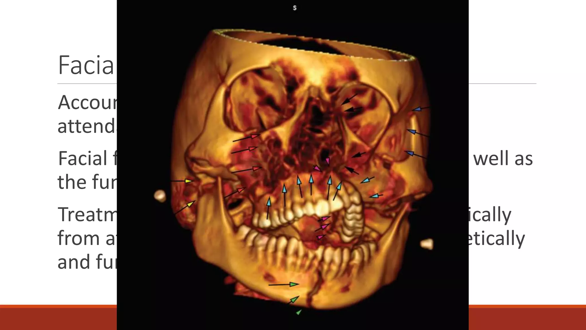
Facial fractures- Etiology,
Clinical features, Evaluation
and Management
PRESENTER: DR SREENIVAS
Facial Fractures
Accounts for 10% of all accidents and EMD
attendance
Facial fractures impairs the facial aesthetics as well as
the functions
Treatment of facial fracture has changed drastically
from attaining mere dental occlusion to aesthetically
and functionally normality.
Primary care
Initial Survey
Primary
management
Secondary
management
Initial survey
Airway
Breathing
Circulation
Disability
Exposure
Principles of Primary Management
Once stabilized
Categorize the extent of injury
Structural and functional assessment of facial bones, muscle , nerve
and soft tissue to be done
Visual acuity and movement to assessed ( retrobulbar haemorrhage
and retinal detachment)
Palpate all the bones
Dental occlusion
Radiographic Evaluation
All cases should have chest, cervical, spine and pelvic xrays
Facial xray is need when fractures are suspected
Once the specific fracture is identified, Secondary and
specific management of the fracture is done.
Nasal fractures
Nasal fractures recorded since the time of ancient Egypt
Edwin Smith Described correction
Little force 25-75lb?in
Men>women
15-30yrs
Classification
1. Nature of injury
2. Extent of deformity
3. Pattern of fracture
Nature of injury
Laterally applied force – 66%
Frontal force – 13% (more force is required since cartilage has cushion effect)
Extent of deformity
• Grade 0: bones perfectly straight
• Grade 1: bones deviated less than half of the width of the bridge of the
nose
• Grade 2: bones deviated half to one full width of the bridge of the nose
• Grade 3: bones deviated greater than one full width of the bridge of the
nose
• Grade 4: bones almost touching the cheek.
Pattern of Fracture
•Class 1 fractures
•Class 2 fractures
•Class 3 fractures
Class 1 Fracture
Low force
Lesser extent of deformity
Simplest form – depressed nasal bone
Another classic example is Chavallet frature
Class 2 fracture
Greater force
More cosmetic deformity
Nasal bone, frontal bone , maxillary bone are involved
Example is Jarjway Fracture
Class 3 fractures
Most severe nasal injuries
High velocity traumas
NASO-ORBITO ETHMOIOD fracture
PIG like appearance
2 types
◦ Type 1 and Type 2
◦ anterior skull base, posterior wall of the frontal sinus and optic canal remain intact.
Clinical Presentation
h/o of sustained trauma or assault.
Nasal obstruction
Persisting pain ??? Septal hematoma
Shape change (compare with old photos )
Diplopia, visual disturbance and epiphora suggest orbital
trauma. Loose teeth, an altered bite or trismus
Examination
External deformity
Palpate- deformity and crepitus
r/o septal hematoma
Investigation
Radiological – Xray only for documentation
In case of suspected serious facial fractures- CT scan to be done
Treatment
•80% of cases don’t need active management in acute phase
•In acute phase the soft tissue swelling may be misleading
•Re-examination to be done after 5 days
•Septal hematoma needs active intervention
•Optimal time of correction is 4-7 days
Reduction
LA/GA
LA- ? Tissue swelling with infiltration – under correction (Courtney
et al.)
Closed reduction
◦ INCREASE THEN DECREASE THE DEFORMITY
◦ Ash forceps/ walshams forceps
◦ Class 1 and 2 can be managed with this
◦ Splints- Internal/External- if placed usually kept for 7 days
Open reduction
◦ bilateral fractures with dislocation of the nasal dorsum and
significant (pre-existent or recent) septal deformity
◦ fractures of the cartilaginous pyramid, with or without dislocation
of the upper laterals
◦ K-wire can be placed and removed after 2 weeks
? Septal correction
46.9% cases concomitant septal fractures are noted
Reduce at the same sitting
Reduction/ Septoplasty/Quilting suture
Complication
Poor Cosmetic results
Nasal obstruction
Epistaxis
Septal- Hematoma, abscess, perforation
Parabolic shaped bone with complex articulation that
sonsist of paired synovial joints- TMJ
The traditional method of mandibular fracture treatment
was to immobilize using intermaxillary fixations (IMF).
Now miniplates, extended subperiosteal approach.
Mandibular fracture
Point of weakness – relatively thin.
Edentulous makes it more
vulnerable.
Signs and symptoms
•Step deformity palpable
•Asymmetry of the lower dental arch and
derangement of the occlusion
•Pain and paradoxical movement and crepitus on
distraction of the fractured segments
•Haematomas in the buccal sulcus or floor of the
mouth
•Blood-stained saliva
•Anaesthesia of the lower lip.
Tenderness
Trismus
Deviation - injured side on opening the
mouth
Inability to move -side opposite the
fracture
Symmetrical anterior open bite in
bilateral fractures of the necks of the
condyles.
Treatment
Closed reduction
◦ Intact dental arch – eyelet wires,Lenard buttons.
◦ Incomplete dental arch –arch bars, intermaxillary bones and pins.
External fixations- Gross tissue loss – external bars, mini pennings
Internal fixation- intra oral or extraoral
Principles of mandibular fixations
For simple mandibular fracture – Mono cortical 2 mm plates will be adequate. – Load sharing
and osteosynthesis
Anterior fracture- 2 plates posterior fracture- 1 plate
6mm screws are used.
Care taken not to injure the mental nerve.
Gross dimunition – internal fixations with bicortical screw is used.
Condylar neck fracture ?
Controversial topic on its management
Conservative
Surgical ?
When to intervene ?
Unilateral fractures that are significantly displaced and associated with a malocclusion
◦ IMF- 10–21 days
Open reduction of Condylar fracture
Absolute indications: Relative indications:
• Displacement of condyle into middle cranial
fossa
• Impossibility of restoring occlusion without
ORIF
• Lateral extra-capsular displacement.
• Invasion by foreign body (e.g. missile)
• Bilateral fracture with associated mid-face
fracture (particularly where one condylar
fracture is dislocated or angulated)
• Bilateral fracture with severe open bite
deformity
• Unilateral fracture with dislocation, overlap
or significant angulation of the condylar head
• When inter-maxillary fixation is
contraindicated for medical reasons
Fracture of Maxilla
One of the common fracture involved in the
mid facial trauma.
Signs and symptoms depend on the level of
fracture
The classical features of a midfacial fracture
are circum-orbital ecchymosis (panda facies),
facial oedema and emphysema, lengthening
of the face and an anterior open bite.
Le Fort classification
1. Le fort 1
2. Le fort 2
3. Le fort 3
Clinical feature
• epistaxis
• circumorbital ecchymosis
• facial oedema
• surgical emphysema
• lengthening of the face
• infraorbital anaesthesia.
Management of maxillary fracture
Emergency treatment
Reduction – traction and counter traction , if impacted-Rowe maxillary disimpaction forceps
Fixation- Internal fixation with 1.3-1.5mm plates
Zygomatic fracture
Zygomatic bone makes up the lateral 1/3 of face.
Previously called the tripod fracture.
1. Fronto-zygomatic
2. Infraorbital rim
3. Zygomaticomaxillary buttress.
Zygomatic arch, zygomaticosphenoid are the other articulations that are often involved.
Signs and symptoms
Examination done from front, above and behind.- zygomatic area will be swollen.
Subconjunctival bleeding.
Eyes movements will be restricted.
Associated with blow out fractureof floor of orbit.
Step deformity on palpation
Restricted mouth opening .
Xray- 15 and 30 degree occipitomental view.
CT scanning – standard of care.
Ophthal evaluation to rule out subjective diplopia.
Management
Minimal displaced and undisplaced fracture- Conservative management
Reduction with or without fixation for grossly displaced fractures.
Orbital floor fracture
Blunt trauma to globe and adjacent structure
Form and function of globe will be compromised.
Cardinal signs are-
1. Enophthamos
2. Hypoglobus
3. Supratarsal hollowing
4. Hood of eye, narrowing of palpebral fissure.
5. Diploplia in upper gaze ( trap door phenomenon).
Investigation
CT imaging
Management
All soft tissue should be mobilised and defect is supported by grafting
◦ -Polydimethylsiloxane
◦ Titanium alloplast
Absolute Relative
Indications
1 Significant restriction of eye movement
(diplopia) with CT confirmation of
entrapment
2 Significant enophthalmos
3 Large ‘blowout’ defect
4 Significant orbital dystopia
1 Visual impairment
2 Anticoagulant medication
3 Patient unconcerned
4 Proptosis
5 An already ‘at risk’ globe
Complications of reduction
1 Intraorbital haemorrhage
2 Lower eyelid retraction and ectropion
3 Persistent oedema of lower eyelid
4 Persistent enophthalmos
5 Persistent globe depression
6 Persistent diplopia in vertical gaze
7 Tissue reaction to implant
8 Extrusion of implant
9 Infection and chronic fistula formation
10 Dacryocystitis
11 Blindness
Naso orbito ethmoid complex fracture
Naso-orbito-ethmoid (NOE) fractures involve the anatomical confluence of the nose, orbits and
ethmoids.
Signs and symptoms
Loss of nasal projection
Tipping upof the end of the nose
Splaying of the nasal root
Telecanthus.
Management
Type I fractures can be stabilized using miniplates
Type II and III fractures are also repaired with miniplates,
but require a transnasal canthopexy to reduce the
telecanthus and hold the position of the medial canthal
ligaments.
Frontal sinus fracture
Classified into
◦Fracture involving anterior table
◦Fracture involving posterior table.
◦Fracture affecting nao-frontal duct
Aim of the treatment is have functional sinus with no/
minimal deformity.
Fractures of the anterior table may be treated
conservatively if there is no cosmetic deformity, while
displaced fractures require reduction and fixation.
Fractures of the posterior table demand a neurosurgical
opinion and may need an obliterative procedure or
cranialization with obliteration ofthe frontonasal recess and
its lining
Reference
Scott brown 8th ed
Ballengers 17th ed
Facial fracture by wileys
Thank you

Facial fracture

  • 1.
    Facial fractures- Etiology, Clinicalfeatures, Evaluation and Management PRESENTER: DR SREENIVAS
  • 2.
    Facial Fractures Accounts for10% of all accidents and EMD attendance Facial fractures impairs the facial aesthetics as well as the functions Treatment of facial fracture has changed drastically from attaining mere dental occlusion to aesthetically and functionally normality.
  • 3.
  • 4.
  • 5.
    Principles of PrimaryManagement Once stabilized Categorize the extent of injury Structural and functional assessment of facial bones, muscle , nerve and soft tissue to be done Visual acuity and movement to assessed ( retrobulbar haemorrhage and retinal detachment) Palpate all the bones Dental occlusion
  • 6.
    Radiographic Evaluation All casesshould have chest, cervical, spine and pelvic xrays Facial xray is need when fractures are suspected Once the specific fracture is identified, Secondary and specific management of the fracture is done.
  • 7.
    Nasal fractures Nasal fracturesrecorded since the time of ancient Egypt Edwin Smith Described correction Little force 25-75lb?in Men>women 15-30yrs
  • 8.
    Classification 1. Nature ofinjury 2. Extent of deformity 3. Pattern of fracture
  • 9.
    Nature of injury Laterallyapplied force – 66% Frontal force – 13% (more force is required since cartilage has cushion effect)
  • 10.
    Extent of deformity •Grade 0: bones perfectly straight • Grade 1: bones deviated less than half of the width of the bridge of the nose • Grade 2: bones deviated half to one full width of the bridge of the nose • Grade 3: bones deviated greater than one full width of the bridge of the nose • Grade 4: bones almost touching the cheek.
  • 11.
    Pattern of Fracture •Class1 fractures •Class 2 fractures •Class 3 fractures
  • 12.
    Class 1 Fracture Lowforce Lesser extent of deformity Simplest form – depressed nasal bone Another classic example is Chavallet frature
  • 13.
    Class 2 fracture Greaterforce More cosmetic deformity Nasal bone, frontal bone , maxillary bone are involved Example is Jarjway Fracture
  • 14.
    Class 3 fractures Mostsevere nasal injuries High velocity traumas NASO-ORBITO ETHMOIOD fracture PIG like appearance 2 types ◦ Type 1 and Type 2 ◦ anterior skull base, posterior wall of the frontal sinus and optic canal remain intact.
  • 15.
    Clinical Presentation h/o ofsustained trauma or assault. Nasal obstruction Persisting pain ??? Septal hematoma Shape change (compare with old photos ) Diplopia, visual disturbance and epiphora suggest orbital trauma. Loose teeth, an altered bite or trismus
  • 16.
    Examination External deformity Palpate- deformityand crepitus r/o septal hematoma
  • 17.
    Investigation Radiological – Xrayonly for documentation In case of suspected serious facial fractures- CT scan to be done
  • 18.
    Treatment •80% of casesdon’t need active management in acute phase •In acute phase the soft tissue swelling may be misleading •Re-examination to be done after 5 days •Septal hematoma needs active intervention •Optimal time of correction is 4-7 days
  • 19.
    Reduction LA/GA LA- ? Tissueswelling with infiltration – under correction (Courtney et al.) Closed reduction ◦ INCREASE THEN DECREASE THE DEFORMITY ◦ Ash forceps/ walshams forceps ◦ Class 1 and 2 can be managed with this ◦ Splints- Internal/External- if placed usually kept for 7 days
  • 21.
    Open reduction ◦ bilateralfractures with dislocation of the nasal dorsum and significant (pre-existent or recent) septal deformity ◦ fractures of the cartilaginous pyramid, with or without dislocation of the upper laterals ◦ K-wire can be placed and removed after 2 weeks
  • 22.
    ? Septal correction 46.9%cases concomitant septal fractures are noted Reduce at the same sitting Reduction/ Septoplasty/Quilting suture
  • 23.
    Complication Poor Cosmetic results Nasalobstruction Epistaxis Septal- Hematoma, abscess, perforation
  • 24.
    Parabolic shaped bonewith complex articulation that sonsist of paired synovial joints- TMJ The traditional method of mandibular fracture treatment was to immobilize using intermaxillary fixations (IMF). Now miniplates, extended subperiosteal approach. Mandibular fracture
  • 25.
    Point of weakness– relatively thin. Edentulous makes it more vulnerable.
  • 26.
    Signs and symptoms •Stepdeformity palpable •Asymmetry of the lower dental arch and derangement of the occlusion •Pain and paradoxical movement and crepitus on distraction of the fractured segments •Haematomas in the buccal sulcus or floor of the mouth •Blood-stained saliva •Anaesthesia of the lower lip. Tenderness Trismus Deviation - injured side on opening the mouth Inability to move -side opposite the fracture Symmetrical anterior open bite in bilateral fractures of the necks of the condyles.
  • 27.
    Treatment Closed reduction ◦ Intactdental arch – eyelet wires,Lenard buttons. ◦ Incomplete dental arch –arch bars, intermaxillary bones and pins. External fixations- Gross tissue loss – external bars, mini pennings Internal fixation- intra oral or extraoral
  • 28.
    Principles of mandibularfixations For simple mandibular fracture – Mono cortical 2 mm plates will be adequate. – Load sharing and osteosynthesis Anterior fracture- 2 plates posterior fracture- 1 plate 6mm screws are used. Care taken not to injure the mental nerve. Gross dimunition – internal fixations with bicortical screw is used.
  • 29.
    Condylar neck fracture? Controversial topic on its management Conservative Surgical ? When to intervene ?
  • 30.
    Unilateral fractures thatare significantly displaced and associated with a malocclusion ◦ IMF- 10–21 days
  • 31.
    Open reduction ofCondylar fracture Absolute indications: Relative indications: • Displacement of condyle into middle cranial fossa • Impossibility of restoring occlusion without ORIF • Lateral extra-capsular displacement. • Invasion by foreign body (e.g. missile) • Bilateral fracture with associated mid-face fracture (particularly where one condylar fracture is dislocated or angulated) • Bilateral fracture with severe open bite deformity • Unilateral fracture with dislocation, overlap or significant angulation of the condylar head • When inter-maxillary fixation is contraindicated for medical reasons
  • 32.
    Fracture of Maxilla Oneof the common fracture involved in the mid facial trauma. Signs and symptoms depend on the level of fracture The classical features of a midfacial fracture are circum-orbital ecchymosis (panda facies), facial oedema and emphysema, lengthening of the face and an anterior open bite.
  • 33.
    Le Fort classification 1.Le fort 1 2. Le fort 2 3. Le fort 3
  • 34.
    Clinical feature • epistaxis •circumorbital ecchymosis • facial oedema • surgical emphysema • lengthening of the face • infraorbital anaesthesia.
  • 35.
    Management of maxillaryfracture Emergency treatment Reduction – traction and counter traction , if impacted-Rowe maxillary disimpaction forceps Fixation- Internal fixation with 1.3-1.5mm plates
  • 36.
    Zygomatic fracture Zygomatic bonemakes up the lateral 1/3 of face. Previously called the tripod fracture. 1. Fronto-zygomatic 2. Infraorbital rim 3. Zygomaticomaxillary buttress. Zygomatic arch, zygomaticosphenoid are the other articulations that are often involved.
  • 37.
    Signs and symptoms Examinationdone from front, above and behind.- zygomatic area will be swollen. Subconjunctival bleeding. Eyes movements will be restricted. Associated with blow out fractureof floor of orbit. Step deformity on palpation Restricted mouth opening .
  • 38.
    Xray- 15 and30 degree occipitomental view. CT scanning – standard of care. Ophthal evaluation to rule out subjective diplopia.
  • 39.
    Management Minimal displaced andundisplaced fracture- Conservative management Reduction with or without fixation for grossly displaced fractures.
  • 41.
    Orbital floor fracture Blunttrauma to globe and adjacent structure Form and function of globe will be compromised. Cardinal signs are- 1. Enophthamos 2. Hypoglobus 3. Supratarsal hollowing 4. Hood of eye, narrowing of palpebral fissure. 5. Diploplia in upper gaze ( trap door phenomenon).
  • 42.
  • 43.
    Management All soft tissueshould be mobilised and defect is supported by grafting ◦ -Polydimethylsiloxane ◦ Titanium alloplast Absolute Relative Indications 1 Significant restriction of eye movement (diplopia) with CT confirmation of entrapment 2 Significant enophthalmos 3 Large ‘blowout’ defect 4 Significant orbital dystopia 1 Visual impairment 2 Anticoagulant medication 3 Patient unconcerned 4 Proptosis 5 An already ‘at risk’ globe
  • 45.
    Complications of reduction 1Intraorbital haemorrhage 2 Lower eyelid retraction and ectropion 3 Persistent oedema of lower eyelid 4 Persistent enophthalmos 5 Persistent globe depression 6 Persistent diplopia in vertical gaze 7 Tissue reaction to implant 8 Extrusion of implant 9 Infection and chronic fistula formation 10 Dacryocystitis 11 Blindness
  • 46.
    Naso orbito ethmoidcomplex fracture Naso-orbito-ethmoid (NOE) fractures involve the anatomical confluence of the nose, orbits and ethmoids.
  • 47.
    Signs and symptoms Lossof nasal projection Tipping upof the end of the nose Splaying of the nasal root Telecanthus.
  • 48.
    Management Type I fracturescan be stabilized using miniplates Type II and III fractures are also repaired with miniplates, but require a transnasal canthopexy to reduce the telecanthus and hold the position of the medial canthal ligaments.
  • 49.
    Frontal sinus fracture Classifiedinto ◦Fracture involving anterior table ◦Fracture involving posterior table. ◦Fracture affecting nao-frontal duct Aim of the treatment is have functional sinus with no/ minimal deformity.
  • 50.
    Fractures of theanterior table may be treated conservatively if there is no cosmetic deformity, while displaced fractures require reduction and fixation. Fractures of the posterior table demand a neurosurgical opinion and may need an obliterative procedure or cranialization with obliteration ofthe frontonasal recess and its lining
  • 52.
    Reference Scott brown 8thed Ballengers 17th ed Facial fracture by wileys
  • 53.