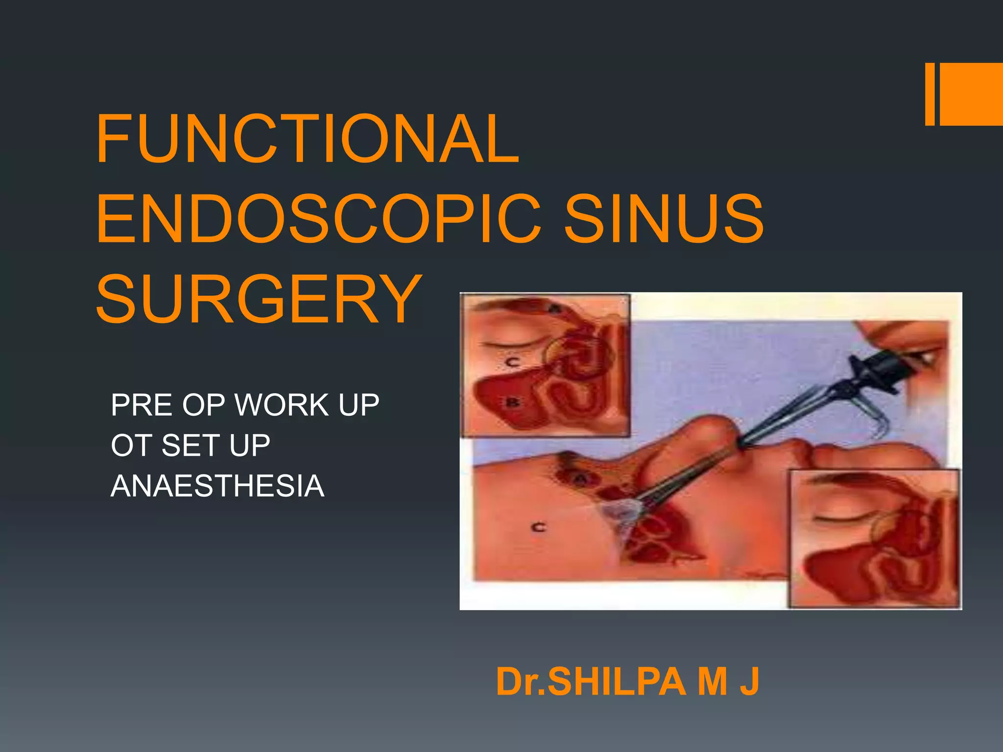
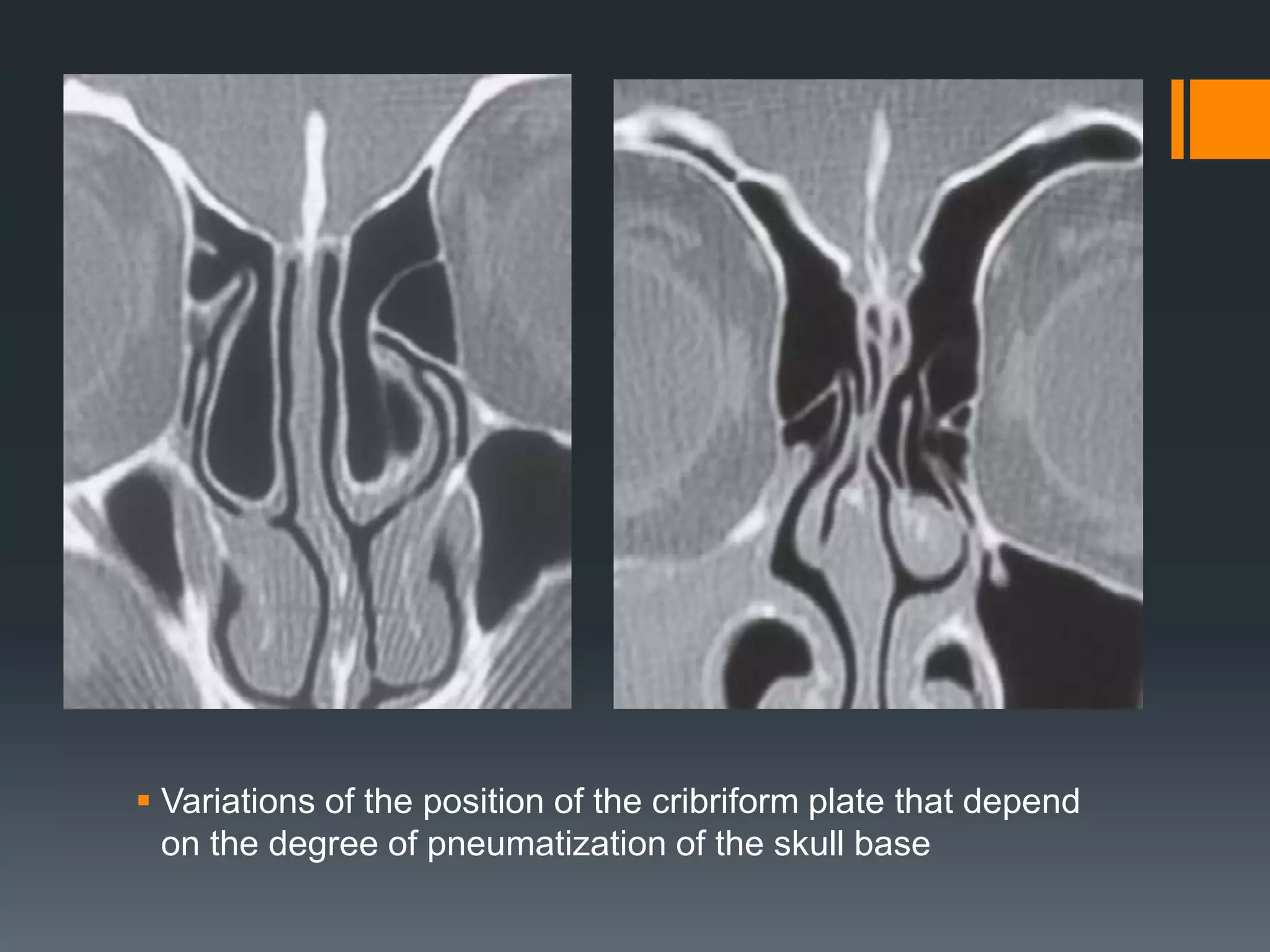
SlideShare a Scribd company logo
FUNCTIONAL
ENDOSCOPIC SINUS
SURGERY
PRE OP WORK UP
OT SET UP
ANAESTHESIA
Dr.SHILPA M J
PRE OPEATIVE CHECKLIST
1 Confirm the diagnosis.
2 Review previous medical treatment.
3 Optimize the immediate preoperative condition.
4 Check that relevant investigations have been done
5 Review the relevant medical history, e.g., drug
allergies , medication.
6 Preoperative CT checklist.
7 Planning and staging the procedure.
8 Informed consent.
CONFIRM THE DIAGNOSIS
Genuine chronic bacterial rhinosinusitis who do
not respond to medical treatment.
Diagnosis that is likely to respond, at least in
part, to surgical intervention.
Surgery will not help
CT is normal - without evidence of sinus disease.
tension-type headache.
allergic rhinitis is very unlikely to help
REVIEW THE PREVIOUS
MEDICAL TREATMENT
A short-lived improvement in the nasal airway with only a
minimum and transient improvement in the sense of smell
after a course of oral steroids will lead the surgeon to give
a more guarded prognosis about the benefits of surgery.
A short-lived response to maximal medical treatment
would also indicate that long-term medical treatment after
surgery may be required.
OPTIMIZE THE IMMEDIATE
PREOPERATIVE CONDITION
Reducing the amount of inflammation
at the time of surgery will make the
operative field easier work in.
In Infective rhinosinusitis - Patient
should have had at least a minimum
of 2 weeks of a broad-spectrum
antibiotic with anaerobic cover.
Allergic Rhinitis - prophylactic nonsedative
antihistamine preoperatively as this will reduce
hyperreactivity and the amount of exudate that occurs
with surgery.
Check compliance with topical nasal steroids
In severe nasal polyposis, and if reduction or absence
of sense of smell, a course of oral steroids in the week
before surgery should help reduce surgical damage to
the olfactory mucosa.
RELEVANT INVESTIGATION
ALLERGY TESTS
IMMUNE STATUS
HEAMATOLOGICAL PARAMETERS
OLFACTION
VISION
ALLERGY TESTS
Skin-prick tests - define the allergens AND
surgery on its own may not be the whole
solution.
Total IgE – Positive in 60% of patients with
allergic rhinitis
Peak flow meter - to check for possible
asthma.
Rhinomanometry and acoustic rhinometry are
mainly used as research tools.
IMMUNE STATUS
Saccharin clearance test - If ciliary
dysmotility is suspected because of
coexisting bronchiectasis or a failure to
respond to medical treatment.
If a patient has had two or more serious
sinus infections within a year or has had
two or more courses of antibiotics with
little effect, then their immunity should
be investigated (Jeffery Modell
Foundation).
The first-line investigations include microbial samples
for cultures;a full blood count with differential white cell
count; immunoglobulins IgG, IgA, IgM; vaccine specific
IgG responses to tetanus, Hib, and pneumococcus;
electrolytes and urea; liver function tests and a fasting
blood glucose; and an HIV test.
Second-line investigations include a nitroblue
tetrazolium reduction test; IgG subclasses; functional
complement assays;and tests for cell-mediated
immunity
Abnormal test results or a strong suspicion justify
referral to an immunologist.
HAEMATOLOGICAL
PARAMETERS
Hemoglobin
differential white cell
count
clotting studies
Electrolytes
liver and kidney
function test
OLFACTION
Document whether it is already
absent or reduced.
Mention to the patient the risks of
surgery to their sense of smell.
OLFACTORY TESTS :
The 20-page UPSIT scratch-and-sniff
test.
A threshold olfactometer is used in
research.
VISION
Asking the patient preoperatively whether they
have any visual problems and whether any
problem is with visual acuity or double vision
Defines any problem preoperatively and will help
to obviate any postoperative recrimination that
surgery has led to a problem that in fact was
already present
look for any alteration in the axis of the pupils from
the front because this is more frequently the cause
of diplopia than is proptosis
 The loss of color discrimination, particularly of red, is a
worrying symptom of pressure on the optic nerve.
Left enophthalmos due to silent sinus syndrome—
involution of the maxillary sinus with collapse of its
roof
REVIEW THE RELEVANT
MEDICAL HISTORY
Document allergies to drugs such as
penicillin and nonsteroidal anti-
inflammatory drugs.
 If the patient is taking aspirin, it is
advisable to ask them to stop taking it
at least 14 days before the day of their
surgery in order to reduce bleeding.
PREOPERATIVE CT CHECKLIST
STEP 1 : orientate the CT scan
sequence from anterior to posterior and
ensure that the sides are marked and
placed as though as you are looking at
the patient.
Follow the cuts anterior to posterior;
follow the septum, note any deviation,
and look for the size and extent of the
ethmoidal bulla, which is a relatively
consistent landmark
 STEP 2 : Examine the lamina papyracea, uncinate
process, and middle turbinate.
Define the site and insertion of the uncinate
process and its proximity to the lamina papyracea.
Examine whether there is a middle turbinate, as its
absence is one of the main factors associated with
complications involving the orbit and the skull
base.
Look for an infraorbital cell.
Localize the uncinate process (arrow) from its free margin posteriorly
and follow it anteriorly and upward
 a The uncinate process inserting into the
skull base (arrow) on the right.
 b The uncinate process inserting into
the middle turbinate (arrow) on the right.
 c The uncinate process inserting into
the lateral nasal wall (arrow) on the right
and the middle turbinate on the left.
 The right uncinate process
originating from the lamina
papyracea.
An absent middle turbinate is a
significant risk factor
 Hypoplasia of the maxillary
sinuses
A left infraorbital cell (arrow),
whose roof looks thin
STEP 3 :
Examine the area of the frontal recess.
The infundibulum expands superiorly to form the
frontal recess, but the way it does so varies.
If an agger nasi cell is well pneumatized and it
extends into the frontal sinus,it is called a bulla
frontalis.
 Serial CT scans in the same patient.
 a Bilateral bulla frontalis.
 b Bulla frontalis and the start of agger nasi cells below
 c The attachment of the uncinate process.
 d Bilateral suprabullar recesses and supraorbital cells.
STEP 4 :
Determine the height of the skull base.
A well pneumatized supraorbital cell will cause the
anterior ethmoid artery to traverse the ethmoid in a
separate channel below the ethmoid roof.
Approximately 18% of patients have an Onodi
(sphenoethmoid) air cell, which increases the risk
to the optic nerve. An Onodi cell is a posterior
ethmoid cell that is lateral to the sphenoid sinus.
 Variations of the position of the cribriform plate that depend
on the degree of pneumatization of the skull base
 Variations of the position of the cribriform plate that depend
on the degree of pneumatization of the skull base
 Sequential CT scans showing the anterior ethmoid artery
leaving the orbit across the ethmoid sinuses to the septum.
 These two CT scans show how the degree of pneumatization
between the left and right side affects the exposure of the
anterior ethmoid artery. The presence of a supraorbital cell (*)
makes it more likely that the anterior ethmoid artery is
dehiscent (arrow).
 a Axial, b coronal, and c
sagittal CT scans of
sphenoethmoid air cells
(arrows).
STEP 5 :
Examine the sphenoid sinus.
The sphenoid septum is frequently asymmetrical
and may attach laterally to the prominence created
by the internal carotid artery or the optic canal.
It is important to note whether the carotid artery is
medially placed or dehiscent.
 Sphenoid intersinus septum going to the carotid artery on
both sides.
 An axial CT scan showing dehiscent optic nerves (arrow) in
the sphenoid sinus. b Coronal CT scan showing dehiscent
optic nerves (arrow) and thin bone over both carotid arteries
 a Soft-tissue window settings fail to show the medially placed
orbital contents revealed in the high-resolution image.
INFORMED CONSENT
Informed consent to be taken when surgery is
considered both when the patient is visiting as an
outpatient and whenever possible on admission
before their surgery.
The following issues need to be addressed
Benefits and risk of surgery
Time off work
Specific Complications - External Incision, Local
Osteitis causing a dull, severe nagging ache that
lasts for 10 days before abating.
Infection, Recurrent Polyposis,Visual
Complications
OPERATING ROOM SET UP
Operating Table :
The patient should be prone and the operating
table tilted to 30 degrees anti-Trendelenburg.
The patient’s head should be in a neutral position
(neither flexed or extended).
This allows the surgeon to operate in a plane
parallel to the skull base, which diminishes the risk
to skull base by decreasing the angle of approach.
Surgeon’s Seat :
An adjustable seat allows the surgeon to find a
comfortable position that will reduce fatigue and
neck problems.
The surgeon may stand and operate
The surgeon is seated, with arm support to reduce
fatigue and stabilize the arm holding the camera.
Standing position
Video Stack/Cameras :
operate from the screen on the video stand that is
positioned directly opposite the surgeon at a level
just above the plane of the surgeon’s visual axis.
This is good for posture, in particular for the neck
.
The screen should be positioned at a level that
makes the surgeon raise their head just a little, as
this will encourage good posture.
A good light source is one of the most critical
factors in obtaining a good image
 A three-chip camera gives a much better image. The stack
may also hold the drive for powered instrumentation and an
image recording system.
The stack system is positioned in
front of the surgeon.
Cables :
The light cable and camera lead should be clipped
to the drapes so that their weight going to the stack
does not pull on the surgeon’s supporting hand.
Other cables from powered instrumentation such
as suction and navigation should be lined up
parallel on the surface top.
 The cables can be arched over the surgeon’s operating hand or
held toward the patient’s head so that they are not in the way.
Lighting :
For optimal viewing, the screen should have a
relatively dim background.
Radiograph Screen :
The CT scans must be placed in order on a screen
for the surgeon to inspect before and during the
procedure.
Ancillary Staff :
The value of a good surgical assistant cannot be
overestimated.To be passed the correct instrument
to fit the hand at the right moment saves time.
Having the correct instrument placed in the hand
saves the surgeon turning their head to look away
from the screen.
A conscientious assistant who looks out for any
movement of the eye when the surgeon is
operating on the lateral nasal wall can save
damage to the patient’s eye.
INTRUMENTS :
Endoscopes
The majority of surgery is best done using a 0°
4mm endoscope, which allows good illumination.
Even in a child or a narrow nose it is easier to
operate with this than with a 2.7mm scope. A 4mm
scope is also more robust. The use of a 0° scope
means that the operator is working in line with their
normal visual axis and is less likely to veer off
course.
Most of the paranasal sinuses can be visualized
with a 0° scope, with the exception of the lateral,
medial, and inferior walls of the maxillary sinus.
 A = 30-degree telescope. B = 70-degree telescope (optional). C = 360-
degree backbiting forceps. D = 360-degree sphenoid punch or forceps.
E = 3.5-mm upbiting through-cut forceps. F = 3.5-mm straight through-
cut forceps. G = 4 mm long curved suction. H = calibrated straight
(Frazier) suction. I = Cottle periosteal elevator. J = ostium seeker or ball
probe, which is angled at one end and curved at the other
 These instruments can be useful when performing an extended
frontal or sphenoid sinusotomy. A = giraffe forceps. B = frontal
curette. C = frontal rasp. D = angled cervical spine curette.
ANAESTHESIA
LOCAL ANAESTHESIA :
ADVANTAGES:
It can be done as a day-stay procedure.
 The surgeon has to be gentle with tissue, thereby
encouraging preservation of mucosa.
 Whether owing to the instillation of local
anesthetic agents with a vasoconstrictor or to
endogenous epinephrine, there is less
peroperative bleeding.
.

 In a patient who is deemed to be unfit for a
general anesthetic, a moderate amount of
endoscopic surgery can be done under local
anesthesia.
It is possible to monitor vision, and the risk of
orbital complications is reduced as the patient will
notice if the surgeon enters the orbit.
DISADVANTAGES :
More extensive procedures such as surgery in the
posterior ethmoids, frontal recess, or sphenoid are
uncomfortable with local anesthesia.
Under local anesthesia a moderate degree of
sedation may be needed, it may result in coughing
and spluttering if the cough reflex has partially
been supressed.
For most patients having extensive surgery done
under local anesthesia, an anesthetist, is required
to provide controlled sedation.
GENERAL ANAESTHESIA :
ADVANTAGES :
The surgeon is freed from worrying about whether
the patient feels discomfort from the surgery.
 It is possible to access areas that are not readily
anaesthetized with local anesthetic, for example,
the frontal recess and the sphenoid.
 It is worth giving local anesthesia at the same time
in order to help reduce the amount of general
anesthetic required and limit immediate
postoperative pain. It also helps reduce bleeding
as it usually also contains vasoconstrictor.
If bleeding is moderate or marked, it can be
sucked out as the patient is not distressed by
having to repeatedly spit out blood.
In infected cases, local anesthetic works poorly
and general anesthesia has a distinct advantage;
for example, it is difficult to anesthetize a patient
with acute sinusitis using local anesthetic even for
a minor procedure such as a maxillary antral
washout.
 The patient is unaware of unpleasant
sensations,even when these do not cause pain, for
example, vibrations from a drill
DISADVANTAGES :
Attention to detail and respect for the mucosa is no
longer encouraged by the complaints of a
conscious patient.
 The patient no longer provides an early warning to
indicate that the orbit has been breached or
entered.
 There are general risks associated with a general
anesthetic, immediate postoperative recovery, and
the likelihood of requiring an overnight stay.
There tends to be more mucosal bleeding than
under local anesthesia. This may in part be due to
less local vasoconstrictor having been used, or to
a reduction in the amount of endogenous
epinephrine.
GENERAL ANAESTHETIC
TECHNIQUE TO PROVIDE A
BETTER OPERATIVE FIELD
A smooth induction, peroperative anesthesia, and
reversal leads to less bleeding. Coughing on a
cuffed tube will result in a great deal of bleeding. If
this happens, it is best to wait until the patient has
settled.
Position the patient 20° body-up during surgery.
The use of topical vasoconstrictors (and allowing
sufficient time for them to work).
Do not rely on vasodilatation to induce hypotension
as this will result in more bleeding.
If hypotensive techniques are used, it is best to
keep the mean blood pressure between 65 and 75
mmHg.
 The use of a laryngeal mask reduces stimulation
to the airway, particularly on extubation.
 Beta blockers need to be used with extreme care,
and not in asthmatics.
THANK YOU

More Related Content

What's hot

Spaces of middle ear and their surgical importance
Spaces of middle ear  and their surgical importanceSpaces of middle ear  and their surgical importance
Spaces of middle ear and their surgical importance
Dr Soumya Singh
 
surgical management of sinusitis
surgical management of sinusitissurgical management of sinusitis
surgical management of sinusitis
hitesh verma
 
Nasal Cavity and Sphenoid Sinus Anatomy
Nasal Cavity and Sphenoid Sinus AnatomyNasal Cavity and Sphenoid Sinus Anatomy
Nasal Cavity and Sphenoid Sinus Anatomy
Farrukh Javeed
 
Chloesteatoma surgery mukace
Chloesteatoma surgery mukaceChloesteatoma surgery mukace
Chloesteatoma surgery mukace
Mukesh Sah
 
Septoplasty
SeptoplastySeptoplasty
Septoplasty
Dr. Nitin taba
 
Mucosal folds of the middle ear
Mucosal folds of the middle earMucosal folds of the middle ear
Mucosal folds of the middle ear
Dʀ Smruti Ranjan Samal
 
Thyroplasty
ThyroplastyThyroplasty
SURGICAL ANATOMY OF DEEP NECK SPACES
SURGICAL ANATOMY OF DEEP NECK SPACESSURGICAL ANATOMY OF DEEP NECK SPACES
SURGICAL ANATOMY OF DEEP NECK SPACES
Ajay Manickam
 
Complications of rhinosinusitis(Dr ravindra daggupati)
Complications of rhinosinusitis(Dr ravindra daggupati)Complications of rhinosinusitis(Dr ravindra daggupati)
Complications of rhinosinusitis(Dr ravindra daggupati)
Ravindra Daggupati
 
Hadad.bassagasteguy flap
Hadad.bassagasteguy flap Hadad.bassagasteguy flap
Hadad.bassagasteguy flap
Chandra Veer Suryavanshi
 
Facial nerve decompression
Facial nerve decompressionFacial nerve decompression
Facial nerve decompression
Mamoon Ameen
 
Cortical mastoidectomy
Cortical mastoidectomy Cortical mastoidectomy
Cortical mastoidectomy
Mamoon Ameen
 
Measurements of nasal airway(dr ravindra daggupati),rhinomanometry,acoustic r...
Measurements of nasal airway(dr ravindra daggupati),rhinomanometry,acoustic r...Measurements of nasal airway(dr ravindra daggupati),rhinomanometry,acoustic r...
Measurements of nasal airway(dr ravindra daggupati),rhinomanometry,acoustic r...
Ravindra Daggupati
 
Ossiculoplasty
OssiculoplastyOssiculoplasty
Ossiculoplasty
Ajay Manickam
 
Juvenile nasopharyngeal angiofibroma
Juvenile nasopharyngeal angiofibromaJuvenile nasopharyngeal angiofibroma
Juvenile nasopharyngeal angiofibroma
praneeth koduru
 
JNA
JNAJNA
Rhinoplasty
RhinoplastyRhinoplasty
Rhinoplasty
Disha Sharma
 

What's hot (20)

Spaces of middle ear and their surgical importance
Spaces of middle ear  and their surgical importanceSpaces of middle ear  and their surgical importance
Spaces of middle ear and their surgical importance
 
surgical management of sinusitis
surgical management of sinusitissurgical management of sinusitis
surgical management of sinusitis
 
Nasal Cavity and Sphenoid Sinus Anatomy
Nasal Cavity and Sphenoid Sinus AnatomyNasal Cavity and Sphenoid Sinus Anatomy
Nasal Cavity and Sphenoid Sinus Anatomy
 
Chloesteatoma surgery mukace
Chloesteatoma surgery mukaceChloesteatoma surgery mukace
Chloesteatoma surgery mukace
 
Septoplasty
SeptoplastySeptoplasty
Septoplasty
 
Mastoidectomy
MastoidectomyMastoidectomy
Mastoidectomy
 
Mucosal folds of the middle ear
Mucosal folds of the middle earMucosal folds of the middle ear
Mucosal folds of the middle ear
 
Thyroplasty
ThyroplastyThyroplasty
Thyroplasty
 
SURGICAL ANATOMY OF DEEP NECK SPACES
SURGICAL ANATOMY OF DEEP NECK SPACESSURGICAL ANATOMY OF DEEP NECK SPACES
SURGICAL ANATOMY OF DEEP NECK SPACES
 
Complications of rhinosinusitis(Dr ravindra daggupati)
Complications of rhinosinusitis(Dr ravindra daggupati)Complications of rhinosinusitis(Dr ravindra daggupati)
Complications of rhinosinusitis(Dr ravindra daggupati)
 
Fungal sinusitis
Fungal sinusitis Fungal sinusitis
Fungal sinusitis
 
Hadad.bassagasteguy flap
Hadad.bassagasteguy flap Hadad.bassagasteguy flap
Hadad.bassagasteguy flap
 
Facial nerve decompression
Facial nerve decompressionFacial nerve decompression
Facial nerve decompression
 
Cortical mastoidectomy
Cortical mastoidectomy Cortical mastoidectomy
Cortical mastoidectomy
 
Measurements of nasal airway(dr ravindra daggupati),rhinomanometry,acoustic r...
Measurements of nasal airway(dr ravindra daggupati),rhinomanometry,acoustic r...Measurements of nasal airway(dr ravindra daggupati),rhinomanometry,acoustic r...
Measurements of nasal airway(dr ravindra daggupati),rhinomanometry,acoustic r...
 
Chronic Rhinosinusitis
Chronic  RhinosinusitisChronic  Rhinosinusitis
Chronic Rhinosinusitis
 
Ossiculoplasty
OssiculoplastyOssiculoplasty
Ossiculoplasty
 
Juvenile nasopharyngeal angiofibroma
Juvenile nasopharyngeal angiofibromaJuvenile nasopharyngeal angiofibroma
Juvenile nasopharyngeal angiofibroma
 
JNA
JNAJNA
JNA
 
Rhinoplasty
RhinoplastyRhinoplasty
Rhinoplasty
 

Similar to FESS - Functional endoscopic sinus surgery

Advanced trauma and life support (atls)
Advanced trauma and life support (atls)Advanced trauma and life support (atls)
Advanced trauma and life support (atls)anu_sandhya
 
Spleenectomy
Spleenectomy Spleenectomy
Spleenectomy
zohaib nadeem
 
Anaesthesia management of patient posted for scoliosis correction
Anaesthesia management of patient posted for scoliosis correctionAnaesthesia management of patient posted for scoliosis correction
Anaesthesia management of patient posted for scoliosis correction
Naveen Kumar Ch
 
Facial trauma and neck trauma
Facial trauma and neck traumaFacial trauma and neck trauma
Facial trauma and neck trauma
EM OMSB
 
Facial trauma and neck trauma
Facial trauma and neck traumaFacial trauma and neck trauma
Facial trauma and neck trauma
EM OMSB
 
Perioperative Nursing Presentation
Perioperative Nursing PresentationPerioperative Nursing Presentation
Perioperative Nursing Presentation
shenell delfin
 
Complications in endoscopic sinus surgery
Complications in endoscopic sinus surgeryComplications in endoscopic sinus surgery
Complications in endoscopic sinus surgery
Ahmed Al-zubiadi
 
Dacrocysstography and Sailography
Dacrocysstography and Sailography Dacrocysstography and Sailography
Dacrocysstography and Sailography
suman duwal
 
Copd clinical cases for anesthesia
Copd clinical cases for anesthesiaCopd clinical cases for anesthesia
Copd clinical cases for anesthesia
Abdallah Alsailamy
 
1ry survey
1ry survey1ry survey
1ry survey
Mohamed ELSAYED
 
1 complications of rhinosonusitis
1 complications of rhinosonusitis1 complications of rhinosonusitis
1 complications of rhinosonusitisAmar Thumma
 
Primary trauma care
Primary trauma carePrimary trauma care
Primary trauma care
Josephine Shamira
 
Rare presentation of left maxillary sinusitis: A Case Report
Rare presentation of left maxillary sinusitis: A Case ReportRare presentation of left maxillary sinusitis: A Case Report
Rare presentation of left maxillary sinusitis: A Case Report
iosrphr_editor
 
Drs. Potter and Richardson's CMC Pediatric X-Ray Mastery week 1
Drs. Potter and Richardson's CMC Pediatric X-Ray Mastery week 1Drs. Potter and Richardson's CMC Pediatric X-Ray Mastery week 1
Drs. Potter and Richardson's CMC Pediatric X-Ray Mastery week 1
Sean M. Fox
 
Pituitary Macroadenoma.pptx
Pituitary Macroadenoma.pptxPituitary Macroadenoma.pptx
Pituitary Macroadenoma.pptx
Dr-Atiqul islam Atique
 
Principle of oral biopsy
Principle of oral biopsy Principle of oral biopsy
Principle of oral biopsy
Abdusalam Alrmali
 
Medicine In Remote Areas MIRA Manual
Medicine In Remote Areas MIRA ManualMedicine In Remote Areas MIRA Manual
Medicine In Remote Areas MIRA Manual
SentinelCourse
 
Ultrasound guided procedures
Ultrasound guided proceduresUltrasound guided procedures
Ultrasound guided proceduresairwave12
 
Dacrocystography and sialography
Dacrocystography and sialographyDacrocystography and sialography
Dacrocystography and sialography
Yashawant Yadav
 
Airway and Anesthetic Management of the Traumatized Patient.pptx
Airway and Anesthetic Management of the Traumatized Patient.pptxAirway and Anesthetic Management of the Traumatized Patient.pptx
Airway and Anesthetic Management of the Traumatized Patient.pptx
Hadi Munib
 

Similar to FESS - Functional endoscopic sinus surgery (20)

Advanced trauma and life support (atls)
Advanced trauma and life support (atls)Advanced trauma and life support (atls)
Advanced trauma and life support (atls)
 
Spleenectomy
Spleenectomy Spleenectomy
Spleenectomy
 
Anaesthesia management of patient posted for scoliosis correction
Anaesthesia management of patient posted for scoliosis correctionAnaesthesia management of patient posted for scoliosis correction
Anaesthesia management of patient posted for scoliosis correction
 
Facial trauma and neck trauma
Facial trauma and neck traumaFacial trauma and neck trauma
Facial trauma and neck trauma
 
Facial trauma and neck trauma
Facial trauma and neck traumaFacial trauma and neck trauma
Facial trauma and neck trauma
 
Perioperative Nursing Presentation
Perioperative Nursing PresentationPerioperative Nursing Presentation
Perioperative Nursing Presentation
 
Complications in endoscopic sinus surgery
Complications in endoscopic sinus surgeryComplications in endoscopic sinus surgery
Complications in endoscopic sinus surgery
 
Dacrocysstography and Sailography
Dacrocysstography and Sailography Dacrocysstography and Sailography
Dacrocysstography and Sailography
 
Copd clinical cases for anesthesia
Copd clinical cases for anesthesiaCopd clinical cases for anesthesia
Copd clinical cases for anesthesia
 
1ry survey
1ry survey1ry survey
1ry survey
 
1 complications of rhinosonusitis
1 complications of rhinosonusitis1 complications of rhinosonusitis
1 complications of rhinosonusitis
 
Primary trauma care
Primary trauma carePrimary trauma care
Primary trauma care
 
Rare presentation of left maxillary sinusitis: A Case Report
Rare presentation of left maxillary sinusitis: A Case ReportRare presentation of left maxillary sinusitis: A Case Report
Rare presentation of left maxillary sinusitis: A Case Report
 
Drs. Potter and Richardson's CMC Pediatric X-Ray Mastery week 1
Drs. Potter and Richardson's CMC Pediatric X-Ray Mastery week 1Drs. Potter and Richardson's CMC Pediatric X-Ray Mastery week 1
Drs. Potter and Richardson's CMC Pediatric X-Ray Mastery week 1
 
Pituitary Macroadenoma.pptx
Pituitary Macroadenoma.pptxPituitary Macroadenoma.pptx
Pituitary Macroadenoma.pptx
 
Principle of oral biopsy
Principle of oral biopsy Principle of oral biopsy
Principle of oral biopsy
 
Medicine In Remote Areas MIRA Manual
Medicine In Remote Areas MIRA ManualMedicine In Remote Areas MIRA Manual
Medicine In Remote Areas MIRA Manual
 
Ultrasound guided procedures
Ultrasound guided proceduresUltrasound guided procedures
Ultrasound guided procedures
 
Dacrocystography and sialography
Dacrocystography and sialographyDacrocystography and sialography
Dacrocystography and sialography
 
Airway and Anesthetic Management of the Traumatized Patient.pptx
Airway and Anesthetic Management of the Traumatized Patient.pptxAirway and Anesthetic Management of the Traumatized Patient.pptx
Airway and Anesthetic Management of the Traumatized Patient.pptx
 

More from Dr. Shilpa M J

OSSICULOPLASTY.pptx
OSSICULOPLASTY.pptxOSSICULOPLASTY.pptx
OSSICULOPLASTY.pptx
Dr. Shilpa M J
 
Bekesy Audiometry
Bekesy AudiometryBekesy Audiometry
Bekesy Audiometry
Dr. Shilpa M J
 
Anatomy of skull base
Anatomy of skull baseAnatomy of skull base
Anatomy of skull base
Dr. Shilpa M J
 
Rguhs ms papers
Rguhs ms papersRguhs ms papers
Rguhs ms papers
Dr. Shilpa M J
 
Vestibular rehabilitation
Vestibular rehabilitationVestibular rehabilitation
Vestibular rehabilitation
Dr. Shilpa M J
 
Anatomy of the Inner ear
Anatomy of the Inner earAnatomy of the Inner ear
Anatomy of the Inner ear
Dr. Shilpa M J
 
Physiology of Equilibrium
Physiology of EquilibriumPhysiology of Equilibrium
Physiology of Equilibrium
Dr. Shilpa M J
 
Ethmoid bone anatomy
Ethmoid bone anatomyEthmoid bone anatomy
Ethmoid bone anatomy
Dr. Shilpa M J
 
Vestibular rehabilitation
Vestibular rehabilitationVestibular rehabilitation
Vestibular rehabilitation
Dr. Shilpa M J
 
Mucoceles of the Paranasal Sinuses
Mucoceles of the Paranasal SinusesMucoceles of the Paranasal Sinuses
Mucoceles of the Paranasal Sinuses
Dr. Shilpa M J
 
Nasal Polyps
Nasal PolypsNasal Polyps
Nasal Polyps
Dr. Shilpa M J
 

More from Dr. Shilpa M J (11)

OSSICULOPLASTY.pptx
OSSICULOPLASTY.pptxOSSICULOPLASTY.pptx
OSSICULOPLASTY.pptx
 
Bekesy Audiometry
Bekesy AudiometryBekesy Audiometry
Bekesy Audiometry
 
Anatomy of skull base
Anatomy of skull baseAnatomy of skull base
Anatomy of skull base
 
Rguhs ms papers
Rguhs ms papersRguhs ms papers
Rguhs ms papers
 
Vestibular rehabilitation
Vestibular rehabilitationVestibular rehabilitation
Vestibular rehabilitation
 
Anatomy of the Inner ear
Anatomy of the Inner earAnatomy of the Inner ear
Anatomy of the Inner ear
 
Physiology of Equilibrium
Physiology of EquilibriumPhysiology of Equilibrium
Physiology of Equilibrium
 
Ethmoid bone anatomy
Ethmoid bone anatomyEthmoid bone anatomy
Ethmoid bone anatomy
 
Vestibular rehabilitation
Vestibular rehabilitationVestibular rehabilitation
Vestibular rehabilitation
 
Mucoceles of the Paranasal Sinuses
Mucoceles of the Paranasal SinusesMucoceles of the Paranasal Sinuses
Mucoceles of the Paranasal Sinuses
 
Nasal Polyps
Nasal PolypsNasal Polyps
Nasal Polyps
 

Recently uploaded

heat stroke and heat exhaustion in children
heat stroke and heat exhaustion in childrenheat stroke and heat exhaustion in children
heat stroke and heat exhaustion in children
SumeraAhmad5
 
Lung Cancer: Artificial Intelligence, Synergetics, Complex System Analysis, S...
Lung Cancer: Artificial Intelligence, Synergetics, Complex System Analysis, S...Lung Cancer: Artificial Intelligence, Synergetics, Complex System Analysis, S...
Lung Cancer: Artificial Intelligence, Synergetics, Complex System Analysis, S...
Oleg Kshivets
 
The Normal Electrocardiogram - Part I of II
The Normal Electrocardiogram - Part I of IIThe Normal Electrocardiogram - Part I of II
The Normal Electrocardiogram - Part I of II
MedicoseAcademics
 
TEST BANK for Operations Management, 14th Edition by William J. Stevenson, Ve...
TEST BANK for Operations Management, 14th Edition by William J. Stevenson, Ve...TEST BANK for Operations Management, 14th Edition by William J. Stevenson, Ve...
TEST BANK for Operations Management, 14th Edition by William J. Stevenson, Ve...
kevinkariuki227
 
Maxilla, Mandible & Hyoid Bone & Clinical Correlations by Dr. RIG.pptx
Maxilla, Mandible & Hyoid Bone & Clinical Correlations by Dr. RIG.pptxMaxilla, Mandible & Hyoid Bone & Clinical Correlations by Dr. RIG.pptx
Maxilla, Mandible & Hyoid Bone & Clinical Correlations by Dr. RIG.pptx
Dr. Rabia Inam Gandapore
 
Alcohol_Dr. Jeenal Mistry MD Pharmacology.pdf
Alcohol_Dr. Jeenal Mistry MD Pharmacology.pdfAlcohol_Dr. Jeenal Mistry MD Pharmacology.pdf
Alcohol_Dr. Jeenal Mistry MD Pharmacology.pdf
Dr Jeenal Mistry
 
Ocular injury ppt Upendra pal optometrist upums saifai etawah
Ocular injury  ppt  Upendra pal  optometrist upums saifai etawahOcular injury  ppt  Upendra pal  optometrist upums saifai etawah
Ocular injury ppt Upendra pal optometrist upums saifai etawah
pal078100
 
Cervical & Brachial Plexus By Dr. RIG.pptx
Cervical & Brachial Plexus By Dr. RIG.pptxCervical & Brachial Plexus By Dr. RIG.pptx
Cervical & Brachial Plexus By Dr. RIG.pptx
Dr. Rabia Inam Gandapore
 
ARTIFICIAL INTELLIGENCE IN HEALTHCARE.pdf
ARTIFICIAL INTELLIGENCE IN  HEALTHCARE.pdfARTIFICIAL INTELLIGENCE IN  HEALTHCARE.pdf
ARTIFICIAL INTELLIGENCE IN HEALTHCARE.pdf
Anujkumaranit
 
Triangles of Neck and Clinical Correlation by Dr. RIG.pptx
Triangles of Neck and Clinical Correlation by Dr. RIG.pptxTriangles of Neck and Clinical Correlation by Dr. RIG.pptx
Triangles of Neck and Clinical Correlation by Dr. RIG.pptx
Dr. Rabia Inam Gandapore
 
Report Back from SGO 2024: What’s the Latest in Cervical Cancer?
Report Back from SGO 2024: What’s the Latest in Cervical Cancer?Report Back from SGO 2024: What’s the Latest in Cervical Cancer?
Report Back from SGO 2024: What’s the Latest in Cervical Cancer?
bkling
 
ANATOMY AND PHYSIOLOGY OF URINARY SYSTEM.pptx
ANATOMY AND PHYSIOLOGY OF URINARY SYSTEM.pptxANATOMY AND PHYSIOLOGY OF URINARY SYSTEM.pptx
ANATOMY AND PHYSIOLOGY OF URINARY SYSTEM.pptx
Swetaba Besh
 
New Drug Discovery and Development .....
New Drug Discovery and Development .....New Drug Discovery and Development .....
New Drug Discovery and Development .....
NEHA GUPTA
 
Flu Vaccine Alert in Bangalore Karnataka
Flu Vaccine Alert in Bangalore KarnatakaFlu Vaccine Alert in Bangalore Karnataka
Flu Vaccine Alert in Bangalore Karnataka
addon Scans
 
Charaka Samhita Sutra sthana Chapter 15 Upakalpaniyaadhyaya
Charaka Samhita Sutra sthana Chapter 15 UpakalpaniyaadhyayaCharaka Samhita Sutra sthana Chapter 15 Upakalpaniyaadhyaya
Charaka Samhita Sutra sthana Chapter 15 Upakalpaniyaadhyaya
Dr KHALID B.M
 
How to Give Better Lectures: Some Tips for Doctors
How to Give Better Lectures: Some Tips for DoctorsHow to Give Better Lectures: Some Tips for Doctors
How to Give Better Lectures: Some Tips for Doctors
LanceCatedral
 
Superficial & Deep Fascia of the NECK.pptx
Superficial & Deep Fascia of the NECK.pptxSuperficial & Deep Fascia of the NECK.pptx
Superficial & Deep Fascia of the NECK.pptx
Dr. Rabia Inam Gandapore
 
ARTHROLOGY PPT NCISM SYLLABUS AYURVEDA STUDENTS
ARTHROLOGY PPT NCISM SYLLABUS AYURVEDA STUDENTSARTHROLOGY PPT NCISM SYLLABUS AYURVEDA STUDENTS
ARTHROLOGY PPT NCISM SYLLABUS AYURVEDA STUDENTS
Dr. Vinay Pareek
 
Tom Selleck Health: A Comprehensive Look at the Iconic Actor’s Wellness Journey
Tom Selleck Health: A Comprehensive Look at the Iconic Actor’s Wellness JourneyTom Selleck Health: A Comprehensive Look at the Iconic Actor’s Wellness Journey
Tom Selleck Health: A Comprehensive Look at the Iconic Actor’s Wellness Journey
greendigital
 
Are There Any Natural Remedies To Treat Syphilis.pdf
Are There Any Natural Remedies To Treat Syphilis.pdfAre There Any Natural Remedies To Treat Syphilis.pdf
Are There Any Natural Remedies To Treat Syphilis.pdf
Little Cross Family Clinic
 

Recently uploaded (20)

heat stroke and heat exhaustion in children
heat stroke and heat exhaustion in childrenheat stroke and heat exhaustion in children
heat stroke and heat exhaustion in children
 
Lung Cancer: Artificial Intelligence, Synergetics, Complex System Analysis, S...
Lung Cancer: Artificial Intelligence, Synergetics, Complex System Analysis, S...Lung Cancer: Artificial Intelligence, Synergetics, Complex System Analysis, S...
Lung Cancer: Artificial Intelligence, Synergetics, Complex System Analysis, S...
 
The Normal Electrocardiogram - Part I of II
The Normal Electrocardiogram - Part I of IIThe Normal Electrocardiogram - Part I of II
The Normal Electrocardiogram - Part I of II
 
TEST BANK for Operations Management, 14th Edition by William J. Stevenson, Ve...
TEST BANK for Operations Management, 14th Edition by William J. Stevenson, Ve...TEST BANK for Operations Management, 14th Edition by William J. Stevenson, Ve...
TEST BANK for Operations Management, 14th Edition by William J. Stevenson, Ve...
 
Maxilla, Mandible & Hyoid Bone & Clinical Correlations by Dr. RIG.pptx
Maxilla, Mandible & Hyoid Bone & Clinical Correlations by Dr. RIG.pptxMaxilla, Mandible & Hyoid Bone & Clinical Correlations by Dr. RIG.pptx
Maxilla, Mandible & Hyoid Bone & Clinical Correlations by Dr. RIG.pptx
 
Alcohol_Dr. Jeenal Mistry MD Pharmacology.pdf
Alcohol_Dr. Jeenal Mistry MD Pharmacology.pdfAlcohol_Dr. Jeenal Mistry MD Pharmacology.pdf
Alcohol_Dr. Jeenal Mistry MD Pharmacology.pdf
 
Ocular injury ppt Upendra pal optometrist upums saifai etawah
Ocular injury  ppt  Upendra pal  optometrist upums saifai etawahOcular injury  ppt  Upendra pal  optometrist upums saifai etawah
Ocular injury ppt Upendra pal optometrist upums saifai etawah
 
Cervical & Brachial Plexus By Dr. RIG.pptx
Cervical & Brachial Plexus By Dr. RIG.pptxCervical & Brachial Plexus By Dr. RIG.pptx
Cervical & Brachial Plexus By Dr. RIG.pptx
 
ARTIFICIAL INTELLIGENCE IN HEALTHCARE.pdf
ARTIFICIAL INTELLIGENCE IN  HEALTHCARE.pdfARTIFICIAL INTELLIGENCE IN  HEALTHCARE.pdf
ARTIFICIAL INTELLIGENCE IN HEALTHCARE.pdf
 
Triangles of Neck and Clinical Correlation by Dr. RIG.pptx
Triangles of Neck and Clinical Correlation by Dr. RIG.pptxTriangles of Neck and Clinical Correlation by Dr. RIG.pptx
Triangles of Neck and Clinical Correlation by Dr. RIG.pptx
 
Report Back from SGO 2024: What’s the Latest in Cervical Cancer?
Report Back from SGO 2024: What’s the Latest in Cervical Cancer?Report Back from SGO 2024: What’s the Latest in Cervical Cancer?
Report Back from SGO 2024: What’s the Latest in Cervical Cancer?
 
ANATOMY AND PHYSIOLOGY OF URINARY SYSTEM.pptx
ANATOMY AND PHYSIOLOGY OF URINARY SYSTEM.pptxANATOMY AND PHYSIOLOGY OF URINARY SYSTEM.pptx
ANATOMY AND PHYSIOLOGY OF URINARY SYSTEM.pptx
 
New Drug Discovery and Development .....
New Drug Discovery and Development .....New Drug Discovery and Development .....
New Drug Discovery and Development .....
 
Flu Vaccine Alert in Bangalore Karnataka
Flu Vaccine Alert in Bangalore KarnatakaFlu Vaccine Alert in Bangalore Karnataka
Flu Vaccine Alert in Bangalore Karnataka
 
Charaka Samhita Sutra sthana Chapter 15 Upakalpaniyaadhyaya
Charaka Samhita Sutra sthana Chapter 15 UpakalpaniyaadhyayaCharaka Samhita Sutra sthana Chapter 15 Upakalpaniyaadhyaya
Charaka Samhita Sutra sthana Chapter 15 Upakalpaniyaadhyaya
 
How to Give Better Lectures: Some Tips for Doctors
How to Give Better Lectures: Some Tips for DoctorsHow to Give Better Lectures: Some Tips for Doctors
How to Give Better Lectures: Some Tips for Doctors
 
Superficial & Deep Fascia of the NECK.pptx
Superficial & Deep Fascia of the NECK.pptxSuperficial & Deep Fascia of the NECK.pptx
Superficial & Deep Fascia of the NECK.pptx
 
ARTHROLOGY PPT NCISM SYLLABUS AYURVEDA STUDENTS
ARTHROLOGY PPT NCISM SYLLABUS AYURVEDA STUDENTSARTHROLOGY PPT NCISM SYLLABUS AYURVEDA STUDENTS
ARTHROLOGY PPT NCISM SYLLABUS AYURVEDA STUDENTS
 
Tom Selleck Health: A Comprehensive Look at the Iconic Actor’s Wellness Journey
Tom Selleck Health: A Comprehensive Look at the Iconic Actor’s Wellness JourneyTom Selleck Health: A Comprehensive Look at the Iconic Actor’s Wellness Journey
Tom Selleck Health: A Comprehensive Look at the Iconic Actor’s Wellness Journey
 
Are There Any Natural Remedies To Treat Syphilis.pdf
Are There Any Natural Remedies To Treat Syphilis.pdfAre There Any Natural Remedies To Treat Syphilis.pdf
Are There Any Natural Remedies To Treat Syphilis.pdf
 

FESS - Functional endoscopic sinus surgery

  • 1. FUNCTIONAL ENDOSCOPIC SINUS SURGERY PRE OP WORK UP OT SET UP ANAESTHESIA Dr.SHILPA M J
  • 2. PRE OPEATIVE CHECKLIST 1 Confirm the diagnosis. 2 Review previous medical treatment. 3 Optimize the immediate preoperative condition. 4 Check that relevant investigations have been done 5 Review the relevant medical history, e.g., drug allergies , medication. 6 Preoperative CT checklist. 7 Planning and staging the procedure. 8 Informed consent.
  • 3. CONFIRM THE DIAGNOSIS Genuine chronic bacterial rhinosinusitis who do not respond to medical treatment. Diagnosis that is likely to respond, at least in part, to surgical intervention. Surgery will not help CT is normal - without evidence of sinus disease. tension-type headache. allergic rhinitis is very unlikely to help
  • 4. REVIEW THE PREVIOUS MEDICAL TREATMENT A short-lived improvement in the nasal airway with only a minimum and transient improvement in the sense of smell after a course of oral steroids will lead the surgeon to give a more guarded prognosis about the benefits of surgery. A short-lived response to maximal medical treatment would also indicate that long-term medical treatment after surgery may be required.
  • 5. OPTIMIZE THE IMMEDIATE PREOPERATIVE CONDITION Reducing the amount of inflammation at the time of surgery will make the operative field easier work in. In Infective rhinosinusitis - Patient should have had at least a minimum of 2 weeks of a broad-spectrum antibiotic with anaerobic cover.
  • 6. Allergic Rhinitis - prophylactic nonsedative antihistamine preoperatively as this will reduce hyperreactivity and the amount of exudate that occurs with surgery. Check compliance with topical nasal steroids In severe nasal polyposis, and if reduction or absence of sense of smell, a course of oral steroids in the week before surgery should help reduce surgical damage to the olfactory mucosa.
  • 7. RELEVANT INVESTIGATION ALLERGY TESTS IMMUNE STATUS HEAMATOLOGICAL PARAMETERS OLFACTION VISION
  • 8. ALLERGY TESTS Skin-prick tests - define the allergens AND surgery on its own may not be the whole solution. Total IgE – Positive in 60% of patients with allergic rhinitis Peak flow meter - to check for possible asthma. Rhinomanometry and acoustic rhinometry are mainly used as research tools.
  • 9. IMMUNE STATUS Saccharin clearance test - If ciliary dysmotility is suspected because of coexisting bronchiectasis or a failure to respond to medical treatment. If a patient has had two or more serious sinus infections within a year or has had two or more courses of antibiotics with little effect, then their immunity should be investigated (Jeffery Modell Foundation).
  • 10. The first-line investigations include microbial samples for cultures;a full blood count with differential white cell count; immunoglobulins IgG, IgA, IgM; vaccine specific IgG responses to tetanus, Hib, and pneumococcus; electrolytes and urea; liver function tests and a fasting blood glucose; and an HIV test. Second-line investigations include a nitroblue tetrazolium reduction test; IgG subclasses; functional complement assays;and tests for cell-mediated immunity Abnormal test results or a strong suspicion justify referral to an immunologist.
  • 11. HAEMATOLOGICAL PARAMETERS Hemoglobin differential white cell count clotting studies Electrolytes liver and kidney function test
  • 12. OLFACTION Document whether it is already absent or reduced. Mention to the patient the risks of surgery to their sense of smell. OLFACTORY TESTS : The 20-page UPSIT scratch-and-sniff test. A threshold olfactometer is used in research.
  • 13. VISION Asking the patient preoperatively whether they have any visual problems and whether any problem is with visual acuity or double vision Defines any problem preoperatively and will help to obviate any postoperative recrimination that surgery has led to a problem that in fact was already present look for any alteration in the axis of the pupils from the front because this is more frequently the cause of diplopia than is proptosis
  • 14.  The loss of color discrimination, particularly of red, is a worrying symptom of pressure on the optic nerve. Left enophthalmos due to silent sinus syndrome— involution of the maxillary sinus with collapse of its roof
  • 15. REVIEW THE RELEVANT MEDICAL HISTORY Document allergies to drugs such as penicillin and nonsteroidal anti- inflammatory drugs.  If the patient is taking aspirin, it is advisable to ask them to stop taking it at least 14 days before the day of their surgery in order to reduce bleeding.
  • 16. PREOPERATIVE CT CHECKLIST STEP 1 : orientate the CT scan sequence from anterior to posterior and ensure that the sides are marked and placed as though as you are looking at the patient. Follow the cuts anterior to posterior; follow the septum, note any deviation, and look for the size and extent of the ethmoidal bulla, which is a relatively consistent landmark
  • 17.  STEP 2 : Examine the lamina papyracea, uncinate process, and middle turbinate. Define the site and insertion of the uncinate process and its proximity to the lamina papyracea. Examine whether there is a middle turbinate, as its absence is one of the main factors associated with complications involving the orbit and the skull base. Look for an infraorbital cell.
  • 18. Localize the uncinate process (arrow) from its free margin posteriorly and follow it anteriorly and upward
  • 19.  a The uncinate process inserting into the skull base (arrow) on the right.  b The uncinate process inserting into the middle turbinate (arrow) on the right.  c The uncinate process inserting into the lateral nasal wall (arrow) on the right and the middle turbinate on the left.
  • 20.  The right uncinate process originating from the lamina papyracea. An absent middle turbinate is a significant risk factor
  • 21.  Hypoplasia of the maxillary sinuses A left infraorbital cell (arrow), whose roof looks thin
  • 22. STEP 3 : Examine the area of the frontal recess. The infundibulum expands superiorly to form the frontal recess, but the way it does so varies. If an agger nasi cell is well pneumatized and it extends into the frontal sinus,it is called a bulla frontalis.
  • 23.  Serial CT scans in the same patient.  a Bilateral bulla frontalis.  b Bulla frontalis and the start of agger nasi cells below
  • 24.  c The attachment of the uncinate process.  d Bilateral suprabullar recesses and supraorbital cells.
  • 25. STEP 4 : Determine the height of the skull base. A well pneumatized supraorbital cell will cause the anterior ethmoid artery to traverse the ethmoid in a separate channel below the ethmoid roof. Approximately 18% of patients have an Onodi (sphenoethmoid) air cell, which increases the risk to the optic nerve. An Onodi cell is a posterior ethmoid cell that is lateral to the sphenoid sinus.
  • 26.  Variations of the position of the cribriform plate that depend on the degree of pneumatization of the skull base
  • 27.  Variations of the position of the cribriform plate that depend on the degree of pneumatization of the skull base
  • 28.  Sequential CT scans showing the anterior ethmoid artery leaving the orbit across the ethmoid sinuses to the septum.
  • 29.  These two CT scans show how the degree of pneumatization between the left and right side affects the exposure of the anterior ethmoid artery. The presence of a supraorbital cell (*) makes it more likely that the anterior ethmoid artery is dehiscent (arrow).
  • 30.  a Axial, b coronal, and c sagittal CT scans of sphenoethmoid air cells (arrows).
  • 31. STEP 5 : Examine the sphenoid sinus. The sphenoid septum is frequently asymmetrical and may attach laterally to the prominence created by the internal carotid artery or the optic canal. It is important to note whether the carotid artery is medially placed or dehiscent.
  • 32.  Sphenoid intersinus septum going to the carotid artery on both sides.
  • 33.  An axial CT scan showing dehiscent optic nerves (arrow) in the sphenoid sinus. b Coronal CT scan showing dehiscent optic nerves (arrow) and thin bone over both carotid arteries
  • 34.  a Soft-tissue window settings fail to show the medially placed orbital contents revealed in the high-resolution image.
  • 35. INFORMED CONSENT Informed consent to be taken when surgery is considered both when the patient is visiting as an outpatient and whenever possible on admission before their surgery. The following issues need to be addressed Benefits and risk of surgery Time off work Specific Complications - External Incision, Local Osteitis causing a dull, severe nagging ache that lasts for 10 days before abating. Infection, Recurrent Polyposis,Visual Complications
  • 36. OPERATING ROOM SET UP Operating Table : The patient should be prone and the operating table tilted to 30 degrees anti-Trendelenburg. The patient’s head should be in a neutral position (neither flexed or extended). This allows the surgeon to operate in a plane parallel to the skull base, which diminishes the risk to skull base by decreasing the angle of approach.
  • 37. Surgeon’s Seat : An adjustable seat allows the surgeon to find a comfortable position that will reduce fatigue and neck problems. The surgeon may stand and operate The surgeon is seated, with arm support to reduce fatigue and stabilize the arm holding the camera.
  • 39. Video Stack/Cameras : operate from the screen on the video stand that is positioned directly opposite the surgeon at a level just above the plane of the surgeon’s visual axis. This is good for posture, in particular for the neck . The screen should be positioned at a level that makes the surgeon raise their head just a little, as this will encourage good posture. A good light source is one of the most critical factors in obtaining a good image
  • 40.  A three-chip camera gives a much better image. The stack may also hold the drive for powered instrumentation and an image recording system. The stack system is positioned in front of the surgeon.
  • 41. Cables : The light cable and camera lead should be clipped to the drapes so that their weight going to the stack does not pull on the surgeon’s supporting hand. Other cables from powered instrumentation such as suction and navigation should be lined up parallel on the surface top.
  • 42.  The cables can be arched over the surgeon’s operating hand or held toward the patient’s head so that they are not in the way.
  • 43. Lighting : For optimal viewing, the screen should have a relatively dim background. Radiograph Screen : The CT scans must be placed in order on a screen for the surgeon to inspect before and during the procedure.
  • 44. Ancillary Staff : The value of a good surgical assistant cannot be overestimated.To be passed the correct instrument to fit the hand at the right moment saves time. Having the correct instrument placed in the hand saves the surgeon turning their head to look away from the screen. A conscientious assistant who looks out for any movement of the eye when the surgeon is operating on the lateral nasal wall can save damage to the patient’s eye.
  • 45. INTRUMENTS : Endoscopes The majority of surgery is best done using a 0° 4mm endoscope, which allows good illumination. Even in a child or a narrow nose it is easier to operate with this than with a 2.7mm scope. A 4mm scope is also more robust. The use of a 0° scope means that the operator is working in line with their normal visual axis and is less likely to veer off course. Most of the paranasal sinuses can be visualized with a 0° scope, with the exception of the lateral, medial, and inferior walls of the maxillary sinus.
  • 46.  A = 30-degree telescope. B = 70-degree telescope (optional). C = 360- degree backbiting forceps. D = 360-degree sphenoid punch or forceps. E = 3.5-mm upbiting through-cut forceps. F = 3.5-mm straight through- cut forceps. G = 4 mm long curved suction. H = calibrated straight (Frazier) suction. I = Cottle periosteal elevator. J = ostium seeker or ball probe, which is angled at one end and curved at the other
  • 47.  These instruments can be useful when performing an extended frontal or sphenoid sinusotomy. A = giraffe forceps. B = frontal curette. C = frontal rasp. D = angled cervical spine curette.
  • 48. ANAESTHESIA LOCAL ANAESTHESIA : ADVANTAGES: It can be done as a day-stay procedure.  The surgeon has to be gentle with tissue, thereby encouraging preservation of mucosa.  Whether owing to the instillation of local anesthetic agents with a vasoconstrictor or to endogenous epinephrine, there is less peroperative bleeding. . 
  • 49.  In a patient who is deemed to be unfit for a general anesthetic, a moderate amount of endoscopic surgery can be done under local anesthesia. It is possible to monitor vision, and the risk of orbital complications is reduced as the patient will notice if the surgeon enters the orbit.
  • 50. DISADVANTAGES : More extensive procedures such as surgery in the posterior ethmoids, frontal recess, or sphenoid are uncomfortable with local anesthesia. Under local anesthesia a moderate degree of sedation may be needed, it may result in coughing and spluttering if the cough reflex has partially been supressed. For most patients having extensive surgery done under local anesthesia, an anesthetist, is required to provide controlled sedation.
  • 51. GENERAL ANAESTHESIA : ADVANTAGES : The surgeon is freed from worrying about whether the patient feels discomfort from the surgery.  It is possible to access areas that are not readily anaesthetized with local anesthetic, for example, the frontal recess and the sphenoid.  It is worth giving local anesthesia at the same time in order to help reduce the amount of general anesthetic required and limit immediate postoperative pain. It also helps reduce bleeding as it usually also contains vasoconstrictor.
  • 52. If bleeding is moderate or marked, it can be sucked out as the patient is not distressed by having to repeatedly spit out blood. In infected cases, local anesthetic works poorly and general anesthesia has a distinct advantage; for example, it is difficult to anesthetize a patient with acute sinusitis using local anesthetic even for a minor procedure such as a maxillary antral washout.  The patient is unaware of unpleasant sensations,even when these do not cause pain, for example, vibrations from a drill
  • 53. DISADVANTAGES : Attention to detail and respect for the mucosa is no longer encouraged by the complaints of a conscious patient.  The patient no longer provides an early warning to indicate that the orbit has been breached or entered.  There are general risks associated with a general anesthetic, immediate postoperative recovery, and the likelihood of requiring an overnight stay.
  • 54. There tends to be more mucosal bleeding than under local anesthesia. This may in part be due to less local vasoconstrictor having been used, or to a reduction in the amount of endogenous epinephrine.
  • 55. GENERAL ANAESTHETIC TECHNIQUE TO PROVIDE A BETTER OPERATIVE FIELD A smooth induction, peroperative anesthesia, and reversal leads to less bleeding. Coughing on a cuffed tube will result in a great deal of bleeding. If this happens, it is best to wait until the patient has settled. Position the patient 20° body-up during surgery. The use of topical vasoconstrictors (and allowing sufficient time for them to work).
  • 56. Do not rely on vasodilatation to induce hypotension as this will result in more bleeding. If hypotensive techniques are used, it is best to keep the mean blood pressure between 65 and 75 mmHg.  The use of a laryngeal mask reduces stimulation to the airway, particularly on extubation.  Beta blockers need to be used with extreme care, and not in asthmatics.