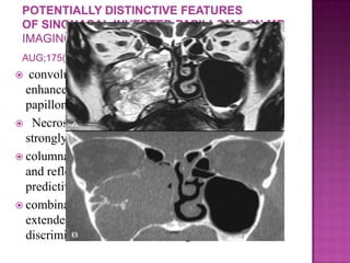
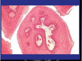
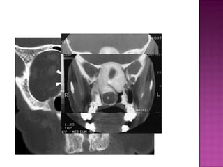
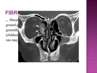
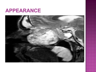
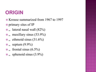
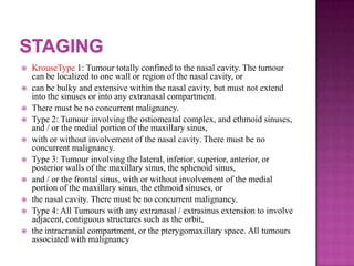
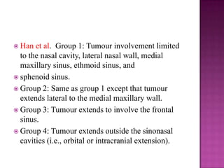
This document discusses various fibro-osseous tumors of the sinonasal region, including fibrous dysplasia. Fibrous dysplasia is a benign condition caused by a defect in osteoblast differentiation and maturation, resulting in the replacement of normal bone by fibrous connective tissue. It is caused by mutations in the GNAS1 gene. Histologically, there is slow replacement of medullary bone by abnormal fibrous tissue at different stages of bone metaplasia. Fibrous dysplasia typically presents in children and adolescents, with females being affected more often than males.
This is a presentation I used for my seminar on 'Phonosurgery' on 4th November, 2015. I hope they are useful to you. Constructive as well as Destructive criticism welcomed.
Cavity obliteration is a procedure done at the end of Mastoidectomy to get a cavity-less mastoid cavity thus solving the problem of discharging post-operative cavity.
Maxillectomy and craniofacial resection Mamoon Ameen
all maxillectomy types in detail and maxillofacial resection ,indications ,contraindications ,preoperative asssessment and detail techniques and rehabilitations
Imaging of paranasal sinuses (including anatomy and varaints)pk1 pdf pptDr pradeep Kumar
This is very good powerpoint presentation of imaging anatomy and variants of paranasal sinuses and imaging pathology as well as multiple pathological imaging findings and images.it will helps for radiologist and radiology resident and even ent resident. our references is CT and mri whole body by Haaga and various internet sources. THANKS.
Report Back from SGO 2024: What’s the Latest in Cervical Cancer?bkling
Are you curious about what’s new in cervical cancer research or unsure what the findings mean? Join Dr. Emily Ko, a gynecologic oncologist at Penn Medicine, to learn about the latest updates from the Society of Gynecologic Oncology (SGO) 2024 Annual Meeting on Women’s Cancer. Dr. Ko will discuss what the research presented at the conference means for you and answer your questions about the new developments.
Ozempic: Preoperative Management of Patients on GLP-1 Receptor Agonists Saeid Safari
Preoperative Management of Patients on GLP-1 Receptor Agonists like Ozempic and Semiglutide
ASA GUIDELINE
NYSORA Guideline
2 Case Reports of Gastric Ultrasound
TEST BANK for Operations Management, 14th Edition by William J. Stevenson, Ve...kevinkariuki227
TEST BANK for Operations Management, 14th Edition by William J. Stevenson, Verified Chapters 1 - 19, Complete Newest Version.pdf
micro teaching on communication m.sc nursing.pdfAnurag Sharma
Microteaching is a unique model of practice teaching. It is a viable instrument for the. desired change in the teaching behavior or the behavior potential which, in specified types of real. classroom situations, tends to facilitate the achievement of specified types of objectives.
Lung Cancer: Artificial Intelligence, Synergetics, Complex System Analysis, S...Oleg Kshivets
RESULTS: Overall life span (LS) was 2252.1±1742.5 days and cumulative 5-year survival (5YS) reached 73.2%, 10 years – 64.8%, 20 years – 42.5%. 513 LCP lived more than 5 years (LS=3124.6±1525.6 days), 148 LCP – more than 10 years (LS=5054.4±1504.1 days).199 LCP died because of LC (LS=562.7±374.5 days). 5YS of LCP after bi/lobectomies was significantly superior in comparison with LCP after pneumonectomies (78.1% vs.63.7%, P=0.00001 by log-rank test). AT significantly improved 5YS (66.3% vs. 34.8%) (P=0.00000 by log-rank test) only for LCP with N1-2. Cox modeling displayed that 5YS of LCP significantly depended on: phase transition (PT) early-invasive LC in terms of synergetics, PT N0—N12, cell ratio factors (ratio between cancer cells- CC and blood cells subpopulations), G1-3, histology, glucose, AT, blood cell circuit, prothrombin index, heparin tolerance, recalcification time (P=0.000-0.038). Neural networks, genetic algorithm selection and bootstrap simulation revealed relationships between 5YS and PT early-invasive LC (rank=1), PT N0—N12 (rank=2), thrombocytes/CC (3), erythrocytes/CC (4), eosinophils/CC (5), healthy cells/CC (6), lymphocytes/CC (7), segmented neutrophils/CC (8), stick neutrophils/CC (9), monocytes/CC (10); leucocytes/CC (11). Correct prediction of 5YS was 100% by neural networks computing (area under ROC curve=1.0; error=0.0).
CONCLUSIONS: 5YS of LCP after radical procedures significantly depended on: 1) PT early-invasive cancer; 2) PT N0--N12; 3) cell ratio factors; 4) blood cell circuit; 5) biochemical factors; 6) hemostasis system; 7) AT; 8) LC characteristics; 9) LC cell dynamics; 10) surgery type: lobectomy/pneumonectomy; 11) anthropometric data. Optimal diagnosis and treatment strategies for LC are: 1) screening and early detection of LC; 2) availability of experienced thoracic surgeons because of complexity of radical procedures; 3) aggressive en block surgery and adequate lymph node dissection for completeness; 4) precise prediction; 5) adjuvant chemoimmunoradiotherapy for LCP with unfavorable prognosis.
Acute scrotum is a general term referring to an emergency condition affecting the contents or the wall of the scrotum.
There are a number of conditions that present acutely, predominantly with pain and/or swelling
A careful and detailed history and examination, and in some cases, investigations allow differentiation between these diagnoses. A prompt diagnosis is essential as the patient may require urgent surgical intervention
Testicular torsion refers to twisting of the spermatic cord, causing ischaemia of the testicle.
Testicular torsion results from inadequate fixation of the testis to the tunica vaginalis producing ischemia from reduced arterial inflow and venous outflow obstruction.
The prevalence of testicular torsion in adult patients hospitalized with acute scrotal pain is approximately 25 to 50 percent
Pulmonary Thromboembolism - etilogy, types, medical- Surgical and nursing man...VarunMahajani
Disruption of blood supply to lung alveoli due to blockage of one or more pulmonary blood vessels is called as Pulmonary thromboembolism. In this presentation we will discuss its causes, types and its management in depth.
New Drug Discovery and Development .....NEHA GUPTA
The "New Drug Discovery and Development" process involves the identification, design, testing, and manufacturing of novel pharmaceutical compounds with the aim of introducing new and improved treatments for various medical conditions. This comprehensive endeavor encompasses various stages, including target identification, preclinical studies, clinical trials, regulatory approval, and post-market surveillance. It involves multidisciplinary collaboration among scientists, researchers, clinicians, regulatory experts, and pharmaceutical companies to bring innovative therapies to market and address unmet medical needs.
NVBDCP.pptx Nation vector borne disease control programSapna Thakur
NVBDCP was launched in 2003-2004 . Vector-Borne Disease: Disease that results from an infection transmitted to humans and other animals by blood-feeding arthropods, such as mosquitoes, ticks, and fleas. Examples of vector-borne diseases include Dengue fever, West Nile Virus, Lyme disease, and malaria.
HOT NEW PRODUCT! BIG SALES FAST SHIPPING NOW FROM CHINA!! EU KU DB BK substit...GL Anaacs
Contact us if you are interested:
Email / Skype : kefaya1771@gmail.com
Threema: PXHY5PDH
New BATCH Ku !!! MUCH IN DEMAND FAST SALE EVERY BATCH HAPPY GOOD EFFECT BIG BATCH !
Contact me on Threema or skype to start big business!!
Hot-sale products:
NEW HOT EUTYLONE WHITE CRYSTAL!!
5cl-adba precursor (semi finished )
5cl-adba raw materials
ADBB precursor (semi finished )
ADBB raw materials
APVP powder
5fadb/4f-adb
Jwh018 / Jwh210
Eutylone crystal
Protonitazene (hydrochloride) CAS: 119276-01-6
Flubrotizolam CAS: 57801-95-3
Metonitazene CAS: 14680-51-4
Payment terms: Western Union,MoneyGram,Bitcoin or USDT.
Deliver Time: Usually 7-15days
Shipping method: FedEx, TNT, DHL,UPS etc.Our deliveries are 100% safe, fast, reliable and discreet.
Samples will be sent for your evaluation!If you are interested in, please contact me, let's talk details.
We specializes in exporting high quality Research chemical, medical intermediate, Pharmaceutical chemicals and so on. Products are exported to USA, Canada, France, Korea, Japan,Russia, Southeast Asia and other countries.
Ethanol (CH3CH2OH), or beverage alcohol, is a two-carbon alcohol
that is rapidly distributed in the body and brain. Ethanol alters many
neurochemical systems and has rewarding and addictive properties. It
is the oldest recreational drug and likely contributes to more morbidity,
mortality, and public health costs than all illicit drugs combined. The
5th edition of the Diagnostic and Statistical Manual of Mental Disorders
(DSM-5) integrates alcohol abuse and alcohol dependence into a single
disorder called alcohol use disorder (AUD), with mild, moderate,
and severe subclassifications (American Psychiatric Association, 2013).
In the DSM-5, all types of substance abuse and dependence have been
combined into a single substance use disorder (SUD) on a continuum
from mild to severe. A diagnosis of AUD requires that at least two of
the 11 DSM-5 behaviors be present within a 12-month period (mild
AUD: 2–3 criteria; moderate AUD: 4–5 criteria; severe AUD: 6–11 criteria).
The four main behavioral effects of AUD are impaired control over
drinking, negative social consequences, risky use, and altered physiological
effects (tolerance, withdrawal). This chapter presents an overview
of the prevalence and harmful consequences of AUD in the U.S.,
the systemic nature of the disease, neurocircuitry and stages of AUD,
comorbidities, fetal alcohol spectrum disorders, genetic risk factors, and
pharmacotherapies for AUD.
7. In 1600s C. Victor Schneider demonstrated nasal
mucosa :
Produces catarrh not csf
Ectodermal origin
1854 Ward first described Schneiderian papillomas
(SPs) of the nose (i.e., sinonasal papilloma)
Kramer and Som classified SPs as true nasal
neoplasms and described them as true papillomas
Ringertz was the first to identify the tendency of
SPs to invert into the underlying connective tissue
stroma, which differs from other types of
papillomas
9. arise in the nasal septum
„ exophytic growth pattern
„ do not grow down into the underlying normal
stroma
„ associated with human papillomavirus
„ do not have a tendency to recur
>57% HPV DNA positivity
10. ragged, beefy appearance and
„ histologically : composed of columnar cells
„ pink cytoplasm
„ nuclei are oval or round
„ many areas of mucin cysts
„ actually act very similar to inverted papillomas
More chances of malignancy
11. The US National Cancer Institute has defined inverted
papilloma as a type of tumor in which surface
epithelial cells grow downward into the underlying
supportive tissue
Inverted papillomas arise from the Schneiderian
membrane, which is an invagination of the olfactory
ectoderm that occurs during the fourth week of
embryonic development. The mucosa creates a
transitional zone between the endo-dermally derived
respiratory epithelium of the nasopharynx and
keratinizing squamous epithelium with the nasal
vestibule
12. Locally aggressive sinonasal tumor
„ Arises from nasal respiratory epithelium,,
which undergoes metaplastic change and
proliferation
„ characterized best by its local invasion,
tendency for recurrence, and association with
malignancy
16. appearance of the epithelium inverting into the
stroma
with a distinct and intact basement membrane
that separates and defines the epithelial component
from the underlying connective tissue
May be associated with:
Atypia
Ca in situ
Carcinoma:
Scc, transitional cell carcinoma, adenocarcinoma
Mucoepidermoid ca, verrucous ca
17. Etiology : association between common
papillomas and the human papillomavirus (HPV)
„ HPV types 6 and 11
Kusiak and Hudson described the presence of
intracytoplasmic and intranuclear inclusion bodies
in SPs
air pollution and industrial carcinogens
„ male: female ratio of 3:1. .
„ average age was 54.3 years
„ most common in the fifth and sixth decades
Respler DS, Jahn A, Pater A, Pater MM.
Isolation and characterization of papillomavirus
DNA from nasal inverting (schneiderian)
papillomas. Ann Otol Rhinol Laryngol. Mar-
Apr 1987;96(2 Pt 1):170-3
Weber RS, Shillitoe EJ, Robbins KT, Luna
MA, Batsakis JG, Donovan DT, et al.
Prevalence of human papillomavirus in
inverted nasal papillomas. Arch Otolaryngol
Head Neck Surg. Jan 1988;114(1):23-6
18. Grossly :: exophytic and polypoid
„ more vascular than an inflammatory polyp
„ gray to pink
„ frond--like projections that extend from the
main bulk of the specimen
„ Epithelium inverts into underlying stroma
19. epithelium distinct from the respiratory
(pseudostratified
ciliated columnar)
mucosa of the sinonasal cavity
-- may be squamous, transitional
or respiratory
-- epithelium proliferative and
thickened
-- lacks mucus--secreting cells
and eosinophils
20. Tendency to recur
Destructive capacity to surrounding structures
Propensity to be associated with malignancy
21. They are associated with malignancy, 5%
to 15% malignancy rates are most
generally accepted. Inverted papillomas are
more commonly associated with squamous
cell carcinomas.
There are four types of association:
Metachronous squamous cell carcinoma.
Carcinoma in situ within the IP
Synchronous lesions
Malignant transformation
24. KrouseType 1: Tumour totally confined to the nasal cavity. The tumour
can be localized to one wall or region of the nasal cavity, or
can be bulky and extensive within the nasal cavity, but must not extend
into the sinuses or into any extranasal compartment.
There must be no concurrent malignancy.
Type 2: Tumour involving the ostiomeatal complex, and ethmoid sinuses,
and / or the medial portion of the maxillary sinus,
with or without involvement of the nasal cavity. There must be no
concurrent malignancy.
Type 3: Tumour involving the lateral, inferior, superior, anterior, or
posterior walls of the maxillary sinus, the sphenoid sinus,
and / or the frontal sinus, with or without involvement of the medial
portion of the maxillary sinus, the ethmoid sinuses, or
the nasal cavity. There must be no concurrent malignancy.
Type 4: All Tumours with any extranasal / extrasinus extension to involve
adjacent, contiguous structures such as the orbit,
the intracranial compartment, or the pterygomaxillary space. All tumours
associated with malignancy
25. Han et al. Group 1: Tumour involvement limited
to the nasal cavity, lateral nasal wall, medial
maxillary sinus, ethmoid sinus, and
sphenoid sinus.
Group 2: Same as group 1 except that tumour
extends lateral to the medial maxillary wall.
Group 3: Tumour extends to involve the frontal
sinus.
Group 4: Tumour extends outside the sinonasal
cavities (i.e., orbital or intracranial extension).
26. Kamel et al. Type 1: Tumour originating from the
nasal septum or lateral nasal wall.
Type 2: Tumour originating from the maxillary
sinus.
[This classification focuses on origin and not size /
extent of tumour-does not analyze frontal sinus
separately]
27. Oikawa et al. T1: Tumour limited to nasal cavity.
T2: Tumour limited to ethmoid sinus and / or medial
and superior portions of maxillary sinus.
T3: Tumour involves lateral, inferior, anterior, or
posterior walls of maxillary sinus, sphenoid sinus, or
frontal sinus.
T3-A: without extension to frontal sinus or supraorbital
recess.
T3-B: involving frontal sinus or supraorbital recess.
T4: Tumour extends outside sinonasal cavities (orbital
or intracranial extension) or associated with
malignancy
28. cannady et al. Group A: Inverted papilloma
confined to the nasal cavity, ethmoid sinuses, or
medial maxillary wall.
Group B: Inverted papilloma with involvement of
any maxillary wall (other than the medial wall), or
frontal sinus, or
sphenoid sinus.
Group C: Inverted papilloma with extension
beyond the paranasal sinuses
30. „ CT and MRI are the common modalities
„ CT is the most widely performed study
„ bony changes
„ bowing of the bones near the soft tissue
mass
31. convoluted cerebriform pattern on T2- or
enhanced T1-weighted images suggests inverted
papilloma as a histologic diagnosis
Necrosis in a mass with such an appearance
strongly suggests coexistent carcinoma
columnar pattern is a reliable MRI indicator of IP
and reflects its histological architecture (positive
predictive value of 95.8%)
combination of this finding with the absence of
extended bone erosion allows for the confident
discrimination of IPs from malignant tumors
34. Surgical approaches:
• Headlight intranasal polpectomy
• Caldwell Luc procedure
• Medial maxillectomy
• Lateral rhinotomy
• External procedures of the frontal sinus
• Endoscopic intranasal polypectomy
• Endoscopic maxillary antrostomy/ethmoidectomy,
Sphenoidotomy
• Endoscopic medial maxillectomy
• Median drainage procedure to gain access to the
frontal sinus
35. Stammberger reported the first purely endoscopic
approach for treatment of SP, which was
performed in 15 patients in 1981
Most reported endoscopic resections have involved
piecemeal resection of the inverting papilloma
followed by piecemeal resection of the lateral
nasal wall
36. Contraindicated in:
massive involvement of the mucosa of the frontal
sinus
and/or of a supraorbital cells
intradural extension or transorbital extension
concomitant presence of a malignancy
presence of abundant scar tissue from previous
surgery
37. Key point:
to dissect the involved mucosa along the
subperiosteal plane
to drill the underlying bone whenever required by
imaging and/or intraoperative finding
Extent surgery:
site of the lesion
area of mucosa involved by the lesion
38. Initially via transnasal resection:
50-80% recurrence
Medial Maxillectomy via lateral rhinotomy:
Gold Standard
10-20%
Endoscopic medial maxillectomy:
Key concepts:
Identify the origin of the papilloma
Bony removal of this region
Recurrent lesions:
Via medial maxillectomy vs. Endoscopic resection
22%
39. Predisposing factors:
Tumor location
Histology
Multicentricity
Method of removal
Length of follow up
Main factor: thoroughness of removal
40. Bielamowicz et al:
found a statistically significant difference between
the recurrence rates in patients treated with medial
maxillectomy (20%) versus those treated with a
conservative resection (47%).
Mirza et al:
found that recurrence rates in 63 case series was
12.8% for endoscopic procedures (n = 484), 17.0%
for lateral rhinotomy with medial maxillectomy (n
= 1025)
34.2% forlimited resections such as nasal
polypectomy (n = 600)
41. Surgical approach Recurrence/case
series
Percentage Mean –follow up
Endoscopic
resection
173/1190 14.5% 3 years 1month
Lateral rhinotomy
with medial
maxillectomy
207/1239 16.7% 5 years 2 month
Limited resection
such as nasal
polypectomy
208/606 34.4% Inadequate
44. Definition :
Benign condition in which normal bone is replaced
by fibrous connective tissue due to a defect in
osteoblast differentiation and maturation
45. Pensler et al:
investigated cultures derived from the involved
bone in two children with MFD and in one child
with Albright’s syndrome
showed a two- to threefold increased level of
estrogen and progesterone receptors by
radioimmunoassay and immunocytochemical
assay
concluded that estrogen may play a major role in
the bony metabolism of FD
46. Actual cause:
set of mutations in the GNAS1 gene
located on chromosome 20q13.2
that normally codes for the alpha subunit of the G-
protein
47. Genetically based developmental anomaly
Defect in osteoblastic differentiation and
maturation
replacement of normal bony tissue by fibrous
tissue of variable cellularity and immature woven
bone
Histology:
slow replacement of medullary bone by abnormal
fibrous tissue with different stages of bone
metaplasia
48. Incidence not known
Females > males
No race predilection
Initial symptoms manifest age 3-15
Not heritable
Questionable genetic transformation
Malignant transformation in < 1%
49. Cystic (21%)
- Radiolucency surrounded by solid rim
Sclerotic (23%)
- Dense and homogenous
Mixed (56%)
- “Ground glass appearance”
50. Monostotic
- Most common
- 25% involve head and neck
Polyostotic
- 15% of cases
- 50% involve head and neck
54. „ fibrous dysplasia characteristically has a
ground glass appearance on CT images. . It
generally presents in younger patients
(children and adolescents), and its growth
rate may decrease or stop after puberty
55. Surgery:
intended to relieve symptoms :
visual impairment due to compression of the optic
nerve
to correct aesthetic deformities
but not to remove the entire lesion
Radiotherapy is contraindicated
Medical treatment :
bisphosphonates (i.e., pamidronate)
with success in patients with extensive lesions
associated with significant disfigurement and pain
Octreotide, anti hyperthyroid agents, antiandrogens
56. a true benign neoplasm
occurs in the third and fourth decades of life
preferentially in black women
Manifestation:
space-occupying lesion in the nasal cavity
evident at endoscopic examination
57. Aetiology:
so far not been clarified.
Trauma has been reported to play a major role in
the development of OF,
especially of the cemento-ossifying fibroma
Histology:
demarcated margins consisting of fibrous tissue with
varying amounts of mineralized or calcified
psammomatoid
bodies
58. present between the second and fourth decades of life
female incidence is more frequent than male incidence
a male to female ratio of 1:5
Ossifying fibroma of the mandible is usually
asymptomatic
Depending on its extent:
facial pain
Swelling
nasal obstruction
Rhinosinusitis
ocular symptoms
59.
60. Differs from common OF by age (younger), location
(maxilla) & biologic behavior (sometime aggressive and/or
recurrent).
Two microscopic types; trabecular & psammomatoid; age at
diagnosis 11 and 22 respectively.
Maxilla often with impingement on sinuses, orbit, nose and
cranium.
61. Treatment of choice is dependent on the site of tumour
location
Tumours invading the midfacial portion and the
sinonasal tract:
more aggressive behavior
need surgical resection
surgical treatment :
requires radical resection/when possible a total removal
of the tumour:
high rate of relapses
aggressive behavior of recurring tumors :with local
destruction and potential invasion of adjacent vital
structures
62. Since OF is very rare, larger series on pure
endonasal resection are not reported
In case reports, some authors have achieved
tumour resection via an endoscopic approach
Draf et al. (480) reported on endonasal
microendoscopic resection of four OF without any
complications
OF with a large extension, most authors
recommend a combined approach including
craniofacial resection
63. Introduction:
Sinonasal osteomas are common entity seen in
patients. Rhinosinus osteomas are commonly asymptomatic and
are incidental findings on imaging studies.
History:
Viega was the first to document sinus osteoma in 1506.
In 1733, Vallisnieri described frontal sinus osteoma that
protruded into the brain.
Epidemiology:
Ostomas are the most common benign tumors of paranasal
sinuses.
This condition is more common in males.
It is common in patients between their second and third
decades of life.
64. Three accepted theories of etiology of osteomas
are:
1. Developmental
2. Traumatic
3. Infectious
Developmental :
Adult tissue contains embryonic remnants usually
remaining dormant
Could be the reason for occurrence near
frontoethmoidal suture lines
65. Traumatic :
Inflammatory process as the initiating force
Bony trauma could be the root cause
Could account for male preponderance
Infectious :
This theory suggests that ostietis resulting from
chronic infections could lead to osteoma formation
in paranasal sinuses.
66. possible aetio-pathogenetic role of traumatic and
inflammatory factors:
Moretti A, Croce A, Leone O, D'Agostino L. Osteoma
of maxillary sinus: case report. Acta
Otorhinolaryngologica Italica2004;24(4):219-22
Naraghi M, Kashfi A. Endonasal endoscopic resection
of ethmoido-orbital osteoma compressing the optic
nerve. American Journal of Otolaryngology
2003;24(6):408-12.
Sayan NB, Ucok C, Karasu HA, Gunhan O. Peripheral
osteoma of the oral and maxillofacial region: a study of
35 new cases. Journal of Oral & Maxillofacial Surgery
2002;60(11):1299-301.
67. Fu and Perzin classification:
1. Ivory / compact osteoma:
Otherwise also known as eburnated osteoma.
In this type of osteoma the bone is very dense and lacks
haversian canals.
These osteomas develop from membranous elements.
2. Osteoma spongiosum:
Also known as mature osteoma is composed of softer bone.
This type of osteomas are known to arise from cartilaginous
elements.
These osteomas have little medullary component containing
fibrofatty tissue.
3.Mixed
68. Behavior: Osteomas are very slow growing and are mostly
asymptomatic. They always stay benign and don't recur
after excision.
The growth rate of osteomas of the paranasal sinuses
Koivunen P et.al. Clin. Otolaryng Allied Sci
1997Apr;22(2):111-4
mean growth rate: 1.61mm/yr range 0.44 to 6.0 mm/yr
Common sites:
95% osteomas in the sinonasal region arise from the
frontoethmoidal region.
80% of osteomas in the sinus region arise from the floor of
frontal sinus
69. Plain sinus radiographs are adequate for detecting
osteomas of paranasal sinuses.
70. most commonly localized to the frontal
sinus
„ slow growing and well circumscribed
„ symptoms attributable obstruction of the
drainage pathway of nearby sinuses
„ On endoscopy : firm masses with a smooth
mucosal covering
71. Wait ,watch n scan
Surgery:
Indications:
Rapid growth of tumor
Symptomatic patients with chronic rhinosinusitis due to
tumor obstruction
Massive headache
Facial deformity
Smith & Calcaterra et al:
Surgery if tumor occupies more than 50% of frontal sinus
Approaches :
Endonasal
External
Combined approach
72. Frontoethmoidal and intraorbital osteomas: exploring
the limits of the endoscopic approach
(Turri-Zannoni et. al Arch. Otolar Head Neck Surg.2012
May)
Conclusion:
The size of the osteoma, far lateral extension of the tumor
in the frontal sinus beyond the lamina papyracea, and
intraorbital involvement are no longer absolute
contraindications for purely transnasal endoscopic
resection. What is important is that the surgeon should
not be dogmatic but rather be ready to change his or her
mind during surgery, shifting to an external approach
when required.
73. Exceptions:
scarring of the frontal recess
Distortion from previous surgery,
very advanced disease and associated malignancy
75. made of neuroglial elements consisting of
glial cells in a connective tissue matrix
with or without a fibrous connection to the dura
„ no fluid filled space connected to the
subarachnoid space
76. „ present as a red or bluish lump at or along the
nasomaxillary suture, or as an intranasal mass.
„ firm, noncompressible
„ not increase in size with crying, and do not
transilluminate
„ not enlarge with bilateral compression of the
internal jugular veins (Furstenberg test)
„ associated with a widened nose or with
hypertelorism secondary to growth of the mass
77. arise from the lateral wall of the nose or less
often from the nasal septum.
„ 60%extranasal
„ 30% intranasal
„ 10% are both
„ 15% are connected to the dura
„ intranasal type associated with dural
attachment (35%) > extranasal type (9%).
78. development similar to nasal dermoids.
„ Abnormal closure of the fonticulus
Develop from extracranial rests of glial tissue
Abnormal closure of fonticulus nasofrontalis, possibly
encephaloceles which have lost CSF connection
tissue being left extracranially.
„ not always an intracranial connection to a
glioma
79. Bluish, soft, compressible, transilluminate,
pulsatile
Enlarge with crying
Positive Furstenberg test (bilateral compression
of internal jugular veins)
Originate medially in the nose
May have associated CSF leak
80. 4% are found within the paranasal sinuses
Schwannoma
Neurofibroma
Treatment via surgical resection
When associated with Von Recklinghausen’s
syndrome: more aggressive (30% 5yr survival).
81. Shrinivas SC, Deshmukh S, Pawar V, et al. Case
Study of Clinicopathological correlation of benign
sinonasal masses. World Articles in Ear, Nose and
Throat 2012;5(1)
:showed nasal schwannoma accounting for 2.4% of
benign nasal masses
Vaideeswar P et al :
schwannoma formed 2.5% (three cases) of all
Sinonasal tumors
82. slowly growing mass
arise more frequently from the ophthalmic and
maxillary divisions of the trigeminal nerves
also originate from sympathetic fibers of the
carotid plexus or parasympathetic fibers of the
pterygopalatine ganglion
Symptoms depend on the location :
progressive nasal obstruction ± epistaxis
anosmia / hyposmia
headache
facial swelling and proptosis
83. Macroscopically:
well-delineated but nonencapsulated globular
firm to rubbery yellow-tan mass
Histologically:
cellular Antoni A areas with Verocay bodies
hypocellular myxoid Antoni B areas
Immunocytohistochemistry:
strongly and diffusely immunoreactive for S-100
protein
84.
85. Mey KH et al in a case series of five nasal
schwannoma
Buob et al in a series of nasal schwannoma cases
Unlike other areas nasal schwannoma is usually
not encapsulated
According to these authors this peculiarity could
be explained by the development of these tumours
from Sinonasal mucosal autonomic nervous
system fibres
which are devoid of perineural cells similar to the
case of gastric Schwannoma
86. Immunohistochemistry :
Positive stains: S100
Negative stains: EMA, CD34, Ki-67/MIB1 (1-
5%)
Treatment: radical surgical resection; no
recurrence or metastases
87. Rare, frequently misclassified
Mean age 48 years, range 13-88 years; no gender
preference
Usually primary intranasal or paranasal mass; may
also be an intracranial tumor invading sphenoid or
frontal sinuses
Primary extracranial tumors have a good
prognosis; intracranial lesions invading into upper
respiratory tract are difficult to excise
Positive stains: EMA, vimentin
Negative stains: S100 (usually)
89. The term hemangioma has traditionally been used
to describe a variety of developmental vascular
anomalies.
Currently, hemangiomas are considered to be
benign tumors of infancy that are characterized by
a rapid growth phase with endothelial cell
proliferation, followed by a gradual involution.
Most cannot be recognized at birth but make their
appearance in most instances during the first 8
weeks of life.
Vascular malformations are structural anomalies of
blood vessels without endothelial proliferation and
are present at birth and persist throughout life.
90. These lesions are present from birth and persist
throughout life.
As with hemangiomas, these lesions tend to
darken with age.
Low-flow venous malformations typically have
a blue color and are easily compressible.
Arteriovenous malformations are high-flow
lesions that result from persistent direct arterial
and venous communications. A palpable thrill
or bruit is often noticeable and the overlying
skin typically feels warmer to the touch.
91. Hemangiomas are the most common tumors of
infancy.
They have a female gender predilection (3:1)
and are most common in the White population.
60% of the hemangiomas occur in the head and
neck region with 80% of them occurring as
single lesions. Multiple lesions may be part of
a syndrome.
About 50 % of all hemangiomas will show
complete resolution by 5 years of age.
92. Types : depending on size of vessels
Capillary
Cavernous
Mixed
Multifocal angiomatosis:
when multiple or affecting a large segment of the
body
93. frequently occur in the head and neck region,
• uncommon in the nasal cavity and paranasal sinuses,
• cavernous and capillary types have been reported in the
sinonasal region,
• represent less than 20% of all benign nasal cavity tumors,
• 80% arise from the nasal septum (Kiesselbach area), 15%
arise from the lateral wall of the nasal cavity.
• originate from the soft tissues of the nasal cavity
• also occur in bones of the inferior or middle turbinate (>
1% bone tumors).
• cavernous hemangiomas usually do not present until
adulthood (adults)
• in both sex with equal frequency,
• incidence peaks in the 4th decade of life.
94. Trauma, viral oncogenes, inflammatory conditions and
the production of
angiogenic growth factors may play a role in the
development
hyperplastic, neovascular response to an angiogenic
stimulus with imbalance of promoters and inhibitors
The occurrence and growth of these lesions during
pregnancy may be
related to increased blood volume or hormonal
influences.
Cavernous hemangioma in adults not undergo
spontaneous involution
such as head and neck hemangioma in the paediatric
population
95. Clinical presentation and symptoms:
• epistaxis and/or hemoptysis
• enlarging lesion in the nasal cavity
Macroscopy:
Red-blue, soft, spongy mass or nodule
96. Differential Diagnosis
• Antrochoanal polyp
•Inverted papilloma
• Hemangiopericytoma
• Esthesioneuroblastoma
Treatment and prognosis:
• surgical resection via transnasal endoscopic approach,
• for extensive lesions: preoperative selective
embolization,
• radiation therapy: for unresectable or inaccessible
lesions,
• no recurrences after surgical removal have been
reported.
97. misnamed; they are neither infectious nor
granulomatous
usually occurs in children and young adults
solitary, glistening red papule or nodule that is
prone to bleeding and ulceration
98. precise mechanism for the development of
pyogenic granuloma is unknown
Postulated: Trauma, hormonal influences, viral
oncogenes, underlying microscopic arteriovenous
malformations, the production of angiogenic
growth factors, and cytogenetic
Trauma: Nose picking, nasal packing,
cauterization, shaving/hair removal, and
nonspecific microtrauma
Isolated cases: Sturge-Weber or von Hippel-
Lindau syndrome
100. Cumming’s 5th edition
Scott-Brown’s 7th edition
Rhinology journal(Official Journal of the
International and European Rhinologic Societies)
European Position Paper on Endoscopic
Management of Tumours of the Nose, Paranasal
Sinuses and Skull Base(Professor Stammberger,
Professor Lund)
Pubmed
medscap