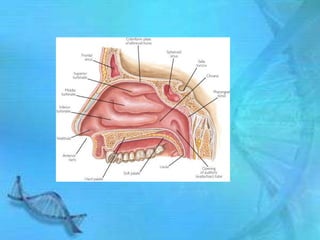
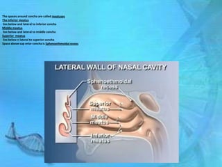
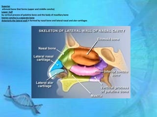
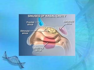
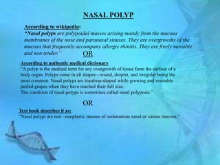
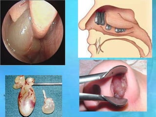
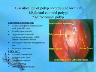
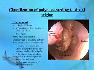
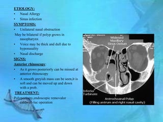
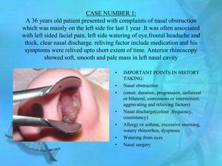
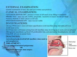
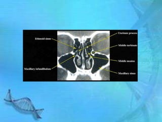
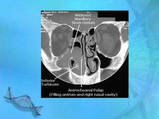
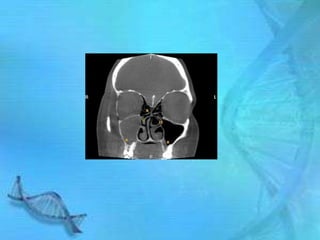
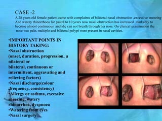
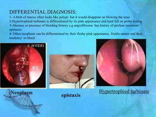
Welcome
   To The
Presentation
Of group “A”
“Table of Contents”
        1st part
  “Anatomy of nasal
cavity in relationship
     with polp”
       2nd part
Main topic :nasal polpi
       3rd part
     Case study
PART-1
“ANATOMY OF NASAL CAVITY IN
    RELATION TO POLYP”
MEDIAL WALL OF NASAL SEPTUM
Skeleton of nasal septum is partly bone and partly cartilage.
The bony part is formed by perpendicular plate of the ethmoid bone ,the vomer and small
vertical ridges from the superior surfaces of the palatine bone and maxilla
Anterior septum is formed by septal vomerine and alar cartilage
LATERAL WALL OF NASAL SEPTUM
The lateral wall of the nasal cavity consist of prominent elevation
Superior concha
 Middle concha
Inferior concha
Conchas narrow the nasal passage create large surface area foricng inhaled air to pass around and over them which helps in warming n moisting of air
Chonca are projections of bones from lateral wall of the nasal cavity covered by mucous membrane
The spaces around concha are called meatuses
The inferior meatus
lies below and lateral to inferior concha
Middle meatus
lies below and lateral to middle concha
Superior meatus
lies below n lateral to superior concha
Space above sup erior concha is Sphenoethmoidal recess
Superior
 ethmoid bone that forms (upper and middle concha)
Lower half
by vertical process of palatine bone and the body of maxillary bone
Inerior concha is a separate bone
Anteriorly the lateral wall is formed by nasal bone and lateral nasal and alar cartilages
The openings or ostia through which the sinuses communicate with nasal cavity are coved with overl ining concha but after removal of concha can be seen
Frontal sinus
drains into infundibulum , a furnel like turnel that opens into the upper end of hiatus semilunaris .
Ethmoidal sinus:
Can be divided into 3 parts
Anterior part drain ant to hiatus semilunaris
Middle air cells drain into one or more openings in bubble like structure ethmoidal bulla
Posterior air cells drain by one or more opening into the superior meatus
Sphenoidal sinus:
One or more openings may be presnt drains into sphenoethmoidal sinus
Maxillary sinus
drains into middle meatus by an opening in the inferior part of hiatus semilunaris
Nasolacrimal duct
Opening at inferior meatus
Carries tears from eyes
MUCOUS MEMBRANE

•Cilia and mucus along the inside wall of the nasal cavity trap remove dust and pathogens from the air
 as it flows through the nasal cavity.
• The cilia move the mucus down the nasal cavity to the pharynx, where it can be swallowed.
 •The nasal mucous membrane lines the nasal cavities, and is adherent to
 the periosteum or perichondrium.

   The epithelium is divided into :
   • Respiratory epithelium:
   (consisting of mucous secreting goblet cells and ciliated cells)
   • Olfactory epithelium:
   (bipolar nerve cells the olfactory cells)
Nasal cavities
The nasal cavities consist of two extensive chambers and their
associated nasal sinuses.
 The two main chambers are separated by midline wall the nasal
septum.
The cavities are lined by mucus membrane,contains sebaceous glands
hair follicles called VIBRISSAE
PART -2
NASAL POLYP
NASAL POLYP
  According to wikipedia:
  “Nasal polyps are polypoidal masses arising mainly from the mucous
  membranes of the nose and paranasal sinuses. They are overgrowths of the
  mucosa that frequently accompany allergic rhinitis. They are freely movable
  and non tender.”          OR
According to authentic medical dictionary
―A polyp is the medical term for any overgrowth of tissue from the surface of a
body organ. Polyps come in all shapes—round, droplet, and irregular being the
most common. Nasal polyps are teardrop-shaped while growing and resemble
peeled grapes when they have reached their full size.
The condition of nasal polyps is sometimes called nasal polyposis.‖

                               OR
Text book describes it as:
―Nasal polypi are non –neoplastic masses of oedematous nasal or sinous mucosa.‖
Classification of polyp according to location
                1.Bilateral ethmoid polypi
                   2.antrochoanal polyp
1.Bilateral Ethmoidal polypi
• Bilateral,multiple in number,usually
    small grape like mass
•    Usually found in adults.
• Originate from ethmoidal
    sinuses,uncinate process, middle
    turbinate and middle meatus
• Mostly grow anteriorly may present at
    nares
• Reoccurence common
ETIOLOGY:
• A)chronic rhinosinusitis
• B)Asthma (risk factor)
• C)Asprin intolerence
• D)cystic fibrosis
• E)Allergic fungal sinusitus
F)Kartagener’s syndrome
 G)young’s syndrome
H)Churg-Strauss syndrome
I)nasal mastocytosis
PATHOGENESIS:
Nasal mucosa ,perticularly in the region of
     middle meatus and turbinate becomes
     oedematus due to collection of ECF.
PATHOLOGY:
A) Early :surface of nasal polp I is covered
     byciliated coloumnar epithelium (normal
     nasal mucosa)
B) Late : it undergoes metaplastic change to
     transitional and squamous type on exposure
     to atmospheric irritation
Submucosa shows large intercellular spaces
     filled with serous fluid.
Infiltration with eosinophills
SYMPTIOMS:
Nasal stiffness that leads to nasal obstruction
Partial or total loss of smell
Headache due to associated sinusitis
•  Sneezing and watery nasal discharge
   due to associated allergy
• Mass protruding from the nostrils.
SIGNS:
Anterior rhinoscopy :
 Polpi appears as smooth, glistering,
   grape-like mass often pale in colour .
 May be sessile or penduculated.
 Insensitive to probing.
 Do not bleed on touch.
DIAGNOSIS:
1) Clinical examination
2) CT scan for correct analysis of extent
   and also helps to plan surgery
TREATMENT

• CONGESTIVE TREATMENT
 ―That is designed to avoid radical medical therapeutic measures or operative procedures.‖
• Control of allergy
• Anti histaminics
Short course of steroids (for those who cant tolerate anti histaminics or asthma)
CONTRAINDICATION OF STEROIDS
1. Hypertension
2. peptic ulcer
3. Diabetes
4. Pregnancy
5. Tuberculosis
TREATMENT
• SURGERICAL TREATMENT
“Surgery is an ancient medical specialty that uses operative manual and instrumental techniques
    on a patient to investigate and/or treat a pathological condition such as disease or injury, or to
    help improve bodily function or appearance.‖
For removal of nasal polyps:
1.Polypectomy
2.Intranasal ethomoidectomy
3.Extranasal ethmoidectomy
4.Transnasal ethmoidectomy
5.Endoscopic sinus surgery
Polypectomy
• 1 or 2 polyps which
  are pedunculated are
  removed with snare.
• Multiple and sessile
  polypi reqire special
  forceps.
Intranasal ethmoidectomy
• Done for multiple and
  sessile polypi
• Uncapping of
  ethmoidal air cells by
  intranasal route
  required
External nasal ethmoidectomy
• Done if reoccurance of
  polyps occur after
  surgery
• Approach is through
  the medial wall of the
  orbitby an external
  incision ,medial to
  medial canthus
Transnasal ethmoidectomy
• Done if infection and
  polypoidal changes are
  also seen in maxillary
  antrum
• Caldwell-luc approach
  is used
Endoscopic sinus surgery
   FESS(functional endoscopic sinus
               surgery
• Presently used
• Polypi can be removed
  more accurately when
  ethmoidal cells are
  removed, and drainage
  and ventilation
  provided to the othe
  involved sinuses.
• Done with endoscope
  of 0,30,70 degree
Classification of polyps according to site of
                       origion
•    1. Antrochoanal
      – a. Single, Unilateral
      – b. Can originate from maxillary
         sinus near ostium
      – It has 3 parts
      Antral which is a thin stalk
      Choanal which is round and globular
      Nasal which is flat from side to side
      – c. Usually found in children.
      – Grows backward to choana may
         hang down behind the soft palaet.
      – Trilobed with antral, nasal and
         choana & fill the nasopharynx
         obstruction both sides
      – Reoccurrence uncommon, if
         removed completely
ETIOLOGY:
• Nasal Allergy
• Sinus infection
SYMPTOMS:
• Unilateral nasal obstruction
 May be bilateral if polyp grows in
    nasopharynx
• Voice may be thick and dull due to
    hyponasality
• Nasal discharge
SIGNS:
Anterior rhinoscopy:
• As it grows posteriorly can be missed at
    anterior rhinoscopy
• A smooth greyish mass can be seen,it is
    soft and can be moved up and down
    with a prob.
 TREATMENT:
Polypectomy,endoscopic removalor
    caldwell-luc operation
PART-3
CASE STUDY
CASE NUMBER 1:
  A 36 years old patient presented with complaints of nasal obstruction
which was mainly on the left side for last 1 year .It was often associated
with left sided facial pain, left side watering of eye,frontal headache and
 thick, clear nasal discharge. reliving factor include medication and his
 symptoms were relived upto short extent of time. Anterior rhinoscopy
          showed soft, smooth and pale mass in left nasal cavity

                                       •   IMPORTANT POINTS IN HISTORY
                                           TAKING:
                                       •   Nasal obstruction
                                       •   (onset, duration, progression, unilateral
                                           or bilateral, continuous or intermittent,
                                           aggravating and relieving factors)
                                       •   Nasal discharge(colour ,frequency,
                                           consistency)
                                       •   Allergy or asthma, excessive sneezing,
                                           watery rhinorrhea, dyspnoea
                                       •   Watering from eyes
                                       •   Nasal surgery
EXTERNAL EXAMINATION:
external examination of nose, face and eyes (watery eyes positive)
CLINICAL EXAMINATION:
•ANTERIOR RHINOSCOPY : presence of mass in left nasal cavity filling it completely
•PROBE TEST : mass was soft, mobile, polypoidal, insensitive to touch, but did not bleed
•NASAL PATENCY TEST: absent on left side
•POSTERIOR RHINOSCOPY : mass was not visible
INVESTIGATIONS:
1)X-rays PNS(water’s view) will show opacification in left maxillary sinus and with soft tissue
in left nasal cavity.
2) CT Scan show soft tissues arising from left maxillary sinus involving nasal cavity and nasopharynx
3)For general anaesthesia e.g blood CP, prothrombin time, activated partial thromboplastin time and
Urine D/R :all were in normal limits
DIAGNOSIS:
Antrochoanal polp involvinf left maxillary sinus nasal cavity
and nasopharynx
TREATMENT :
Convensional intranasal polypectomy
                  OR
Functional endoscopic sinus surgery
CASE -2
   A 28 years old female patient came with complaints of bilateral nasal obstruction ,excessive sneezing
   And watery rhinorrhoea for past 8 to 10 years now nasal obstruction has increased markedly to
   become almost continuous and she can not breath through her nose. On clinical examination the
   nose was pale, multiple and bilateral polypi were present in nasal cavities.

•IMPORTANT POINTS IN
HISTORY TAKING:
•Nasal obstruction
(onset, duration, progression, u
nilateral or
bilateral, continuous or
intermittent, aggravating and
relieving factors)
•Nasal discharge(colour
,frequency, consistency)
•Allergy or asthma, excessive
sneezing, watery
rhinorrhea, dyspnoea
•Watering from eyes
•Nasal surgery
EXTERNAL EXAMINATION:
external examination of nose, face and eyes (no positive findings)

 CLINICAL EXAMINATION:
 •ANTERIOR RHINOSCOPY: It revealed multiple, pale, smooth and shiny grape like polypi
 completely filling both nasal cavities
 •PROBE TEST : mass was soft, mobile, polypoidal, insensitive to touch, but did not bleed
 •NASAL PATENCY TEST: absent on both side
 •POSTERIOR RHINOSCOPY : nasopharynx was clear
 INVESTIGATIONS:
 1) CT Scan shows presence of polypi in both nasal cavities with involvement of both ethmoidal air
 cells and maxillary sinuses
 2)For general anaesthesia e.g blood CP, prothrombin time, activated partial thromboplastin time
 and
 Urine D/R :all were in normal limits
 3)Peripheral eosinophil count and total serumIge level both were increased
 DIAGNOSIS:
 Bilateral ethmoidal nasal polypi
 TREATMENT :
 Convensional intranasal polypectomy
                      OR
 Functional endoscopic sinus surgery
 Histopathological examination of polyp
Bilateral ethmoidal
Point to remember:
1- if polypus is red flshy, friable and has granular surface, especially in older patients think about
                                             MALIGNENCY
2-All polyps should be subjected to histology
3-A simple polp in achild may be a glioma , an encephalocele or a meningoencephalocele.
It should always be aspirated and fluid examination for CSF should be done.careless removal of such
Polyp would cause CSF rhinorrhoea and meningitis .
4-Multiple nasal polyps in children may be associated with mucoviscidosis
5-Epistaxis and orbital syndrome associated with polyp should
always arouse the suspicion of malignancy


                                                                malignancy
DIFFERENTIAL DIAGNOSIS:
1- A blob of mucus often looks like polypi but it would disappear on blowing the nose
2-Hypertrophied turbinate is differentiated by its pink appearance and hard fell on probe testing
3-Absence or presence of bleeding history e.g angiofibroma has history of profuse recurrent
epistaxis.
4- Other neoplasm can be differentiated by their fleshy pink appearance, friable nature and their
tendency to bleed


Neoplasm                                                         Hypertrophied turbinate
                                     epistaxis
Group a presentation 20th feb 2012

Group a presentation 20th feb 2012

  • 2.
    Welcome To The Presentation Of group “A”
  • 3.
    “Table of Contents” 1st part “Anatomy of nasal cavity in relationship with polp” 2nd part Main topic :nasal polpi 3rd part Case study
  • 4.
    PART-1 “ANATOMY OF NASALCAVITY IN RELATION TO POLYP”
  • 5.
    MEDIAL WALL OFNASAL SEPTUM Skeleton of nasal septum is partly bone and partly cartilage. The bony part is formed by perpendicular plate of the ethmoid bone ,the vomer and small vertical ridges from the superior surfaces of the palatine bone and maxilla Anterior septum is formed by septal vomerine and alar cartilage
  • 6.
    LATERAL WALL OFNASAL SEPTUM The lateral wall of the nasal cavity consist of prominent elevation Superior concha Middle concha Inferior concha Conchas narrow the nasal passage create large surface area foricng inhaled air to pass around and over them which helps in warming n moisting of air Chonca are projections of bones from lateral wall of the nasal cavity covered by mucous membrane
  • 8.
    The spaces aroundconcha are called meatuses The inferior meatus lies below and lateral to inferior concha Middle meatus lies below and lateral to middle concha Superior meatus lies below n lateral to superior concha Space above sup erior concha is Sphenoethmoidal recess
  • 9.
    Superior ethmoid bonethat forms (upper and middle concha) Lower half by vertical process of palatine bone and the body of maxillary bone Inerior concha is a separate bone Anteriorly the lateral wall is formed by nasal bone and lateral nasal and alar cartilages
  • 11.
    The openings orostia through which the sinuses communicate with nasal cavity are coved with overl ining concha but after removal of concha can be seen Frontal sinus drains into infundibulum , a furnel like turnel that opens into the upper end of hiatus semilunaris .
  • 12.
    Ethmoidal sinus: Can bedivided into 3 parts Anterior part drain ant to hiatus semilunaris Middle air cells drain into one or more openings in bubble like structure ethmoidal bulla Posterior air cells drain by one or more opening into the superior meatus
  • 13.
    Sphenoidal sinus: One ormore openings may be presnt drains into sphenoethmoidal sinus
  • 14.
    Maxillary sinus drains intomiddle meatus by an opening in the inferior part of hiatus semilunaris
  • 15.
    Nasolacrimal duct Opening atinferior meatus Carries tears from eyes
  • 18.
    MUCOUS MEMBRANE •Cilia andmucus along the inside wall of the nasal cavity trap remove dust and pathogens from the air as it flows through the nasal cavity. • The cilia move the mucus down the nasal cavity to the pharynx, where it can be swallowed. •The nasal mucous membrane lines the nasal cavities, and is adherent to the periosteum or perichondrium. The epithelium is divided into : • Respiratory epithelium: (consisting of mucous secreting goblet cells and ciliated cells) • Olfactory epithelium: (bipolar nerve cells the olfactory cells)
  • 20.
    Nasal cavities The nasalcavities consist of two extensive chambers and their associated nasal sinuses.  The two main chambers are separated by midline wall the nasal septum. The cavities are lined by mucus membrane,contains sebaceous glands hair follicles called VIBRISSAE
  • 21.
  • 22.
    NASAL POLYP According to wikipedia: “Nasal polyps are polypoidal masses arising mainly from the mucous membranes of the nose and paranasal sinuses. They are overgrowths of the mucosa that frequently accompany allergic rhinitis. They are freely movable and non tender.” OR According to authentic medical dictionary ―A polyp is the medical term for any overgrowth of tissue from the surface of a body organ. Polyps come in all shapes—round, droplet, and irregular being the most common. Nasal polyps are teardrop-shaped while growing and resemble peeled grapes when they have reached their full size. The condition of nasal polyps is sometimes called nasal polyposis.‖ OR Text book describes it as: ―Nasal polypi are non –neoplastic masses of oedematous nasal or sinous mucosa.‖
  • 24.
    Classification of polypaccording to location 1.Bilateral ethmoid polypi 2.antrochoanal polyp 1.Bilateral Ethmoidal polypi • Bilateral,multiple in number,usually small grape like mass • Usually found in adults. • Originate from ethmoidal sinuses,uncinate process, middle turbinate and middle meatus • Mostly grow anteriorly may present at nares • Reoccurence common ETIOLOGY: • A)chronic rhinosinusitis • B)Asthma (risk factor) • C)Asprin intolerence • D)cystic fibrosis • E)Allergic fungal sinusitus
  • 25.
    F)Kartagener’s syndrome G)young’ssyndrome H)Churg-Strauss syndrome I)nasal mastocytosis PATHOGENESIS: Nasal mucosa ,perticularly in the region of middle meatus and turbinate becomes oedematus due to collection of ECF. PATHOLOGY: A) Early :surface of nasal polp I is covered byciliated coloumnar epithelium (normal nasal mucosa) B) Late : it undergoes metaplastic change to transitional and squamous type on exposure to atmospheric irritation Submucosa shows large intercellular spaces filled with serous fluid. Infiltration with eosinophills SYMPTIOMS: Nasal stiffness that leads to nasal obstruction Partial or total loss of smell Headache due to associated sinusitis
  • 26.
    • Sneezingand watery nasal discharge due to associated allergy • Mass protruding from the nostrils. SIGNS: Anterior rhinoscopy :  Polpi appears as smooth, glistering, grape-like mass often pale in colour .  May be sessile or penduculated.  Insensitive to probing.  Do not bleed on touch. DIAGNOSIS: 1) Clinical examination 2) CT scan for correct analysis of extent and also helps to plan surgery
  • 27.
    TREATMENT • CONGESTIVE TREATMENT ―That is designed to avoid radical medical therapeutic measures or operative procedures.‖ • Control of allergy • Anti histaminics Short course of steroids (for those who cant tolerate anti histaminics or asthma) CONTRAINDICATION OF STEROIDS 1. Hypertension 2. peptic ulcer 3. Diabetes 4. Pregnancy 5. Tuberculosis
  • 28.
    TREATMENT • SURGERICAL TREATMENT “Surgeryis an ancient medical specialty that uses operative manual and instrumental techniques on a patient to investigate and/or treat a pathological condition such as disease or injury, or to help improve bodily function or appearance.‖ For removal of nasal polyps: 1.Polypectomy 2.Intranasal ethomoidectomy 3.Extranasal ethmoidectomy 4.Transnasal ethmoidectomy 5.Endoscopic sinus surgery
  • 29.
    Polypectomy • 1 or2 polyps which are pedunculated are removed with snare. • Multiple and sessile polypi reqire special forceps.
  • 30.
    Intranasal ethmoidectomy • Donefor multiple and sessile polypi • Uncapping of ethmoidal air cells by intranasal route required
  • 31.
    External nasal ethmoidectomy •Done if reoccurance of polyps occur after surgery • Approach is through the medial wall of the orbitby an external incision ,medial to medial canthus
  • 32.
    Transnasal ethmoidectomy • Doneif infection and polypoidal changes are also seen in maxillary antrum • Caldwell-luc approach is used
  • 33.
    Endoscopic sinus surgery FESS(functional endoscopic sinus surgery • Presently used • Polypi can be removed more accurately when ethmoidal cells are removed, and drainage and ventilation provided to the othe involved sinuses. • Done with endoscope of 0,30,70 degree
  • 35.
    Classification of polypsaccording to site of origion • 1. Antrochoanal – a. Single, Unilateral – b. Can originate from maxillary sinus near ostium – It has 3 parts Antral which is a thin stalk Choanal which is round and globular Nasal which is flat from side to side – c. Usually found in children. – Grows backward to choana may hang down behind the soft palaet. – Trilobed with antral, nasal and choana & fill the nasopharynx obstruction both sides – Reoccurrence uncommon, if removed completely
  • 36.
    ETIOLOGY: • Nasal Allergy •Sinus infection SYMPTOMS: • Unilateral nasal obstruction May be bilateral if polyp grows in nasopharynx • Voice may be thick and dull due to hyponasality • Nasal discharge SIGNS: Anterior rhinoscopy: • As it grows posteriorly can be missed at anterior rhinoscopy • A smooth greyish mass can be seen,it is soft and can be moved up and down with a prob. TREATMENT: Polypectomy,endoscopic removalor caldwell-luc operation
  • 37.
  • 38.
    CASE NUMBER 1: A 36 years old patient presented with complaints of nasal obstruction which was mainly on the left side for last 1 year .It was often associated with left sided facial pain, left side watering of eye,frontal headache and thick, clear nasal discharge. reliving factor include medication and his symptoms were relived upto short extent of time. Anterior rhinoscopy showed soft, smooth and pale mass in left nasal cavity • IMPORTANT POINTS IN HISTORY TAKING: • Nasal obstruction • (onset, duration, progression, unilateral or bilateral, continuous or intermittent, aggravating and relieving factors) • Nasal discharge(colour ,frequency, consistency) • Allergy or asthma, excessive sneezing, watery rhinorrhea, dyspnoea • Watering from eyes • Nasal surgery
  • 39.
    EXTERNAL EXAMINATION: external examinationof nose, face and eyes (watery eyes positive) CLINICAL EXAMINATION: •ANTERIOR RHINOSCOPY : presence of mass in left nasal cavity filling it completely •PROBE TEST : mass was soft, mobile, polypoidal, insensitive to touch, but did not bleed •NASAL PATENCY TEST: absent on left side •POSTERIOR RHINOSCOPY : mass was not visible INVESTIGATIONS: 1)X-rays PNS(water’s view) will show opacification in left maxillary sinus and with soft tissue in left nasal cavity. 2) CT Scan show soft tissues arising from left maxillary sinus involving nasal cavity and nasopharynx 3)For general anaesthesia e.g blood CP, prothrombin time, activated partial thromboplastin time and Urine D/R :all were in normal limits DIAGNOSIS: Antrochoanal polp involvinf left maxillary sinus nasal cavity and nasopharynx TREATMENT : Convensional intranasal polypectomy OR Functional endoscopic sinus surgery
  • 43.
    CASE -2 A 28 years old female patient came with complaints of bilateral nasal obstruction ,excessive sneezing And watery rhinorrhoea for past 8 to 10 years now nasal obstruction has increased markedly to become almost continuous and she can not breath through her nose. On clinical examination the nose was pale, multiple and bilateral polypi were present in nasal cavities. •IMPORTANT POINTS IN HISTORY TAKING: •Nasal obstruction (onset, duration, progression, u nilateral or bilateral, continuous or intermittent, aggravating and relieving factors) •Nasal discharge(colour ,frequency, consistency) •Allergy or asthma, excessive sneezing, watery rhinorrhea, dyspnoea •Watering from eyes •Nasal surgery
  • 44.
    EXTERNAL EXAMINATION: external examinationof nose, face and eyes (no positive findings) CLINICAL EXAMINATION: •ANTERIOR RHINOSCOPY: It revealed multiple, pale, smooth and shiny grape like polypi completely filling both nasal cavities •PROBE TEST : mass was soft, mobile, polypoidal, insensitive to touch, but did not bleed •NASAL PATENCY TEST: absent on both side •POSTERIOR RHINOSCOPY : nasopharynx was clear INVESTIGATIONS: 1) CT Scan shows presence of polypi in both nasal cavities with involvement of both ethmoidal air cells and maxillary sinuses 2)For general anaesthesia e.g blood CP, prothrombin time, activated partial thromboplastin time and Urine D/R :all were in normal limits 3)Peripheral eosinophil count and total serumIge level both were increased DIAGNOSIS: Bilateral ethmoidal nasal polypi TREATMENT : Convensional intranasal polypectomy OR Functional endoscopic sinus surgery Histopathological examination of polyp
  • 45.
  • 46.
    Point to remember: 1-if polypus is red flshy, friable and has granular surface, especially in older patients think about MALIGNENCY 2-All polyps should be subjected to histology 3-A simple polp in achild may be a glioma , an encephalocele or a meningoencephalocele. It should always be aspirated and fluid examination for CSF should be done.careless removal of such Polyp would cause CSF rhinorrhoea and meningitis . 4-Multiple nasal polyps in children may be associated with mucoviscidosis 5-Epistaxis and orbital syndrome associated with polyp should always arouse the suspicion of malignancy malignancy
  • 47.
    DIFFERENTIAL DIAGNOSIS: 1- Ablob of mucus often looks like polypi but it would disappear on blowing the nose 2-Hypertrophied turbinate is differentiated by its pink appearance and hard fell on probe testing 3-Absence or presence of bleeding history e.g angiofibroma has history of profuse recurrent epistaxis. 4- Other neoplasm can be differentiated by their fleshy pink appearance, friable nature and their tendency to bleed Neoplasm Hypertrophied turbinate epistaxis

Editor's Notes

  • #20 Nasal cavitiesThe nasal cavities consist of two extensive chambers and their associated nasal sinuses. The two main chambers are separated by midline wall the nasal septum.