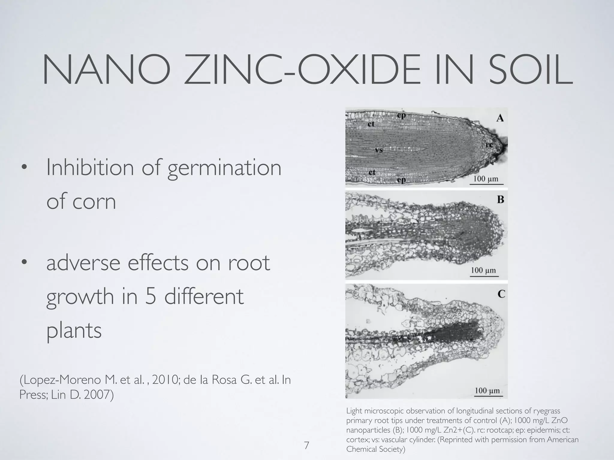
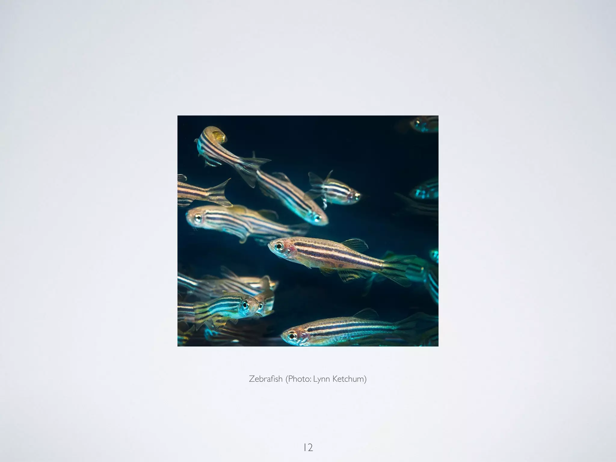
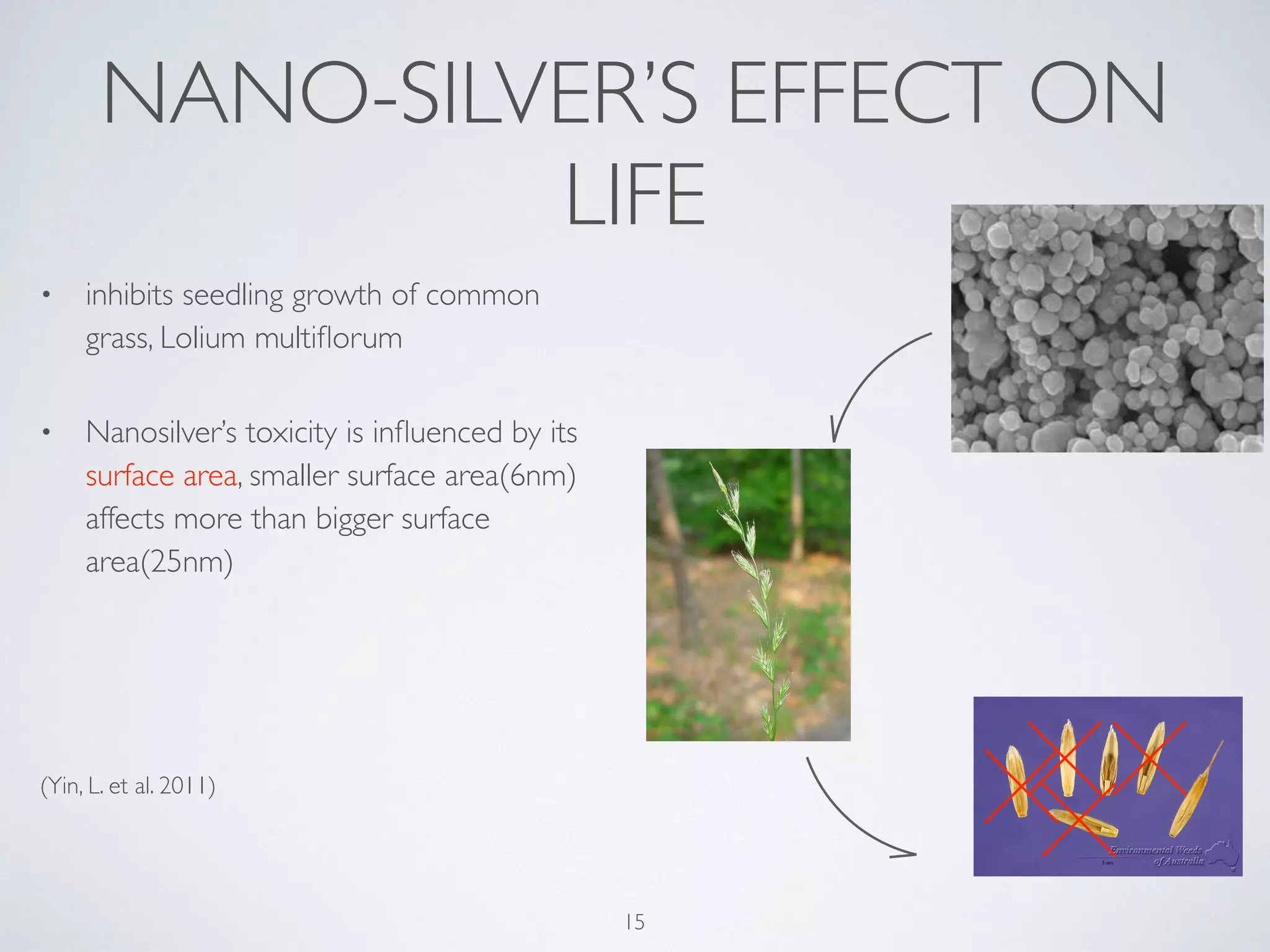
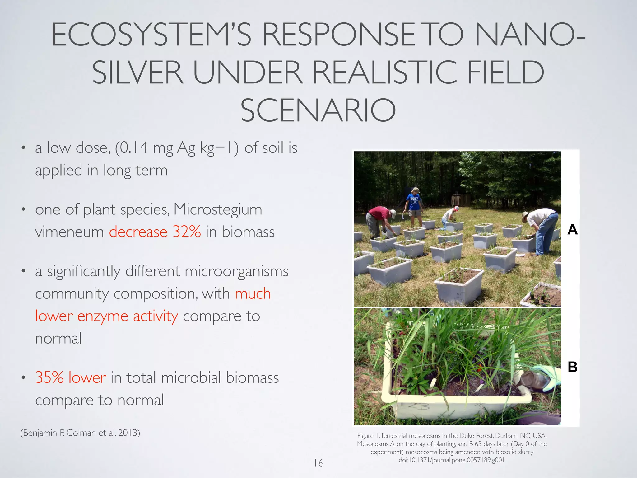
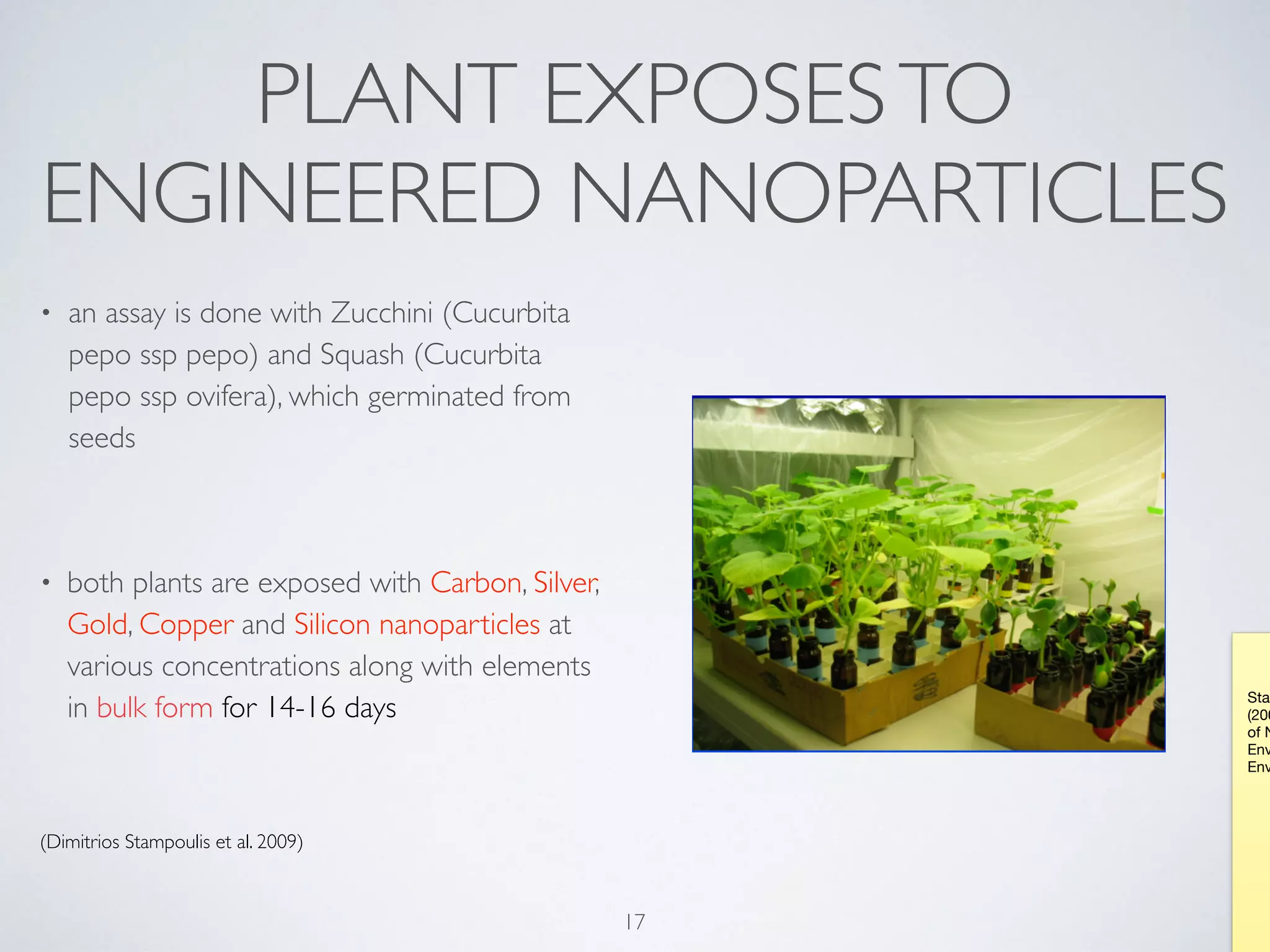
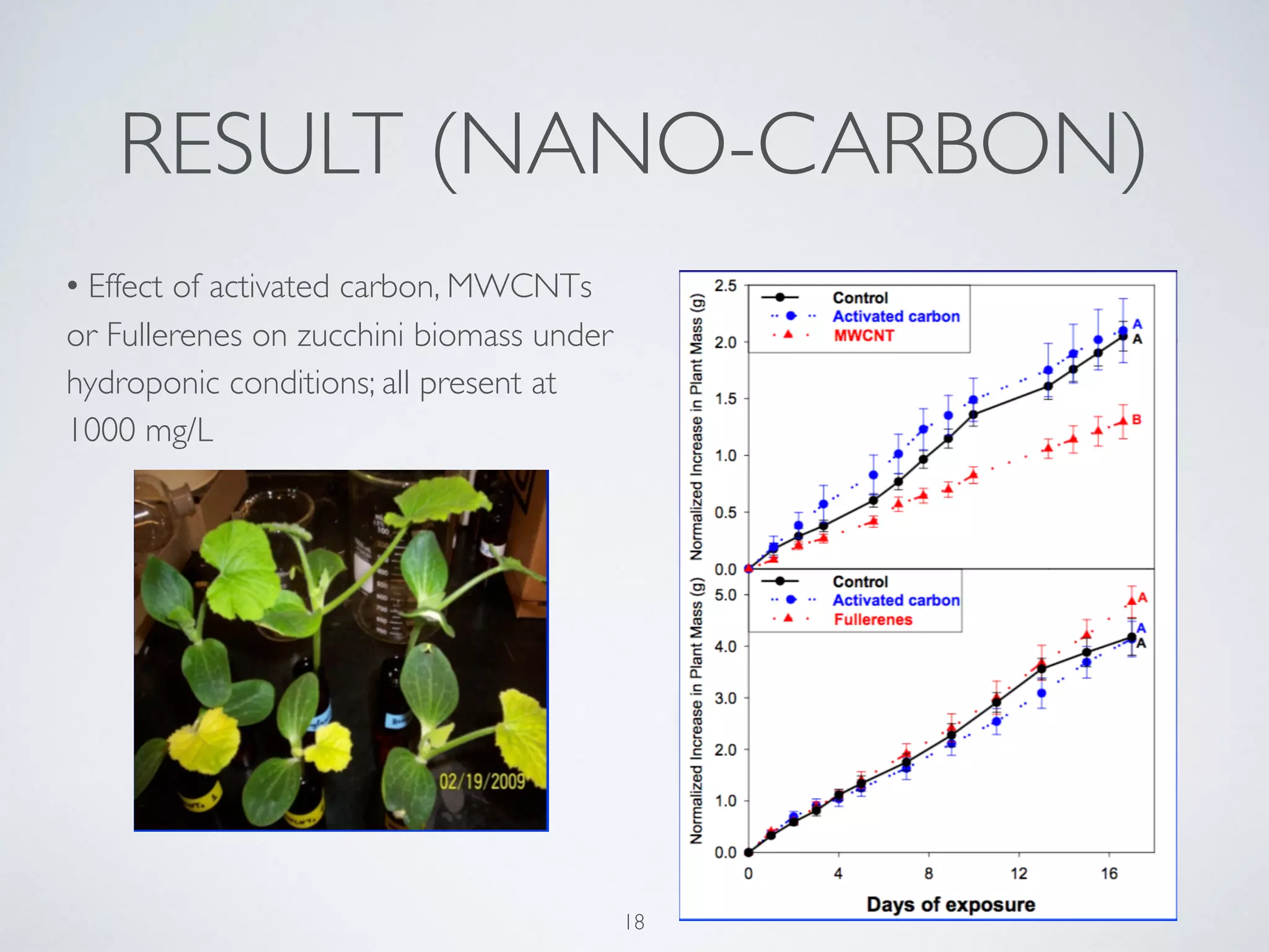
TINYTHINGS FOR BIG PROBLEMS
NANOMATERIAL’S INTHE ENVIRONMENT (WATER AIR AND SOIL) CONTAMINATION
King F.Wong 14817
Rhein Waal University of Applied Sciences
1
NATURAL NANOPARTICLES
2
Photograph courtesy NASA Earth Observatory
3source: http://www.futuretimeline.net/subject/nanotechnology.htm
4
Source: http://sustainable-nano.com/2014/05/13/nano-contaminants-how-nanoparticles-get-into-the-environment/
POSSIBLE FATE OF NEP
5
6
Ball-and-stick model of part of the crystal structure of rutile, one of the mineral forms of titanium dioxide,TiO2. Oxygen atoms are coloured red,
titaniums are grey. X-ray crystallographic data from: R.W. G.Wyckoff (1963) Second edition. Interscience Publishers, NewYork, NewYork. Crystal
Structures 1, 239-444 CIF retrieved fromThe American Mineralogist Crystal Structure Database. See R.T. Downs, M. Hall-Wallace, "The American
Mineralogist Crystal Structure Database.",American Mineralogist (2003) 88, 247-250 for details. Image generated in Accelrys DSVisualizer.
Nano-silica made by chrispotocki in lab
Nano-silver, taken from http://www.mining.com/silver-nano-particles-used-
to-make-hiv-resistant-super-prophylactic-81331/
NANO ZINC-OXIDE IN SOIL
Light microscopic observation of longitudinal sections of ryegrass
primary root tips under treatments of control (A); 1000 mg/L ZnO
nanoparticles (B); 1000 mg/L Zn2+(C). rc: rootcap; ep: epidermis; ct:
cortex; vs: vascular cylinder. (Reprinted with permission from American
Chemical Society)
• Inhibition of germination
of corn
• adverse effects on root
growth in 5 different
plants
(Lopez-Moreno M. et al. , 2010; de la Rosa G. et al. In
Press; Lin D. 2007)
7
8 Jacquin, N.J. von, Icones plantarum
rariorum, vol. 1: t. 145 (1781-1786)
mesquite (Prosopis juliflora?) 1880-1883
edition of F.M. Blanco's Flora de Filipinas
Photo of Cercidium floridum (blue palo verde) at the Springs
Preserve garden in LasVegas, Nevada, Stan Shebs May 1,
2005
KaliTragus, taken from http://www.fireflyforest.com/
flowers/2291/salsola-tragus-prickly-russian-thistle/, visited
on 13/05/2015
NANO ZINC-OXIDE/TITANIUM
DIOXIDE IN WATER 1
9
ZINC-OXIDE/TITANIUM
DIOXIDE IN WATER 2
• dissolution of ZnO triggers sublethal and
cytotoxic effects
• reduction in phytoplankton population
growth rate at concentrations at 223︎-428 ︎g/L
• low photoactivity ofTiO2 in fresh/seawater,
due to
• 1. high ionic strength of seawater
• 2. coating of NOM competes for
photons
(Miller R. et al. 2010; Bennett, S. et al. In Press)
10
Courtesy of Prof. Bridgette Clarkston 11
12
Zebrafish (Photo: Lynn Ketchum)
NANO SILICA EFFECTS ON
AQUATIC LIFE
• zebrafish embryos were treated with SiNPs
(25, 50, 100, 200 µg/mL) during 4–96 hours
post fertilization
• decrease hatching rate with increase
exposure dosage
• increase mortality and cell deaths
• caused embryonic malformations, including
pericardial edema, yolk sac edema, tail and
head malformation
(Duan, J. et al. 2013)
13
(A) Representative optical images of deformed zebrafish. (B)
Pericardial and tail malformation were mainly typically malformation
of embryos induced by silica nanoparticles. (C) Time-course
variations of zebrafish embryos malformations induced by silica
nanoparticles. Scale bar: 500 µm
FATE OF DISCHARGED
NANOSILVER
• 8.8 tonnes per year of AgNPs are
released from consumer
products to wastewater in UK
• A yearly increase of AgNP
concentration in agriculture land
of 36 µg per kg per year
(Whiteley, C et al. 2013)
14
NANO-SILVER’S EFFECT ON
LIFE
• inhibits seedling growth of common
grass, Lolium multiflorum
• Nanosilver’s toxicity is influenced by its
surface area, smaller surface area(6nm)
affects more than bigger surface
area(25nm)
(Yin, L. et al. 2011)
15
ECOSYSTEM’S RESPONSETO NANO-
SILVER UNDER REALISTIC FIELD
SCENARIO
• a low dose, (0.14 mg Ag kg−1) of soil is
applied in long term
• one of plant species, Microstegium
vimeneum decrease 32% in biomass
• a significantly different microorganisms
community composition, with much
lower enzyme activity compare to
normal
• 35% lower in total microbial biomass
compare to normal
(Benjamin P. Colman et al. 2013)
16
Figure 1.Terrestrial mesocosms in the Duke Forest, Durham, NC, USA.
Mesocosms A on the day of planting, and B 63 days later (Day 0 of the
experiment) mesocosms being amended with biosolid slurry
doi:10.1371/journal.pone.0057189.g001
PLANT EXPOSESTO
ENGINEERED NANOPARTICLES
• an assay is done with Zucchini (Cucurbita
pepo ssp pepo) and Squash (Cucurbita
pepo ssp ovifera), which germinated from
seeds
• both plants are exposed with Carbon, Silver,
Gold, Copper and Silicon nanoparticles at
various concentrations along with elements
in bulk form for 14-16 days
(Dimitrios Stampoulis et al. 2009)
17
Stam
(200
of N
Env
Env
RESULT (NANO-CARBON)
18
• Effect of activated carbon, MWCNTs
or Fullerenes on zucchini biomass under
hydroponic conditions; all present at
1000 mg/L
19
Zucchini
dose-uptake
study
(0-1000mg/
L) assessing
effect of NP
or bulk form
of silver on
biomass and
transpiration
20
Silver (Ag) content of zucchini shoots grown in silver nanoparticle or bulk
solutions (1-1000mg/L)
Elemental content of plant tissue was determined using Inductively
Coupled Plasma Mass Spectroscopy (ICP-MS) 

Squash biomass and transpiration upon exposure to 500 mg/L
bulk or nanoparticle silver (Ag) in the presence or absence of
50 mg/L humic acid
21
Squash biomass and transpiration upon exposure to 500 mg/L bulk or
nanoparticle copper (Cu) in the presence or absence of 50 mg/L humic acid
22
Squash biomass and transpiration upon exposure to 100 mg/L bulk or
nanoparticle copper (Cu) in the presence or absence of 50 mg/L humic acid
23
CONCLUSION
• Nano-pollution is an imminent
situation
• an limited knowledge of ecological
fate of NEPs
• a scheme for monitoring NEPs is
needed
24
Photo: NASA
REFERENCES
Slide 7

Lopez-Moreno, M. L.; de La Rosa, G.; Hernandez-Viezcas, J. A.; Castillo-Michel, H.; Botez, C. E.; Peralta-Videa, J. R.; Gardea-Torresdey, J. L. Evidence of the Differential Biotransformation and
Genotoxicity of ZnO and CeO2 Nanoparticles on Soybean (Glycine max) Plants. Environ. Sci. Technol. 2010, 44, 7315–7320. 

de la Rosa, G.; Lopez-Moreno, M. L.; Hernandez-Viezcas, J.; Peralta-Videa,

J. R.; Gardea-Torresdey, J. L. Toxicity and Biotransformation of ZnO Nanoparticles in the Desert Plants Prosopis juliflora- velutina, Salsola tragus and Parkinsonia florida. Int. J. Nanotechnol. In press.

Lin, D.; Xing, B. Phytotoxicity of Nanoparticles: Inhibition of Seed Germination and Root Growth. Environ. 

Pollut. 2007, 150, 243–250. 

Slide 10

Miller, R.; Lenihan, H.; Muller, E.; Tseng, N.; Keller, A. A. Impacts of Metal Oxide Nanoparticles on Marine Phytoplankton. Environ. Sci. Technol. 2010, 44, 7329–7334. 

Slide 13

Duan, J., Yu, Y., Shi, H., Tian, L., Guo, C., Huang, P., . . . Sun, Z. (2013). Toxic Effects of Silica Nanoparticles on Zebrafish Embryos and Larvae. PLoS ONE.

Slide 14

Whiteley, C., Valle, M., Jones, K., & Sweetman, A. (n.d.). Challenges in assessing release, exposure and fate of silver nanoparticles within the UK environment. Environ. Sci.: Processes Impacts
Environmental Science: Processes & Impacts, 2050-2050.

Slide 15

Yin, L., Cheng, Y., Espinasse, B., Colman, B., Auffan, M., Wiesner, M., . . . Bernhardt, E. (2011). More than the Ions: The Effects of Silver Nanoparticles on Lolium multiflorum. Environmental Science &
Technology Environ. Sci. Technol., 2360-2367.

Slide 16

Low Concentrations of Silver Nanoparticles in Biosolids Cause Adverse Ecosystem Responses under Realistic Field Scenario; Benjamin P. Colman , Christina L. Arnaout, Sarah Anciaux, Claudia K.
Gunsch, Michael F. Hochella Jr, Bojeong Kim, Gregory V. Lowry, Bonnie M. McGill, Brian C. Reinsch, Curtis J. Richardson, Jason M. Unrine, Justin P. Wright, Liyan Yin, Emily S. Bernhardt; Published:
February 27, 2013DOI: 10.1371/journal.pone.0057189

Slide 17

Stampoulis, D., Sinha, S., & White, J. (2009). Assay-Dependent Phytotoxicity of Nanoparticles to Plants. Environmental Science & Technology Environ. Sci. Technol., 9473-9479.

25

Nanomaterial environmental contamonation

  • 1.
    TINYTHINGS FOR BIGPROBLEMS NANOMATERIAL’S INTHE ENVIRONMENT (WATER AIR AND SOIL) CONTAMINATION King F.Wong 14817 Rhein Waal University of Applied Sciences 1
  • 2.
  • 3.
  • 4.
  • 5.
  • 6.
    6 Ball-and-stick model ofpart of the crystal structure of rutile, one of the mineral forms of titanium dioxide,TiO2. Oxygen atoms are coloured red, titaniums are grey. X-ray crystallographic data from: R.W. G.Wyckoff (1963) Second edition. Interscience Publishers, NewYork, NewYork. Crystal Structures 1, 239-444 CIF retrieved fromThe American Mineralogist Crystal Structure Database. See R.T. Downs, M. Hall-Wallace, "The American Mineralogist Crystal Structure Database.",American Mineralogist (2003) 88, 247-250 for details. Image generated in Accelrys DSVisualizer. Nano-silica made by chrispotocki in lab Nano-silver, taken from http://www.mining.com/silver-nano-particles-used- to-make-hiv-resistant-super-prophylactic-81331/
  • 7.
    NANO ZINC-OXIDE INSOIL Light microscopic observation of longitudinal sections of ryegrass primary root tips under treatments of control (A); 1000 mg/L ZnO nanoparticles (B); 1000 mg/L Zn2+(C). rc: rootcap; ep: epidermis; ct: cortex; vs: vascular cylinder. (Reprinted with permission from American Chemical Society) • Inhibition of germination of corn • adverse effects on root growth in 5 different plants (Lopez-Moreno M. et al. , 2010; de la Rosa G. et al. In Press; Lin D. 2007) 7
  • 8.
    8 Jacquin, N.J.von, Icones plantarum rariorum, vol. 1: t. 145 (1781-1786) mesquite (Prosopis juliflora?) 1880-1883 edition of F.M. Blanco's Flora de Filipinas Photo of Cercidium floridum (blue palo verde) at the Springs Preserve garden in LasVegas, Nevada, Stan Shebs May 1, 2005 KaliTragus, taken from http://www.fireflyforest.com/ flowers/2291/salsola-tragus-prickly-russian-thistle/, visited on 13/05/2015
  • 9.
  • 10.
    ZINC-OXIDE/TITANIUM DIOXIDE IN WATER2 • dissolution of ZnO triggers sublethal and cytotoxic effects • reduction in phytoplankton population growth rate at concentrations at 223︎-428 ︎g/L • low photoactivity ofTiO2 in fresh/seawater, due to • 1. high ionic strength of seawater • 2. coating of NOM competes for photons (Miller R. et al. 2010; Bennett, S. et al. In Press) 10
  • 11.
    Courtesy of Prof.Bridgette Clarkston 11
  • 12.
  • 13.
    NANO SILICA EFFECTSON AQUATIC LIFE • zebrafish embryos were treated with SiNPs (25, 50, 100, 200 µg/mL) during 4–96 hours post fertilization • decrease hatching rate with increase exposure dosage • increase mortality and cell deaths • caused embryonic malformations, including pericardial edema, yolk sac edema, tail and head malformation (Duan, J. et al. 2013) 13 (A) Representative optical images of deformed zebrafish. (B) Pericardial and tail malformation were mainly typically malformation of embryos induced by silica nanoparticles. (C) Time-course variations of zebrafish embryos malformations induced by silica nanoparticles. Scale bar: 500 µm
  • 14.
    FATE OF DISCHARGED NANOSILVER •8.8 tonnes per year of AgNPs are released from consumer products to wastewater in UK • A yearly increase of AgNP concentration in agriculture land of 36 µg per kg per year (Whiteley, C et al. 2013) 14
  • 15.
    NANO-SILVER’S EFFECT ON LIFE •inhibits seedling growth of common grass, Lolium multiflorum • Nanosilver’s toxicity is influenced by its surface area, smaller surface area(6nm) affects more than bigger surface area(25nm) (Yin, L. et al. 2011) 15
  • 16.
    ECOSYSTEM’S RESPONSETO NANO- SILVERUNDER REALISTIC FIELD SCENARIO • a low dose, (0.14 mg Ag kg−1) of soil is applied in long term • one of plant species, Microstegium vimeneum decrease 32% in biomass • a significantly different microorganisms community composition, with much lower enzyme activity compare to normal • 35% lower in total microbial biomass compare to normal (Benjamin P. Colman et al. 2013) 16 Figure 1.Terrestrial mesocosms in the Duke Forest, Durham, NC, USA. Mesocosms A on the day of planting, and B 63 days later (Day 0 of the experiment) mesocosms being amended with biosolid slurry doi:10.1371/journal.pone.0057189.g001
  • 17.
    PLANT EXPOSESTO ENGINEERED NANOPARTICLES •an assay is done with Zucchini (Cucurbita pepo ssp pepo) and Squash (Cucurbita pepo ssp ovifera), which germinated from seeds • both plants are exposed with Carbon, Silver, Gold, Copper and Silicon nanoparticles at various concentrations along with elements in bulk form for 14-16 days (Dimitrios Stampoulis et al. 2009) 17 Stam (200 of N Env Env
  • 18.
    RESULT (NANO-CARBON) 18 • Effectof activated carbon, MWCNTs or Fullerenes on zucchini biomass under hydroponic conditions; all present at 1000 mg/L
  • 19.
    19 Zucchini dose-uptake study (0-1000mg/ L) assessing effect ofNP or bulk form of silver on biomass and transpiration
  • 20.
    20 Silver (Ag) contentof zucchini shoots grown in silver nanoparticle or bulk solutions (1-1000mg/L) Elemental content of plant tissue was determined using Inductively Coupled Plasma Mass Spectroscopy (ICP-MS) 

  • 21.
    Squash biomass andtranspiration upon exposure to 500 mg/L bulk or nanoparticle silver (Ag) in the presence or absence of 50 mg/L humic acid 21
  • 22.
    Squash biomass andtranspiration upon exposure to 500 mg/L bulk or nanoparticle copper (Cu) in the presence or absence of 50 mg/L humic acid 22
  • 23.
    Squash biomass andtranspiration upon exposure to 100 mg/L bulk or nanoparticle copper (Cu) in the presence or absence of 50 mg/L humic acid 23
  • 24.
    CONCLUSION • Nano-pollution isan imminent situation • an limited knowledge of ecological fate of NEPs • a scheme for monitoring NEPs is needed 24 Photo: NASA
  • 25.
    REFERENCES Slide 7 Lopez-Moreno, M.L.; de La Rosa, G.; Hernandez-Viezcas, J. A.; Castillo-Michel, H.; Botez, C. E.; Peralta-Videa, J. R.; Gardea-Torresdey, J. L. Evidence of the Differential Biotransformation and Genotoxicity of ZnO and CeO2 Nanoparticles on Soybean (Glycine max) Plants. Environ. Sci. Technol. 2010, 44, 7315–7320. de la Rosa, G.; Lopez-Moreno, M. L.; Hernandez-Viezcas, J.; Peralta-Videa,
 J. R.; Gardea-Torresdey, J. L. Toxicity and Biotransformation of ZnO Nanoparticles in the Desert Plants Prosopis juliflora- velutina, Salsola tragus and Parkinsonia florida. Int. J. Nanotechnol. In press.
 Lin, D.; Xing, B. Phytotoxicity of Nanoparticles: Inhibition of Seed Germination and Root Growth. Environ. Pollut. 2007, 150, 243–250. Slide 10 Miller, R.; Lenihan, H.; Muller, E.; Tseng, N.; Keller, A. A. Impacts of Metal Oxide Nanoparticles on Marine Phytoplankton. Environ. Sci. Technol. 2010, 44, 7329–7334. Slide 13 Duan, J., Yu, Y., Shi, H., Tian, L., Guo, C., Huang, P., . . . Sun, Z. (2013). Toxic Effects of Silica Nanoparticles on Zebrafish Embryos and Larvae. PLoS ONE. Slide 14 Whiteley, C., Valle, M., Jones, K., & Sweetman, A. (n.d.). Challenges in assessing release, exposure and fate of silver nanoparticles within the UK environment. Environ. Sci.: Processes Impacts Environmental Science: Processes & Impacts, 2050-2050. Slide 15 Yin, L., Cheng, Y., Espinasse, B., Colman, B., Auffan, M., Wiesner, M., . . . Bernhardt, E. (2011). More than the Ions: The Effects of Silver Nanoparticles on Lolium multiflorum. Environmental Science & Technology Environ. Sci. Technol., 2360-2367. Slide 16 Low Concentrations of Silver Nanoparticles in Biosolids Cause Adverse Ecosystem Responses under Realistic Field Scenario; Benjamin P. Colman , Christina L. Arnaout, Sarah Anciaux, Claudia K. Gunsch, Michael F. Hochella Jr, Bojeong Kim, Gregory V. Lowry, Bonnie M. McGill, Brian C. Reinsch, Curtis J. Richardson, Jason M. Unrine, Justin P. Wright, Liyan Yin, Emily S. Bernhardt; Published: February 27, 2013DOI: 10.1371/journal.pone.0057189 Slide 17 Stampoulis, D., Sinha, S., & White, J. (2009). Assay-Dependent Phytotoxicity of Nanoparticles to Plants. Environmental Science & Technology Environ. Sci. Technol., 9473-9479. 25