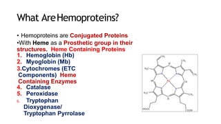
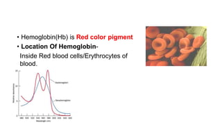
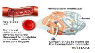
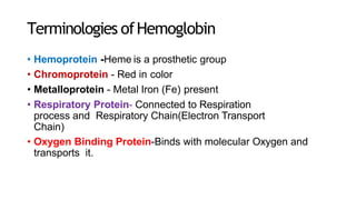
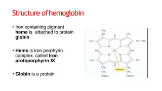
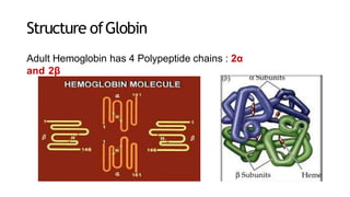
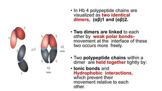
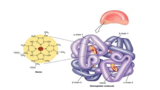
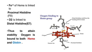
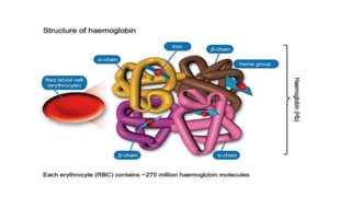
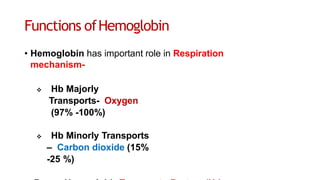
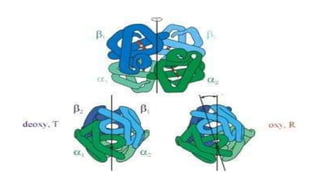
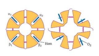
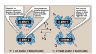
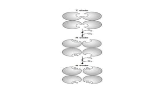
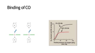
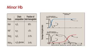
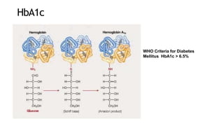
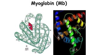
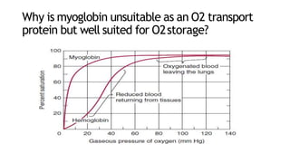
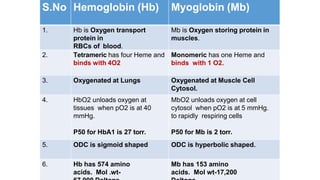
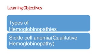
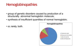
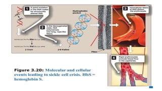
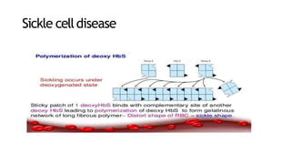
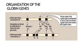
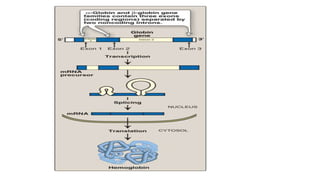
STRUCTURE AND FUNCTIONS
OF
HEMOGLOBIN ANDMYOGLOBIN
• What are
Hemoproteins?
• What is Hemoglobin?
• Structure of Hemoglobin
• Functions of Hemoglobin
• ODC and Factors
affecting it
• Normal Hb Variants
• Hemoglobin Derivatives
• Hemoglobinopathies
Learning objectives
What are
Hemoproteins?
What is Hemoglobin?
Structure of
Hemoglobin
What AreHemoproteins?
• Hemoproteins are Conjugated Proteins
•With Heme as a Prosthetic group in their
structures. Heme Containing Proteins
1. Hemoglobin (Hb)
2. Myoglobin (Mb)
3.Cytochromes (ETC
Components) Heme
Containing Enzymes
4. Catalase
5. Peroxidase
6. Tryptophan
Dioxygenase/
Tryptophan Pyrrolase
What IsHemoglobin?
• Hemoglobin(Hb) is a major Hemoprotein of
Human body
• Hemoglobin Chemically is:
• Conjugated Protein
• In Hemoglobin
• Heme is a Prosthetic group
• Globin is a Protein part
(Hemoglobin = Heme + Globin)
• Hemoglobin(Hb) is Red color pigment
• Location Of Hemoglobin-
Inside Red blood cells/Erythrocytes of
blood.
• Hemoglobin In RBCs Occupies:
• 33% of the RBC volume (1/3)
• 90-95% of the dry weight of RBC is by Hb.
• Normal concentration of Hemoglobin in the Human
Blood:
Adult Males-
13.5–17.5
gm/dL
Adult
Females-
12.5–16.5
Terminologies ofHemoglobin
• Hemoprotein -Heme is a prosthetic group
• Chromoprotein - Red in color
• Metalloprotein - Metal Iron (Fe) present
• Respiratory Protein- Connected to Respiration
process and Respiratory Chain(Electron Transport
Chain)
• Oxygen Binding Protein-Binds with molecular Oxygen and
transports it.
Structure ofhemoglobin
• Iron containing pigment
heme is attached to protein
globin
• Heme is iron porphyrin
complex called Iron
protoporphyrin IX
• Globin is a protein
Iron inHeme
• Functional form Iron in Heme is-
• Ferrous form(Fe++)
• Reduced state
• Fe++ located centrally in Protoporphyrin ring system.
• Fe of Heme is Hexavalent
• 4 bonds linked with each Nitrogen of 4 Pyrrole
rings.
• 5th bond linked with Proximal Histidine (F8) of
Globin chain
•6th bond is with
Oxygen.
Structure Of ProtoporphyrinIX-
• Cyclic substituted Tetrapyrrole ring structure
(I,II,III and IV Tetrapyrrole ).
• Tetrapyrrole rings has substituted groups in systematic
manner-
MV,MV,MP,PM
(M=Methyl ,V=Vinyl, P=Propionyl)
Structure ofGlobin
Adult Hemoglobin has 4 Polypeptide chains : 2α
and 2β
• In Hb 4 polypeptide chains are
visualized as two identical
dimers, (αβ)1 and (αβ)2.
• Two dimers are linked to each
other by weak polar bonds-
movement at the interface of these
two occurs more freely.
• Two polypeptide chains within a
dimer are held together tightly by:
• Ionic bonds and
Hydrophobic interactions,
which prevent their
movement relative to each
other.
Heme Pocket
• Heme Pocket is a
crevice/ hollow
hydrophobic area
• Formed in the interior
of Globin subunits
• Heme group is
tucked between
E and F helices of
Globin subunit.
• Amino acids in Globin chain are identified
by
• The helix name and position of a.a in that
helix.
• E7 His ( Distal His)
• F8 His ( Proximal His)
• Fe++ of Heme is linked
to
Proximal Histidine
(F8)
• O2 is linked to
Distal Histidine(E7).
•Thus to attain
stability Oxygen is
bound to both Heme
and Globin .
Learningobjectives
Functions of
hemoglobin
Features of oxygenation
of Hb
Functions ofHemoglobin
• Hemoglobin has important role in Respiration
mechanism-
 Hb Majorly
Transports- Oxygen
(97% -100%)
 Hb Minorly Transports
– Carbon dioxide (15%
-25 %)
• Oxygen transported by Hb and reached to every cell is
used up in Mitochondrial ETC
(Respiratory Chain/Cellular respiration)
• To generate ATP
(Oxidative Phosphorylation)
• Hemoglobin Plays Role as Buffer-
• (Hb/Hb-H+) in the Erythrocytes
• Resists change in pH
• Imidazole group of amino acid Histidine of Hb
molecule –
• Participates in buffering mechanism of Hb.
SALIENT FEATURES OF OXYGENATION AND
DEOXYGENATION OFHEMOGLOBIN
• Hemoglobin gets Oxygenated
• At Lungs
• At increased pO2 concentration (100-120
mm Hg)
• At decreased pCO2
• Hemoglobin gets deoxygenated
• At Tissues
• With Increased pCO2
• Decreased pO2 levels (40 mm Hg)
• Hemoglobin is 97 % saturated with Oxygen when it leaves the
Lungs- (Arterial Blood-Oxy Hb).
• Under resting conditions Hb is about 75% saturated with
Oxygen
when it returns-(Venous blood- Deoxy Hb).
Pulse Oximeter Is An Instrument
That Measures The Percentage Hb Fully Saturated With
Oxygen In Arterial Blood
Features of Oxygenation of Hb
• Oxygen binds with Hb to form HbO2
• Oxygen links to Ferrous form of Iron, of
Heme
• Non enzymatically, loosely and
reversibly.
• During oxygenation One Hb molecule with 4 Heme can
bind to four O2 molecules
• During Oxygenation Ferrous of Heme is not oxidized to Ferric.
Oxygenation of Hemoglobin causes Considerable
structuralconformational changein Globinsubunits.
T Form of Hb R Form Of Hb
Deoxy Hb is in T form
binds with CO2,H+ and
2,3BPG
Oxy Hb is in R form binds
only with Oxygen
T form has 8 salt bridges
linked in between the
dimer subunits
Salt bridges are broken
in between the dimer
subunits during
oxygenation of Hb.
More constrained form Less constrained
form
T form has low affinity
for Oxygen
R form has higher affinity
for Oxygen
T form of Hb
predominates in low pO2
R form of Hb
predominates at high
COOPERATIVE BINDING MECHANISMOF
OXYGEN WITHHEMOGLOBIN
Allows 100% saturation at po2
much lower than what it actually
requires
Hemoglobin picks up the
largest possible load
of Oxygen in the
lungs, And delivers the
Oxygen where and
when needed.
The cooperative binding ofoxygen
• enhances the efficiency of haemoglobin as an oxygen
transporter.
• Without positive cooperativity, an 81-fold increase of the pO2
would be required to raise the oxygen saturation from 10% to
90%.
• However, about Five fold increase is sufficient to do the same.
• This means that haemoglobin can rapidly bind oxygen in lungs
(where pO2 is high) and then readily liberate it in the tissue
capillaries (where pO2 is low).
• Hill coefficient is an indicator of cooperative binding
• Extent to which the binding of first oxygen molecule to one
subunit of Hb increases the subsequent oxygen binding to
other subunits.
• Max.4, practically 2.5
LearningObjectives
Oxygen dissociation
curve(ODC)
Factors affecting ODC (Allosteric
Effectors)
Oxygen dissociationcurve
Factors affectingODC
Bohr effect
Role of 2,3BPG
• ? Response of 2,3-BPG levels to
chronic hypoxia or anemia
• ? Role of 2,3-BPG in transfused
blood
• ? HbF
Mnemonic for Factors
causing Right Shift
of ODC :
CADET
• C – CO2
• A – Acid (H+)
• D– 2,3-BPG /2,3 DPG
• E – Exercise
• T – Temperature
Binding ofCO
Learningobjectives
Minor Hb
Myoglobi
n
Minor Hb
• Binding of 2,3-BPG to HbF:
weak
• ? Importance
ODC of Fetal Hb F vs. Adult Hb
HbA1c
WHO Criteria for Diabetes
Mellitus HbA1c > 6.5%
Myoglobin (Mb)
Why is myoglobin unsuitable as an O2 transport
protein but well suited for O2storage?
• Myoglobin in Muscle Cells is a:
• Oxygen storing Hemoprotein
• Reservoir of Oxygen
• Myoglobin does not allow Oxygen to remain in
free state:
• Oxygen diffused in muscle cells is used up in
Oxidative phosphorylation.
• If Oxygen remained unused in the cells it immediately binds
with Mb to form MbO2
• Mb present within muscle cells comes out in blood after
damage to muscle cells.
• Mb is found abnormally in blood and urine of MI cases.
• Thus elevated Myoglobin levels in blood/urine is a
marker of Myocardial damage.
Differences of HbAnd Mb
S.No Hemoglobin (Hb) Myoglobin (Mb)
1. Hb is Oxygen transport
protein in
RBCs of blood.
Mb is Oxygen storing protein in
muscles.
2. Tetrameric has four Heme and
binds with 4O2
Monomeric has one Heme and
binds with 1 O2.
3. Oxygenated at Lungs Oxygenated at Muscle Cell
Cytosol.
4. HbO2 unloads oxygen at
tissues when pO2 is at 40
mmHg.
P50 for HbA1 is 27 torr.
MbO2 unloads oxygen at cell
cytosol when pO2 is at 5 mmHg.
to rapidly respiring cells
P50 for Mb is 2 torr.
5. ODC is sigmoid shaped ODC is hyperbolic shaped.
6. Hb has 574 amino
acids. Mol .wt-
Mb has 153 amino
acids. Mol wt-17,200
LearningObjectives
Types of
Hemoglobinopathies
Sickle cell anemia(Qualitative
Hemoglobinopathy)
Hemoglobinopathies
• group of genetic disorders caused by production of a
structurally abnormal hemoglobin molecule;
• synthesis of insufficient quantities of normal hemoglobin;
• or, rarely, both.
Sickle celldisease
Hb Electrophoresis
Treatment
• Hydroxyure
a
Possible selective advantage ofthe
heterozygousstate
• high frequency of the bS mutation among black
Africans
Other types ofQualitative
Hemoglobinopathies
• HbC
• HbS
C
• HbM
Conversion Of MethemoglobinTo
Hemoglobin is
NADPH+H+ Dependent
H2O2 and Oxidant
Drugs
Methemoglobin
Reductase
NADP+ NADPH+ H+
Hemoglobin
(Fe+2)
Methemoglob
in (Fe+3)
Methemoglobinemias
• Oxidation of the heme iron in hemoglobin to the ferric (Fe3+)
state forms methemoglobin, which cannot bind O2.
• Causes
drugs, such as nitrates
endogenous products such as reactive oxygen species
inherited defects: HbM, deficiency of NADH-
cytochrome b5
reductase,
• S/S: chocolate cyanosis
• Treatment: methylene blue
•Dried blood and old meat
have brown color.
•Butchers uses Ascorbic
acid to reduce
Methemoglobin to make
the meat look fresh!!
LearningObjectives
Thalassemia
Quiz on
hemoglobinopathies
ORGANIZATION OFTHE
GLOBIN GENES
Quantitativehemoglobinopathies
Thalassemia(α andβ)
β Thalassemia
β Thalassemia
Anemia
• Anemias,
• reductions in the number of red blood cells or of
hemoglobin in the blood.
• can reflect impaired synthesis of hemoglobin eg, in iron
deficiency;
• or impaired production of erythrocytes (eg, in folic acid or
vitamin B12 deficiency;
• Diagnosis of anemias begins with spectroscopic
measurement of blood hemoglobin levels.
• Thank
you

hemoglobin and myoglobin (1).pptx

  • 1.
  • 2.
    • What are Hemoproteins? •What is Hemoglobin? • Structure of Hemoglobin • Functions of Hemoglobin • ODC and Factors affecting it • Normal Hb Variants • Hemoglobin Derivatives • Hemoglobinopathies
  • 3.
    Learning objectives What are Hemoproteins? Whatis Hemoglobin? Structure of Hemoglobin
  • 4.
    What AreHemoproteins? • Hemoproteinsare Conjugated Proteins •With Heme as a Prosthetic group in their structures. Heme Containing Proteins 1. Hemoglobin (Hb) 2. Myoglobin (Mb) 3.Cytochromes (ETC Components) Heme Containing Enzymes 4. Catalase 5. Peroxidase 6. Tryptophan Dioxygenase/ Tryptophan Pyrrolase
  • 5.
    What IsHemoglobin? • Hemoglobin(Hb)is a major Hemoprotein of Human body • Hemoglobin Chemically is: • Conjugated Protein • In Hemoglobin • Heme is a Prosthetic group • Globin is a Protein part (Hemoglobin = Heme + Globin)
  • 6.
    • Hemoglobin(Hb) isRed color pigment • Location Of Hemoglobin- Inside Red blood cells/Erythrocytes of blood.
  • 8.
    • Hemoglobin InRBCs Occupies: • 33% of the RBC volume (1/3) • 90-95% of the dry weight of RBC is by Hb. • Normal concentration of Hemoglobin in the Human Blood: Adult Males- 13.5–17.5 gm/dL Adult Females- 12.5–16.5
  • 9.
    Terminologies ofHemoglobin • Hemoprotein-Heme is a prosthetic group • Chromoprotein - Red in color • Metalloprotein - Metal Iron (Fe) present • Respiratory Protein- Connected to Respiration process and Respiratory Chain(Electron Transport Chain) • Oxygen Binding Protein-Binds with molecular Oxygen and transports it.
  • 10.
    Structure ofhemoglobin • Ironcontaining pigment heme is attached to protein globin • Heme is iron porphyrin complex called Iron protoporphyrin IX • Globin is a protein
  • 11.
    Iron inHeme • Functionalform Iron in Heme is- • Ferrous form(Fe++) • Reduced state • Fe++ located centrally in Protoporphyrin ring system. • Fe of Heme is Hexavalent • 4 bonds linked with each Nitrogen of 4 Pyrrole rings. • 5th bond linked with Proximal Histidine (F8) of Globin chain •6th bond is with Oxygen.
  • 13.
    Structure Of ProtoporphyrinIX- •Cyclic substituted Tetrapyrrole ring structure (I,II,III and IV Tetrapyrrole ). • Tetrapyrrole rings has substituted groups in systematic manner- MV,MV,MP,PM (M=Methyl ,V=Vinyl, P=Propionyl)
  • 15.
    Structure ofGlobin Adult Hemoglobinhas 4 Polypeptide chains : 2α and 2β
  • 19.
    • In Hb4 polypeptide chains are visualized as two identical dimers, (αβ)1 and (αβ)2. • Two dimers are linked to each other by weak polar bonds- movement at the interface of these two occurs more freely. • Two polypeptide chains within a dimer are held together tightly by: • Ionic bonds and Hydrophobic interactions, which prevent their movement relative to each other.
  • 21.
    Heme Pocket • HemePocket is a crevice/ hollow hydrophobic area • Formed in the interior of Globin subunits • Heme group is tucked between E and F helices of Globin subunit.
  • 22.
    • Amino acidsin Globin chain are identified by • The helix name and position of a.a in that helix. • E7 His ( Distal His) • F8 His ( Proximal His)
  • 23.
    • Fe++ ofHeme is linked to Proximal Histidine (F8) • O2 is linked to Distal Histidine(E7). •Thus to attain stability Oxygen is bound to both Heme and Globin .
  • 25.
  • 26.
    Functions ofHemoglobin • Hemoglobinhas important role in Respiration mechanism-  Hb Majorly Transports- Oxygen (97% -100%)  Hb Minorly Transports – Carbon dioxide (15% -25 %)
  • 27.
    • Oxygen transportedby Hb and reached to every cell is used up in Mitochondrial ETC (Respiratory Chain/Cellular respiration) • To generate ATP (Oxidative Phosphorylation)
  • 28.
    • Hemoglobin PlaysRole as Buffer- • (Hb/Hb-H+) in the Erythrocytes • Resists change in pH • Imidazole group of amino acid Histidine of Hb molecule – • Participates in buffering mechanism of Hb.
  • 29.
    SALIENT FEATURES OFOXYGENATION AND DEOXYGENATION OFHEMOGLOBIN • Hemoglobin gets Oxygenated • At Lungs • At increased pO2 concentration (100-120 mm Hg) • At decreased pCO2 • Hemoglobin gets deoxygenated • At Tissues • With Increased pCO2 • Decreased pO2 levels (40 mm Hg)
  • 30.
    • Hemoglobin is97 % saturated with Oxygen when it leaves the Lungs- (Arterial Blood-Oxy Hb). • Under resting conditions Hb is about 75% saturated with Oxygen when it returns-(Venous blood- Deoxy Hb).
  • 32.
    Pulse Oximeter IsAn Instrument That Measures The Percentage Hb Fully Saturated With Oxygen In Arterial Blood
  • 33.
    Features of Oxygenationof Hb • Oxygen binds with Hb to form HbO2 • Oxygen links to Ferrous form of Iron, of Heme • Non enzymatically, loosely and reversibly. • During oxygenation One Hb molecule with 4 Heme can bind to four O2 molecules • During Oxygenation Ferrous of Heme is not oxidized to Ferric.
  • 34.
    Oxygenation of Hemoglobincauses Considerable structuralconformational changein Globinsubunits.
  • 40.
    T Form ofHb R Form Of Hb Deoxy Hb is in T form binds with CO2,H+ and 2,3BPG Oxy Hb is in R form binds only with Oxygen T form has 8 salt bridges linked in between the dimer subunits Salt bridges are broken in between the dimer subunits during oxygenation of Hb. More constrained form Less constrained form T form has low affinity for Oxygen R form has higher affinity for Oxygen T form of Hb predominates in low pO2 R form of Hb predominates at high
  • 41.
    COOPERATIVE BINDING MECHANISMOF OXYGENWITHHEMOGLOBIN Allows 100% saturation at po2 much lower than what it actually requires Hemoglobin picks up the largest possible load of Oxygen in the lungs, And delivers the Oxygen where and when needed.
  • 42.
    The cooperative bindingofoxygen • enhances the efficiency of haemoglobin as an oxygen transporter. • Without positive cooperativity, an 81-fold increase of the pO2 would be required to raise the oxygen saturation from 10% to 90%. • However, about Five fold increase is sufficient to do the same. • This means that haemoglobin can rapidly bind oxygen in lungs (where pO2 is high) and then readily liberate it in the tissue capillaries (where pO2 is low).
  • 43.
    • Hill coefficientis an indicator of cooperative binding • Extent to which the binding of first oxygen molecule to one subunit of Hb increases the subsequent oxygen binding to other subunits. • Max.4, practically 2.5
  • 44.
  • 45.
  • 46.
  • 47.
  • 50.
  • 51.
    • ? Responseof 2,3-BPG levels to chronic hypoxia or anemia • ? Role of 2,3-BPG in transfused blood • ? HbF
  • 52.
    Mnemonic for Factors causingRight Shift of ODC : CADET • C – CO2 • A – Acid (H+) • D– 2,3-BPG /2,3 DPG • E – Exercise • T – Temperature
  • 53.
  • 54.
  • 55.
  • 58.
    • Binding of2,3-BPG to HbF: weak • ? Importance
  • 59.
    ODC of FetalHb F vs. Adult Hb
  • 60.
    HbA1c WHO Criteria forDiabetes Mellitus HbA1c > 6.5%
  • 61.
  • 62.
    Why is myoglobinunsuitable as an O2 transport protein but well suited for O2storage?
  • 63.
    • Myoglobin inMuscle Cells is a: • Oxygen storing Hemoprotein • Reservoir of Oxygen • Myoglobin does not allow Oxygen to remain in free state: • Oxygen diffused in muscle cells is used up in Oxidative phosphorylation. • If Oxygen remained unused in the cells it immediately binds with Mb to form MbO2
  • 64.
    • Mb presentwithin muscle cells comes out in blood after damage to muscle cells. • Mb is found abnormally in blood and urine of MI cases. • Thus elevated Myoglobin levels in blood/urine is a marker of Myocardial damage.
  • 65.
    Differences of HbAndMb S.No Hemoglobin (Hb) Myoglobin (Mb) 1. Hb is Oxygen transport protein in RBCs of blood. Mb is Oxygen storing protein in muscles. 2. Tetrameric has four Heme and binds with 4O2 Monomeric has one Heme and binds with 1 O2. 3. Oxygenated at Lungs Oxygenated at Muscle Cell Cytosol. 4. HbO2 unloads oxygen at tissues when pO2 is at 40 mmHg. P50 for HbA1 is 27 torr. MbO2 unloads oxygen at cell cytosol when pO2 is at 5 mmHg. to rapidly respiring cells P50 for Mb is 2 torr. 5. ODC is sigmoid shaped ODC is hyperbolic shaped. 6. Hb has 574 amino acids. Mol .wt- Mb has 153 amino acids. Mol wt-17,200
  • 66.
  • 67.
    Hemoglobinopathies • group ofgenetic disorders caused by production of a structurally abnormal hemoglobin molecule; • synthesis of insufficient quantities of normal hemoglobin; • or, rarely, both.
  • 69.
  • 71.
  • 72.
  • 73.
    Possible selective advantageofthe heterozygousstate • high frequency of the bS mutation among black Africans
  • 74.
  • 75.
    Conversion Of MethemoglobinTo Hemoglobinis NADPH+H+ Dependent H2O2 and Oxidant Drugs Methemoglobin Reductase NADP+ NADPH+ H+ Hemoglobin (Fe+2) Methemoglob in (Fe+3)
  • 76.
    Methemoglobinemias • Oxidation ofthe heme iron in hemoglobin to the ferric (Fe3+) state forms methemoglobin, which cannot bind O2. • Causes drugs, such as nitrates endogenous products such as reactive oxygen species inherited defects: HbM, deficiency of NADH- cytochrome b5 reductase, • S/S: chocolate cyanosis • Treatment: methylene blue
  • 77.
    •Dried blood andold meat have brown color. •Butchers uses Ascorbic acid to reduce Methemoglobin to make the meat look fresh!!
  • 78.
  • 79.
  • 81.
  • 82.
  • 83.
  • 87.
    Anemia • Anemias, • reductionsin the number of red blood cells or of hemoglobin in the blood. • can reflect impaired synthesis of hemoglobin eg, in iron deficiency; • or impaired production of erythrocytes (eg, in folic acid or vitamin B12 deficiency; • Diagnosis of anemias begins with spectroscopic measurement of blood hemoglobin levels.
  • 88.