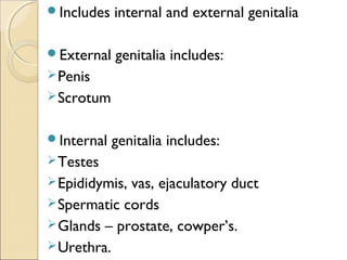
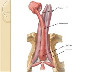
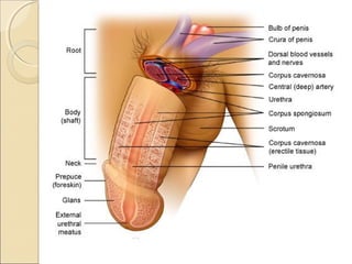
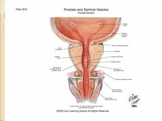
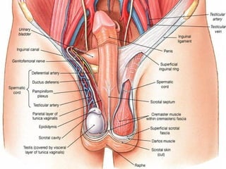
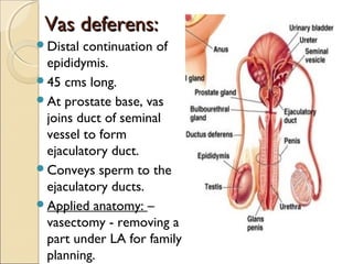
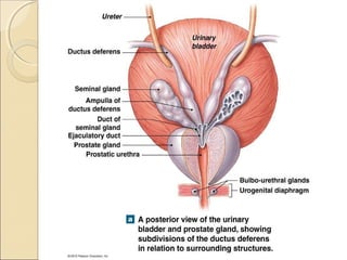
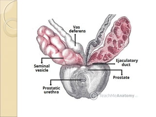
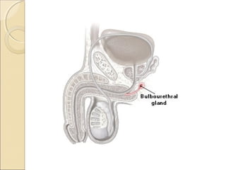
Male genital systemMale genital system
References:
1.V.K.SHARMA
2.IADVL
3.GRAYS ANATOMY
4.B D CHAURASIA
Includes internal and external genitalia
External genitalia includes:
Penis
Scrotum
Internal genitalia includes:
Testes
Epididymis, vas, ejaculatory duct
Spermatic cords
Glands – prostate, cowper’s.
Urethra.
Penis:Penis:
Root
Body
Root:
Superficial perineal pouch.
3 masses of erectile tissue – 2 crura and one
bulb.
Crura – attached to pubic arch – covered by
ischiocavernosus.
Bulb – attached to perineal membrane in b/n
crura – covered by bulbospongiosus.
Pierced by urethra.
Body:
Completely enveloped by skin.
Composed of 3 elongated masses of erectile
tissue – 2 ‘corpora cavernosa’ (continuation of
crura) and a median ‘corpus spongiosum’
(contin. Of bulb).
Terminal part of c.spongiosum expanded –
‘glans penis’.
Rim like raised surface at its proximal end –
‘corona glandis’.
Circular groove, ‘coronal sulcus’ runs along
corona glandis and seperates it from the
shaft of penis – k/a ‘neck’ of the penis.
At the neck, the skin covering the penis is
folded to form the ‘prepuce’ or ‘foreskin’ –
retracted backwards to expose the glans.
Space b/n glans and prepuce – ‘preputial sac’.
prepuce is attached to
the glans at its ventral
surface – ‘frenulum’.
On the corona and
neck of the penis –
numerous small
preputial or sebaceous
glands – secrete
smegma – collected in
the sac.
Lymphatic supply:
External pudendal
lymph vessels 
superficial inguinal
nodes.
Glans  deep inguinal
nodes called ‘gland of
cloquet’.
Applied anatomy:Applied anatomy:
1. PPP – Small pin head sized
projections studded over corona
in one r two rows.
confused with condyloma
acuminata.
2. Preputial sac – due to
occlusive effect over the glans –
vulnerable for inflammation-
‘balanoposthitis’.
3. Tight frenulum – predisposes
to trauma during sexual
intercourse – serves as another
Scrotum:Scrotum:
Cutaneous fibromuscular sac, which contains
testes, epididymis, vas deferens and loose
areolar tissue.
Layers: (o  i)
Skin
Dartos muscle – is prolonged into a median
vertical septum b/n the 2 halves of the
scrotum.
External spermatic fascia.
Cremasteric muscle and fascia.
Internal spermatic fascia.
Divided into right and left halves by a
cutaneous raphe – indicates b/l origin of the
scrotum from the genital swellings.
Left side of the scrotum is usually lower
because the left spermatic cord is longer.
Skin – thin, pigmented and rugous.
Has hairs, sebaceous glands (charac. Odor)
and also sweat glands, pigment cells and
nerve endings.
No subcutaneous adipose tissue.
Drained into superficial inguinal nodes.
Applied anatomy:
Abundance of hair and sebaceous glands –
site of sebaceous cysts.
Vulnerablility to genital ulcer ds. Due to
proximity with penis.
Ulcers over penoscrotal junction and
scrotum are seen in behcet’s ds.
Phthirus pubis (pubic louse) enjoys the
habitat of scrotal and pubic hair.
S.skin can be predisposed to develop nodular
scabies and persistent pruritic nodules of
scabies.
Male urethra:Male urethra:
long membranous canal for discharging
urine and seminal fluid.
extends from bladder neck to external
urinary meatus.
divided into:
Prostatic post.
Membranous urethra
 spongy or penile --- ant. Urethra.
Prostatic urethra:
Tunnels through the subs. of prostate.
3-4 cms is length
Lined by transitional epithelium
Widest and most dilatable part of male
urethra.
Post. Wall has a midline ridge called as
‘urethral crest’.
On each side of crest – shallow depression
called ‘prostatic sinus’ – perforated by orifices
of 15-20 prostatic ducts.
Elevation in the middle
of urethral crest –
‘verumontanum’ (a/k
colliculus seminalis) –
contains slit like orifice
of prostatic utricle.
Openings of
ejaculatory ducts are
present on either side
of this orifice.
Membranous urethra:
1.5 cms – shortest, least dilatable.
Part which passes through the perineal
membrane.
Lined by transitional epithelium.
The wall consists of thin layer of smooth
muscle and prominent outer circular straited
muscle fibers (rhabdosphincter) – form
external urethral sphincter.
Spongy urethra:
16 cms long.
Extends from membranous urethra to
external urethral orifice.
Lined by pseudostratified columnar epi.
2 parts:
Bulbar urethra – surrounded by
bulbospongiosus – widest part.
Penile urethra – dilated part within the glans
– navicular fossa – lined by stratified
squamous epithelium.
Transitional epi.
Transitional epi.
Pseudo. Columnar epi.
Pseudo. columnar
Stratified Squamous epi.
Lymphatic drainage:
Posterior urethra -
internal iliac nodes;
few end in external
iliac nodes.
Anterior urethra +
glans penis – deep
inguinal nodes.
Applied anatomy:Applied anatomy:
Commonly involved structure in urethral
syndromes of gonococcal and non-
gonococcal origin.
Gonococci have predilection for columnar
epithelium.
Inflammation of urethra is known as
‘urethritis’ – regarded as sexually transmitted
unless proven otherwise.
Urethritis often presents with urethral
discharge and dysuria.
Lab: increased PMNL in urethral dis. Smear.
Gonococcal urethritis: Neisseria gonorrhea.
Non-gonococcal:
Chlamydia trachomatis
Ureaplasma urealyticum
Mycoplasma genitalium
Trichomonas vaginalis
Non-infective causes:
Strictures
Traumatic
Neoplastic
Foreign body – catheterisation.
Urethral strictures:
narrowing of the urethra caused by injury or
disease such as UTI.
can occur as complication of gonococcal
(hard strictures) and non gonococcal
urethritis ( soft strictures).
Early Syphilis, chancroid, herpes, TB,
bilharzia.
Trauma - can be physical (eg catheterization,
urethroscopy); Chemical (burns from
podophyllin ,TCA or diathermy).
Rupture Of Urethra:
commonly ruptured beneath the pubis by a
fall astride a sharp object leading to
extravasation of urine into peritoneum.
Hypospadias:
common anamoly in which urethra opens on
the undersurface of penis.
Epispadias:
rare condition in which urethra opens on the
dorsum of penis.
associated with ectopia vesicae
Spermatic cord:Spermatic cord:
As the testis traverses the abdominal wall
into the scrotum during early life, it carries
its vessels, nerves and vas deferens with it.
These meet at the deep inguinal ring to form
the ‘spermatic cord’.
In the canal, the cord acquires coverings
from abd. Wall layers.
Suspends the testis in scrotum and extends
from deep inguinal canal to the posterior
aspect of the testis.
Contents of spermatic cord:
1. ductus deferens
2.testicular and cremasteric arteies and
artery of the ductus deferens.
3. pampiniform plexus of veins.
4. lymph vessels from the testis.
5. ilioinguinal nerve, genital branch of the
genitofemoral nerve, and the plexus of
sympathetic nerves around the artery to
ductus deferens and visceral afferent nerve
fibers.
6.remains of the processus vaginalis.
Testes:Testes:
Primary reproductive organs or gonads.
Covered by 3 coats ( o  i)
Tunica vaginalis
Tunica albuginea
Tunica vasculosa.
Produce sperm and testosterone.
Sperms – produced by seminiferous tubules.
Testosterone – by leydig cells located b/n the
tubules.
400-600 seminiferous tubules in each testes –
contain spermatogenic cells and supporting
sertoli cells.
Tubules join to form ‘rete testis’
Excretory duct system – 5 elements.
Testis
Efferent ducts
Epididymis
Vas
Ejaculatory duct and urethra.
Applied anatomy:Applied anatomy:
Absence of testis:
u/l – monorchism
b/l – anorchism.
Undescended testis or cryptorchidism: may
lie in lumbar, iliac, inguinal, or upper scrotal
region.
May complete after birth.
Spermatogenesis may fail to occur in it.
More prone for malignancy.
Condition is surgically corrected.
Hermaphroditism:
Individual shows some features of male and
some of a female.
True – both testis+ ovary
Pseudo – gonad is of one sex while the
external or internal genitalia are of opposite
sex.
Carcinomas and infections: may be palpated
to check any nodules, or any irregularity or
size or consistency.
Varicocele: dilatation of pampiniform plexus
on veins.
Epididymis:Epididymis:
lies postero-lateral to
the testis.
Has:
1. head or globus
major superiorly,
2. body – corpus
3. tail – cauda or
globus minor.
Vas deferens:Vas deferens:
Distal continuation of
epididymis.
45 cms long.
At prostate base, vas
joins duct of seminal
vessel to form
ejaculatory duct.
Conveys sperm to the
ejaculatory ducts.
Applied anatomy: –
vasectomy - removing a
part under LA for family
planning.
Ejaculatory duct:Ejaculatory duct:
2 cms in length.
Starts from the base of the prostate and
ends on the verumontanum or within
the utricular opening.
Seminal vesicles:Seminal vesicles:
Sacculated tubes located b/n the bladder
and the rectum.
Single coiled tube with irregular
diverticula.
Secrete alkaline, slightly yellowish viscid
fluid which constitutes 60-70% of the
ejaculatory volume.
Prostate:Prostate:
Located b/n bladder neck and the urogenital
diaphragm.
Encircles the urethra completely.
Zones: 3
1. Periurethral zone – surrounding urethra.
2. central zone – wedge-shaped; bounded by
ejaculatory duct, urethra, and base of the
bladder.
Less susceptible to inflammatory,
hyperplastic, or neoplastic ds.
3. peripheral/outer zone – the portion that is
palpable on rectal exmn.
Most frequently involved in carcinoma and
inflammation.
Secretions:
Thin, slightly opaque fluid.
Contributes to 30% of ejaculate volume.
Protects the male lower urinary tract against
infections.
Provides enzymes for ‘liquefying’ the semen
after ejaculation.
Applied anatomyApplied anatomy
Benign prostate hypertrophy:
senile enlargement after 50 years.
Occurs in periurethral zone.
Causes retention of urine due to distortion
of urethra.
Rx: prostatectomy – transvesical
transvesical/
retropubic/
transurethral
resection.
Prostatitis:
Acute or chronic inflammation.
Acute – sec. to gonococcal urethritis.
Chronic – tuberculous infection of
epididymis, seminal vesicles and the bladder.
Ca.prostate:
Occurs after the age of 50-55 years.
Symptoms are urinary obstruction, low
backpain or sciatica.
Rectal exmn – irregular hard prostate.
Mets spread to vertebral column.
Bulbourethral (cowper’s)glands:Bulbourethral (cowper’s)glands:
Paired, pea-sized glands located in urogenital
diaphragm.
Excretory ducts drain into posterior
urethra.
Secrete a thin, mucoid material during the
excitatory stage of sexual response.
Contribute a minimal amount to the
ejaculate.
Immune to hyperplastic and neoplastic ds.
My ppt
My ppt

My ppt

  • 1.
    Male genital systemMalegenital system References: 1.V.K.SHARMA 2.IADVL 3.GRAYS ANATOMY 4.B D CHAURASIA
  • 2.
    Includes internal andexternal genitalia External genitalia includes: Penis Scrotum Internal genitalia includes: Testes Epididymis, vas, ejaculatory duct Spermatic cords Glands – prostate, cowper’s. Urethra.
  • 3.
    Penis:Penis: Root Body Root: Superficial perineal pouch. 3masses of erectile tissue – 2 crura and one bulb. Crura – attached to pubic arch – covered by ischiocavernosus.
  • 5.
    Bulb – attachedto perineal membrane in b/n crura – covered by bulbospongiosus. Pierced by urethra. Body: Completely enveloped by skin. Composed of 3 elongated masses of erectile tissue – 2 ‘corpora cavernosa’ (continuation of crura) and a median ‘corpus spongiosum’ (contin. Of bulb). Terminal part of c.spongiosum expanded – ‘glans penis’.
  • 6.
    Rim like raisedsurface at its proximal end – ‘corona glandis’. Circular groove, ‘coronal sulcus’ runs along corona glandis and seperates it from the shaft of penis – k/a ‘neck’ of the penis. At the neck, the skin covering the penis is folded to form the ‘prepuce’ or ‘foreskin’ – retracted backwards to expose the glans. Space b/n glans and prepuce – ‘preputial sac’.
  • 8.
    prepuce is attachedto the glans at its ventral surface – ‘frenulum’. On the corona and neck of the penis – numerous small preputial or sebaceous glands – secrete smegma – collected in the sac.
  • 9.
    Lymphatic supply: External pudendal lymphvessels  superficial inguinal nodes. Glans  deep inguinal nodes called ‘gland of cloquet’.
  • 10.
    Applied anatomy:Applied anatomy: 1.PPP – Small pin head sized projections studded over corona in one r two rows. confused with condyloma acuminata. 2. Preputial sac – due to occlusive effect over the glans – vulnerable for inflammation- ‘balanoposthitis’. 3. Tight frenulum – predisposes to trauma during sexual intercourse – serves as another
  • 11.
    Scrotum:Scrotum: Cutaneous fibromuscular sac,which contains testes, epididymis, vas deferens and loose areolar tissue. Layers: (o  i) Skin Dartos muscle – is prolonged into a median vertical septum b/n the 2 halves of the scrotum. External spermatic fascia. Cremasteric muscle and fascia.
  • 12.
    Internal spermatic fascia. Dividedinto right and left halves by a cutaneous raphe – indicates b/l origin of the scrotum from the genital swellings. Left side of the scrotum is usually lower because the left spermatic cord is longer.
  • 14.
    Skin – thin,pigmented and rugous. Has hairs, sebaceous glands (charac. Odor) and also sweat glands, pigment cells and nerve endings. No subcutaneous adipose tissue. Drained into superficial inguinal nodes.
  • 15.
    Applied anatomy: Abundance ofhair and sebaceous glands – site of sebaceous cysts. Vulnerablility to genital ulcer ds. Due to proximity with penis. Ulcers over penoscrotal junction and scrotum are seen in behcet’s ds. Phthirus pubis (pubic louse) enjoys the habitat of scrotal and pubic hair. S.skin can be predisposed to develop nodular scabies and persistent pruritic nodules of scabies.
  • 17.
    Male urethra:Male urethra: longmembranous canal for discharging urine and seminal fluid. extends from bladder neck to external urinary meatus. divided into: Prostatic post. Membranous urethra  spongy or penile --- ant. Urethra.
  • 19.
    Prostatic urethra: Tunnels throughthe subs. of prostate. 3-4 cms is length Lined by transitional epithelium Widest and most dilatable part of male urethra. Post. Wall has a midline ridge called as ‘urethral crest’. On each side of crest – shallow depression called ‘prostatic sinus’ – perforated by orifices of 15-20 prostatic ducts.
  • 21.
    Elevation in themiddle of urethral crest – ‘verumontanum’ (a/k colliculus seminalis) – contains slit like orifice of prostatic utricle. Openings of ejaculatory ducts are present on either side of this orifice.
  • 22.
    Membranous urethra: 1.5 cms– shortest, least dilatable. Part which passes through the perineal membrane. Lined by transitional epithelium. The wall consists of thin layer of smooth muscle and prominent outer circular straited muscle fibers (rhabdosphincter) – form external urethral sphincter.
  • 24.
    Spongy urethra: 16 cmslong. Extends from membranous urethra to external urethral orifice. Lined by pseudostratified columnar epi. 2 parts: Bulbar urethra – surrounded by bulbospongiosus – widest part. Penile urethra – dilated part within the glans – navicular fossa – lined by stratified squamous epithelium.
  • 25.
    Transitional epi. Transitional epi. Pseudo.Columnar epi. Pseudo. columnar Stratified Squamous epi.
  • 26.
    Lymphatic drainage: Posterior urethra- internal iliac nodes; few end in external iliac nodes. Anterior urethra + glans penis – deep inguinal nodes.
  • 27.
    Applied anatomy:Applied anatomy: Commonlyinvolved structure in urethral syndromes of gonococcal and non- gonococcal origin. Gonococci have predilection for columnar epithelium. Inflammation of urethra is known as ‘urethritis’ – regarded as sexually transmitted unless proven otherwise. Urethritis often presents with urethral discharge and dysuria. Lab: increased PMNL in urethral dis. Smear.
  • 29.
    Gonococcal urethritis: Neisseriagonorrhea. Non-gonococcal: Chlamydia trachomatis Ureaplasma urealyticum Mycoplasma genitalium Trichomonas vaginalis Non-infective causes: Strictures Traumatic Neoplastic Foreign body – catheterisation.
  • 30.
    Urethral strictures: narrowing ofthe urethra caused by injury or disease such as UTI. can occur as complication of gonococcal (hard strictures) and non gonococcal urethritis ( soft strictures). Early Syphilis, chancroid, herpes, TB, bilharzia. Trauma - can be physical (eg catheterization, urethroscopy); Chemical (burns from podophyllin ,TCA or diathermy).
  • 31.
    Rupture Of Urethra: commonlyruptured beneath the pubis by a fall astride a sharp object leading to extravasation of urine into peritoneum. Hypospadias: common anamoly in which urethra opens on the undersurface of penis. Epispadias: rare condition in which urethra opens on the dorsum of penis. associated with ectopia vesicae
  • 32.
    Spermatic cord:Spermatic cord: Asthe testis traverses the abdominal wall into the scrotum during early life, it carries its vessels, nerves and vas deferens with it. These meet at the deep inguinal ring to form the ‘spermatic cord’. In the canal, the cord acquires coverings from abd. Wall layers. Suspends the testis in scrotum and extends from deep inguinal canal to the posterior aspect of the testis.
  • 34.
    Contents of spermaticcord: 1. ductus deferens 2.testicular and cremasteric arteies and artery of the ductus deferens. 3. pampiniform plexus of veins. 4. lymph vessels from the testis. 5. ilioinguinal nerve, genital branch of the genitofemoral nerve, and the plexus of sympathetic nerves around the artery to ductus deferens and visceral afferent nerve fibers. 6.remains of the processus vaginalis.
  • 36.
    Testes:Testes: Primary reproductive organsor gonads. Covered by 3 coats ( o  i) Tunica vaginalis Tunica albuginea Tunica vasculosa. Produce sperm and testosterone. Sperms – produced by seminiferous tubules. Testosterone – by leydig cells located b/n the tubules.
  • 38.
    400-600 seminiferous tubulesin each testes – contain spermatogenic cells and supporting sertoli cells. Tubules join to form ‘rete testis’ Excretory duct system – 5 elements. Testis Efferent ducts Epididymis Vas Ejaculatory duct and urethra.
  • 40.
    Applied anatomy:Applied anatomy: Absenceof testis: u/l – monorchism b/l – anorchism. Undescended testis or cryptorchidism: may lie in lumbar, iliac, inguinal, or upper scrotal region. May complete after birth. Spermatogenesis may fail to occur in it. More prone for malignancy. Condition is surgically corrected.
  • 41.
    Hermaphroditism: Individual shows somefeatures of male and some of a female. True – both testis+ ovary Pseudo – gonad is of one sex while the external or internal genitalia are of opposite sex. Carcinomas and infections: may be palpated to check any nodules, or any irregularity or size or consistency. Varicocele: dilatation of pampiniform plexus on veins.
  • 42.
    Epididymis:Epididymis: lies postero-lateral to thetestis. Has: 1. head or globus major superiorly, 2. body – corpus 3. tail – cauda or globus minor.
  • 43.
    Vas deferens:Vas deferens: Distalcontinuation of epididymis. 45 cms long. At prostate base, vas joins duct of seminal vessel to form ejaculatory duct. Conveys sperm to the ejaculatory ducts. Applied anatomy: – vasectomy - removing a part under LA for family planning.
  • 44.
    Ejaculatory duct:Ejaculatory duct: 2cms in length. Starts from the base of the prostate and ends on the verumontanum or within the utricular opening.
  • 46.
    Seminal vesicles:Seminal vesicles: Sacculatedtubes located b/n the bladder and the rectum. Single coiled tube with irregular diverticula. Secrete alkaline, slightly yellowish viscid fluid which constitutes 60-70% of the ejaculatory volume.
  • 48.
    Prostate:Prostate: Located b/n bladderneck and the urogenital diaphragm. Encircles the urethra completely. Zones: 3 1. Periurethral zone – surrounding urethra. 2. central zone – wedge-shaped; bounded by ejaculatory duct, urethra, and base of the bladder. Less susceptible to inflammatory, hyperplastic, or neoplastic ds.
  • 49.
    3. peripheral/outer zone– the portion that is palpable on rectal exmn. Most frequently involved in carcinoma and inflammation.
  • 50.
    Secretions: Thin, slightly opaquefluid. Contributes to 30% of ejaculate volume. Protects the male lower urinary tract against infections. Provides enzymes for ‘liquefying’ the semen after ejaculation.
  • 51.
    Applied anatomyApplied anatomy Benignprostate hypertrophy: senile enlargement after 50 years. Occurs in periurethral zone. Causes retention of urine due to distortion of urethra. Rx: prostatectomy – transvesical transvesical/ retropubic/ transurethral resection.
  • 52.
    Prostatitis: Acute or chronicinflammation. Acute – sec. to gonococcal urethritis. Chronic – tuberculous infection of epididymis, seminal vesicles and the bladder. Ca.prostate: Occurs after the age of 50-55 years. Symptoms are urinary obstruction, low backpain or sciatica. Rectal exmn – irregular hard prostate. Mets spread to vertebral column.
  • 53.
    Bulbourethral (cowper’s)glands:Bulbourethral (cowper’s)glands: Paired,pea-sized glands located in urogenital diaphragm. Excretory ducts drain into posterior urethra. Secrete a thin, mucoid material during the excitatory stage of sexual response. Contribute a minimal amount to the ejaculate. Immune to hyperplastic and neoplastic ds.