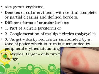
Figurate erythemas
• Aka gyrate erythema.
• Denotes circular erythema with central complete
or partial clearing and defined borders.
• Different forms of annular lesions:
• 1. Part of a circle (arciform) or
• 2. Conglomeration of multiple circles (polycyclic).
• 3. Target – dusky red center surrounded by a
zone of pallor which in turn is surrounded by
peripheral erythematous ring.
• 4. Atypical target – only two zones of color
change.
• 1. Erythema annulare centrifugm
• 2. Erythema chronicum migrans
• 3. Erythema marginatum rheumaticum
• 4. Erythema gyratum repens
• 5. Annular erythema of infancy.
• 6. Necrolytic migratory erythema.
• 7. Annular erythema associated with ENA.
• 8. Neutrophilic and vasculitis annular eruptions.
ERYTHEMA ANNULARE CENTRIFUGUM
• Multiple ring shaped lesions of indurated
erythema.
• Etiology: unknown in most cases.
• Basically a hypersensitivity reaction to a
variety of agents including drugs, arthropod
bites, infections, ingestion, and malignancy.
ETIOLOGIES / TRIGGER FACTORS:
Infection/
Infestation
Fungal, viral, mycobacterial, ascaris,
herpes zoster, HIV
Ingestion Food allergies
Drug Finasteride, piroxicam, hydroxychloride,
amitryptyline, spironolactone, estrogen,
penicillin, salicylate,
hydroxychlorothoazide
Malignancy Mycosis fungoides, blood dyscrasia,
chronic lymphocytic leukemia
Autoimmune SLE, sjogren’s syndrome, grave’s disease.
Others Familial, anthopod bites, liver disease,
appendicitis, hypereosinophilic
• Clinical features:
• Asymmetrical elevated urticarial erythema
• Spreads centrifugally and gradually fades from
the center.
• Trunk, proximal limbs, gluteal region.
• Margin – indurated. ‘Trailing scale’ is present on
the inner aspect of the advancing egde.
• Hyperpigmentation can appear in the centrally
cleared zone.
• Lesions wax and wane.
• Last for months to years.
• Epidemiology: no particular age or sex.
• Histology:
• 1. superficial type: non specific – epidermal
changes like focal spongiosis and focal
parakeratosis and superficial perivascular
lymphohistiocytic infiltrate +.
• 2. classic deep or indurated type: perivascular
“coat-sleeveline” lymphocytic infiltration in mid
and deep dermis.
• Epidermis is unaffected.
• Treatment:
• Identification and eradication of the
underlying cause – important.
• Supportive treatment.
• Systemic antihistamines and NSAIDs.
• Oral /topical corticosteroids
• Topical calcipotriol and tacrolimus.
ERYTHEMA MIGRANS
• Erythema migrans is a lesion that forms at the
location of the tick bite in Lyme disease.
• Caused by spirochete Borrelia burgdoferi and
transmitted by bite of ixodus ticks.
• Clinical features: 3 stages –
• 1. early localized
• 2. early disseminated
• 3. chronic disseminated.
Early localized Early
disseminated
Late
disseminated
Symptoms
/signs
EM
Disseminated
EM
Lymphocytoma
Disseminated
EM
Lymphocytoma
Arthritis
Neuroborreliosis
Cardiac
involvement
Eye changes
ACA
Chronic arthritis
neuroborreliosis
Serology +/- + -
Pathogenesis Direct invasion Direct invasion Immune
mediated
Lymphadenopat
hy
Regional Regional/genera
lized
NIL.
• Average interval b/n bite and app. Of skin
lesions – 9 days.
• Starts as a small papule that spreads
centrifugally, while the central erythema
gradually disappears.
• Lesions are flat, blanches with pressure and
doesn’t desquamate, vesiculate or have
peripheral scales.
• Burning or mild itching +.
• Histology: perivascular infiltrate containing
plasma cells and eosinophils is seen.
• Spirochetes are seen most frequently in the
advancing border of the lesion
• Diagnosis:
• IgM antibody.
• Rising titer of IgG antibody with an ELISA
• Indirect immunofluorescence test.
• Treatment:
• Doxycycline 100mg BD x10-21 days
• Amoxycillin 500mg TID x14-21 days
• Cefuroxime 500mg BD x14-21 days
• Steroids - not helpful.
• Prognosis: disappear with resolution of
underlying ds.
ERYTHEMA MARGINATUM
RHEUMATICUM
• Aka Erythema Circinatum, Erythema Annulare
Rheumaticum.
• Sign seen in about 20% of patients with acute
rheumatic fever.
• One of the major Jones’ criteria.
• Incidence is associated with cardiac involvement
– regarded as sign of acute rheumatic fever and
impending rh.endocarditis.
• Hypersensitivity response to streptococcal
antigens.
• Clinical features:
• Appear with the onset of pyrexia – usually after
fever spike in afternoon.
• Site: trunk esp. periumbilical region.
• Dorsum of hands, face, axillae or buttocks.
• Multiple pinkish-red patches with raised borders
–spread rapidly – ring shaped configuration.
• More prominent on cold exposure.
• Pruritus – absent.
• Also related to psittacosis and occurs before the
attacks of hereditary angioedema.
• Treatment: symptomatic. Penicillin.
ERYTHEMA GYRATUM REPENS
• Erythematous bands spread over the body in
waves - like patterns on wood or the stripes of a
zebra.
• These are faster spreading of the lesions (about 1
cm/day)
• More pronounced desquamation and pruritus.
• Sites: trunk and extremities.
• There is a characteristic collar-like
desquamation.
• Precedes the diagnosis of malignancy – time
range variable.
Wood grain
pattern
• There is an underlying malignancy in about 80%
of the cases of erythema gyratum repens.
• For this reason, the patient must be analyzed
carefully for malignancy.
• Neoplastic conditions: lung, breast, urinary
bladder, uterus/cervix, GI tract (stomach),
prostate.
• Non-neoplastic: pulmonary TB, psoriasis, lupus
erythematosus, PRP, drug reaction to
azathioprine.
• Pathogenesis: cross reaction of tumor Ags with
endogenous skin Ags, tumor Ags inducing
changes – making them susceptible to
autoimmune recog; forming immune complexes.
• Histopathology: focal parakeratosis, exocytosis of
neutrophils and eosinophils.
• Direct immunofluorescence tests shows C3, C4,
and immunoglobulin G at basement membrane
zone.
• Treatment:
• Underlying illness determines the prognosis.
• Disappear with the resolution of underlying
disease.
ANNULAR ERYTHEMA OF
INFANCY
• Resembles EAC.
• Different underlying
causes.
• r/o lupus erythematosus
and infections.
• C.albicans colonization in
intestine and EBV
infection has been
associated.
• Treatment: wait and
watch.
NECROLYTIC MIGRATORY
ERYTHEMA
• Paraneoplastic sign seen when there is an
underlying glucagon secreting malignant
pancreas alpha-cell tumor.
• Idiopathic cases or cases due to other
gastrointestinal causes (chronic
pancreatitis, chronic hepatitis, colon
cancer) – pseudoglucagonoma syndrome.
• Glucagon or its metabolites are thought to
be responsible.
• The lesions start as red-brown macules in
perioral or inguinal regions and later necrotize
and become covered with crusts.
• Glossitis may accompany.
• This appearance is similar to C. albicans
infection.
• The macules may become vesicular, widespread
and may desquamate.
• Symptoms reside after resection of the tumor.
• Recurrence of symptoms is a sensitive indicator
of recurrence of the tumor.
Necrolytic
migratory
erythema
ANNULAR ERYTHEMA WITH
ENAs:
• May occur in anti-
SSA (Ro) – positive
patients with
sjogrens syndrome,
lupus
erythematosus or
both together.
NEUTROPHILIC AND VASCULITIC
ANNULAR ERUPTIONS
• Acute haemmorhaggic edema of infancy
• Erythema elevatum diutinum
• Chronic recurrent annular neutrophilic
dermatosis
• Urticarial vasculitis
• HSP
• Leukocytoclastic vasculitis asso. With
myeloma
• IBD or pregnancy
DD’S
• Mycoses,
• Annular urticaria,
• Granuloma annulare,
• Mycosis fungoides,
• Pseudolymphomas
• Bullous pemphigoid,
pemphigus, dermatitis
herpetiformis, linear
IgA disease.
• Erythema multiforme,
• Sarcoidosis
• Still disease,
• Annular psoriasis,
• Erythrokeratoderma
variabilis,
• Chronic granulomatous
disease,
• Pityriasiform seborrheic
dermatitis,
• Neutrophilic dermatoses,
• Vasculitides,
• Acute hemorrhagic edema
of childhood,
• Lepra, leishmania,
• Trypanosomiasis
?? Diagnostic approach ??
• 1. Are there any signs or symptoms of
malignancy, infection or other systemic disease?
• 2. Are there other findings of tick bite or Lyme
disease?
• 3. Are there lesions of urticaria or angioedema?
Urticaria lasts shorter and itches more than
erythema annulare centrifugum.
• 4. Are there bullous lesions? Bullous pemphigoid
and linear IgA disease also have urticarial
phases.
• 5. Erythema multiforme should be considered if
the lesions have an oral and acral distribution.
• 6. KOH examination should be done.
• 7. If the lesions are psoriasiform, psoriasis and
subacute lupus erythematosus should be
considered.
• Rarely Sjogren syndrome may present with
annulare lesions. Ro/La antibodies should be
investigated.
• 8. Are there any other findings of acute
rheumatismal fever? Erythema marginatum is
the shortest lasting of the figurate erythemas.
• 9. Are the lesions located orally or in
intertriginous locations? Are there any other
signs of glucagon excess?
• 10. Is there family history of similar lesions? Is
there anyone in the family with granulomatous
disease? Are phagocytic functions normal?
Annular lesions may be seen in carrier females
with chronic granulomatous disease.
• 11. Is the patient an infant? Neonatal lupus
erythematosus must be ruled out in this age
group. Although mycoses are not seen commonly
in infants, they should be ruled out.
Fe

Fe

  • 1.
  • 2.
    • Aka gyrateerythema. • Denotes circular erythema with central complete or partial clearing and defined borders. • Different forms of annular lesions: • 1. Part of a circle (arciform) or • 2. Conglomeration of multiple circles (polycyclic). • 3. Target – dusky red center surrounded by a zone of pallor which in turn is surrounded by peripheral erythematous ring. • 4. Atypical target – only two zones of color change.
  • 3.
    • 1. Erythemaannulare centrifugm • 2. Erythema chronicum migrans • 3. Erythema marginatum rheumaticum • 4. Erythema gyratum repens • 5. Annular erythema of infancy. • 6. Necrolytic migratory erythema. • 7. Annular erythema associated with ENA. • 8. Neutrophilic and vasculitis annular eruptions.
  • 4.
    ERYTHEMA ANNULARE CENTRIFUGUM •Multiple ring shaped lesions of indurated erythema. • Etiology: unknown in most cases. • Basically a hypersensitivity reaction to a variety of agents including drugs, arthropod bites, infections, ingestion, and malignancy.
  • 5.
    ETIOLOGIES / TRIGGERFACTORS: Infection/ Infestation Fungal, viral, mycobacterial, ascaris, herpes zoster, HIV Ingestion Food allergies Drug Finasteride, piroxicam, hydroxychloride, amitryptyline, spironolactone, estrogen, penicillin, salicylate, hydroxychlorothoazide Malignancy Mycosis fungoides, blood dyscrasia, chronic lymphocytic leukemia Autoimmune SLE, sjogren’s syndrome, grave’s disease. Others Familial, anthopod bites, liver disease, appendicitis, hypereosinophilic
  • 6.
    • Clinical features: •Asymmetrical elevated urticarial erythema • Spreads centrifugally and gradually fades from the center. • Trunk, proximal limbs, gluteal region. • Margin – indurated. ‘Trailing scale’ is present on the inner aspect of the advancing egde. • Hyperpigmentation can appear in the centrally cleared zone. • Lesions wax and wane. • Last for months to years.
  • 7.
    • Epidemiology: noparticular age or sex. • Histology: • 1. superficial type: non specific – epidermal changes like focal spongiosis and focal parakeratosis and superficial perivascular lymphohistiocytic infiltrate +. • 2. classic deep or indurated type: perivascular “coat-sleeveline” lymphocytic infiltration in mid and deep dermis. • Epidermis is unaffected.
  • 9.
    • Treatment: • Identificationand eradication of the underlying cause – important. • Supportive treatment. • Systemic antihistamines and NSAIDs. • Oral /topical corticosteroids • Topical calcipotriol and tacrolimus.
  • 10.
    ERYTHEMA MIGRANS • Erythemamigrans is a lesion that forms at the location of the tick bite in Lyme disease. • Caused by spirochete Borrelia burgdoferi and transmitted by bite of ixodus ticks. • Clinical features: 3 stages – • 1. early localized • 2. early disseminated • 3. chronic disseminated.
  • 11.
    Early localized Early disseminated Late disseminated Symptoms /signs EM Disseminated EM Lymphocytoma Disseminated EM Lymphocytoma Arthritis Neuroborreliosis Cardiac involvement Eyechanges ACA Chronic arthritis neuroborreliosis Serology +/- + - Pathogenesis Direct invasion Direct invasion Immune mediated Lymphadenopat hy Regional Regional/genera lized NIL.
  • 12.
    • Average intervalb/n bite and app. Of skin lesions – 9 days. • Starts as a small papule that spreads centrifugally, while the central erythema gradually disappears. • Lesions are flat, blanches with pressure and doesn’t desquamate, vesiculate or have peripheral scales. • Burning or mild itching +. • Histology: perivascular infiltrate containing plasma cells and eosinophils is seen. • Spirochetes are seen most frequently in the advancing border of the lesion
  • 14.
    • Diagnosis: • IgMantibody. • Rising titer of IgG antibody with an ELISA • Indirect immunofluorescence test. • Treatment: • Doxycycline 100mg BD x10-21 days • Amoxycillin 500mg TID x14-21 days • Cefuroxime 500mg BD x14-21 days • Steroids - not helpful. • Prognosis: disappear with resolution of underlying ds.
  • 15.
    ERYTHEMA MARGINATUM RHEUMATICUM • AkaErythema Circinatum, Erythema Annulare Rheumaticum. • Sign seen in about 20% of patients with acute rheumatic fever. • One of the major Jones’ criteria. • Incidence is associated with cardiac involvement – regarded as sign of acute rheumatic fever and impending rh.endocarditis. • Hypersensitivity response to streptococcal antigens.
  • 16.
    • Clinical features: •Appear with the onset of pyrexia – usually after fever spike in afternoon. • Site: trunk esp. periumbilical region. • Dorsum of hands, face, axillae or buttocks. • Multiple pinkish-red patches with raised borders –spread rapidly – ring shaped configuration. • More prominent on cold exposure. • Pruritus – absent. • Also related to psittacosis and occurs before the attacks of hereditary angioedema. • Treatment: symptomatic. Penicillin.
  • 18.
    ERYTHEMA GYRATUM REPENS •Erythematous bands spread over the body in waves - like patterns on wood or the stripes of a zebra. • These are faster spreading of the lesions (about 1 cm/day) • More pronounced desquamation and pruritus. • Sites: trunk and extremities. • There is a characteristic collar-like desquamation. • Precedes the diagnosis of malignancy – time range variable.
  • 19.
  • 20.
    • There isan underlying malignancy in about 80% of the cases of erythema gyratum repens. • For this reason, the patient must be analyzed carefully for malignancy. • Neoplastic conditions: lung, breast, urinary bladder, uterus/cervix, GI tract (stomach), prostate. • Non-neoplastic: pulmonary TB, psoriasis, lupus erythematosus, PRP, drug reaction to azathioprine. • Pathogenesis: cross reaction of tumor Ags with endogenous skin Ags, tumor Ags inducing changes – making them susceptible to autoimmune recog; forming immune complexes.
  • 21.
    • Histopathology: focalparakeratosis, exocytosis of neutrophils and eosinophils. • Direct immunofluorescence tests shows C3, C4, and immunoglobulin G at basement membrane zone. • Treatment: • Underlying illness determines the prognosis. • Disappear with the resolution of underlying disease.
  • 22.
    ANNULAR ERYTHEMA OF INFANCY •Resembles EAC. • Different underlying causes. • r/o lupus erythematosus and infections. • C.albicans colonization in intestine and EBV infection has been associated. • Treatment: wait and watch.
  • 23.
    NECROLYTIC MIGRATORY ERYTHEMA • Paraneoplasticsign seen when there is an underlying glucagon secreting malignant pancreas alpha-cell tumor. • Idiopathic cases or cases due to other gastrointestinal causes (chronic pancreatitis, chronic hepatitis, colon cancer) – pseudoglucagonoma syndrome. • Glucagon or its metabolites are thought to be responsible.
  • 24.
    • The lesionsstart as red-brown macules in perioral or inguinal regions and later necrotize and become covered with crusts. • Glossitis may accompany. • This appearance is similar to C. albicans infection. • The macules may become vesicular, widespread and may desquamate. • Symptoms reside after resection of the tumor. • Recurrence of symptoms is a sensitive indicator of recurrence of the tumor.
  • 25.
  • 26.
    ANNULAR ERYTHEMA WITH ENAs: •May occur in anti- SSA (Ro) – positive patients with sjogrens syndrome, lupus erythematosus or both together.
  • 27.
    NEUTROPHILIC AND VASCULITIC ANNULARERUPTIONS • Acute haemmorhaggic edema of infancy • Erythema elevatum diutinum • Chronic recurrent annular neutrophilic dermatosis • Urticarial vasculitis • HSP • Leukocytoclastic vasculitis asso. With myeloma • IBD or pregnancy
  • 28.
    DD’S • Mycoses, • Annularurticaria, • Granuloma annulare, • Mycosis fungoides, • Pseudolymphomas • Bullous pemphigoid, pemphigus, dermatitis herpetiformis, linear IgA disease. • Erythema multiforme, • Sarcoidosis • Still disease, • Annular psoriasis, • Erythrokeratoderma variabilis, • Chronic granulomatous disease, • Pityriasiform seborrheic dermatitis, • Neutrophilic dermatoses, • Vasculitides, • Acute hemorrhagic edema of childhood, • Lepra, leishmania, • Trypanosomiasis
  • 29.
    ?? Diagnostic approach?? • 1. Are there any signs or symptoms of malignancy, infection or other systemic disease? • 2. Are there other findings of tick bite or Lyme disease? • 3. Are there lesions of urticaria or angioedema? Urticaria lasts shorter and itches more than erythema annulare centrifugum. • 4. Are there bullous lesions? Bullous pemphigoid and linear IgA disease also have urticarial phases.
  • 30.
    • 5. Erythemamultiforme should be considered if the lesions have an oral and acral distribution. • 6. KOH examination should be done. • 7. If the lesions are psoriasiform, psoriasis and subacute lupus erythematosus should be considered. • Rarely Sjogren syndrome may present with annulare lesions. Ro/La antibodies should be investigated. • 8. Are there any other findings of acute rheumatismal fever? Erythema marginatum is the shortest lasting of the figurate erythemas. • 9. Are the lesions located orally or in intertriginous locations? Are there any other signs of glucagon excess?
  • 31.
    • 10. Isthere family history of similar lesions? Is there anyone in the family with granulomatous disease? Are phagocytic functions normal? Annular lesions may be seen in carrier females with chronic granulomatous disease. • 11. Is the patient an infant? Neonatal lupus erythematosus must be ruled out in this age group. Although mycoses are not seen commonly in infants, they should be ruled out.