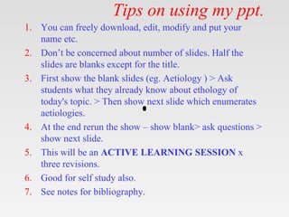
This document provides tips and instructions for using a PowerPoint presentation on penile anatomy and pathology. It recommends showing blank slides to elicit student responses before providing content. Repeating this process of "active learning" three times will reinforce the material. The presentation covers learning objectives, anatomy, embryology, vascular supply, innervation, and pathology. It aims to be useful for both classroom learning and self-study.
This article covers the anatomy of the inguinal canal, including contents, borders,the spermatic cord,the ilioinguinal nerve and related clinical aspects, such as hernias
This is an oblique intermuscular passage in the lower part of the anterior abdominal wall ,
Situated just above the medial half of the inguinal ligament
Anatomy of urinary bladder. surfaces, border of urinary bladder its relation , ligament support, peritoneal relation in male and females, pouches, blood supply of bladder, nerve supply of bladder, true and false ligament of urinary bladder,
Most species have two sexes: male and female. Each sex has its own unique reproductive system. They are different in shape and structure, but both are specifically designed to produce, nourish, and transport either the egg or sperm.
Unlike the female, whose sex organs are located entirely within the pelvis, the male has reproductive organs, or genitals, that are both inside and outside the pelvis. The male genitals include:
the testicles
the duct system, which is made up of the epididymis and the vas deferens
the accessory glands, which include the seminal vesicles and prostate gland
the penis
Title: Sense of Smell
Presenter: Dr. Faiza, Assistant Professor of Physiology
Qualifications:
MBBS (Best Graduate, AIMC Lahore)
FCPS Physiology
ICMT, CHPE, DHPE (STMU)
MPH (GC University, Faisalabad)
MBA (Virtual University of Pakistan)
Learning Objectives:
Describe the primary categories of smells and the concept of odor blindness.
Explain the structure and location of the olfactory membrane and mucosa, including the types and roles of cells involved in olfaction.
Describe the pathway and mechanisms of olfactory signal transmission from the olfactory receptors to the brain.
Illustrate the biochemical cascade triggered by odorant binding to olfactory receptors, including the role of G-proteins and second messengers in generating an action potential.
Identify different types of olfactory disorders such as anosmia, hyposmia, hyperosmia, and dysosmia, including their potential causes.
Key Topics:
Olfactory Genes:
3% of the human genome accounts for olfactory genes.
400 genes for odorant receptors.
Olfactory Membrane:
Located in the superior part of the nasal cavity.
Medially: Folds downward along the superior septum.
Laterally: Folds over the superior turbinate and upper surface of the middle turbinate.
Total surface area: 5-10 square centimeters.
Olfactory Mucosa:
Olfactory Cells: Bipolar nerve cells derived from the CNS (100 million), with 4-25 olfactory cilia per cell.
Sustentacular Cells: Produce mucus and maintain ionic and molecular environment.
Basal Cells: Replace worn-out olfactory cells with an average lifespan of 1-2 months.
Bowman’s Gland: Secretes mucus.
Stimulation of Olfactory Cells:
Odorant dissolves in mucus and attaches to receptors on olfactory cilia.
Involves a cascade effect through G-proteins and second messengers, leading to depolarization and action potential generation in the olfactory nerve.
Quality of a Good Odorant:
Small (3-20 Carbon atoms), volatile, water-soluble, and lipid-soluble.
Facilitated by odorant-binding proteins in mucus.
Membrane Potential and Action Potential:
Resting membrane potential: -55mV.
Action potential frequency in the olfactory nerve increases with odorant strength.
Adaptation Towards the Sense of Smell:
Rapid adaptation within the first second, with further slow adaptation.
Psychological adaptation greater than receptor adaptation, involving feedback inhibition from the central nervous system.
Primary Sensations of Smell:
Camphoraceous, Musky, Floral, Pepperminty, Ethereal, Pungent, Putrid.
Odor Detection Threshold:
Examples: Hydrogen sulfide (0.0005 ppm), Methyl-mercaptan (0.002 ppm).
Some toxic substances are odorless at lethal concentrations.
Characteristics of Smell:
Odor blindness for single substances due to lack of appropriate receptor protein.
Behavioral and emotional influences of smell.
Transmission of Olfactory Signals:
From olfactory cells to glomeruli in the olfactory bulb, involving lateral inhibition.
Primitive, less old, and new olfactory systems with different path
New Directions in Targeted Therapeutic Approaches for Older Adults With Mantl...i3 Health
i3 Health is pleased to make the speaker slides from this activity available for use as a non-accredited self-study or teaching resource.
This slide deck presented by Dr. Kami Maddocks, Professor-Clinical in the Division of Hematology and
Associate Division Director for Ambulatory Operations
The Ohio State University Comprehensive Cancer Center, will provide insight into new directions in targeted therapeutic approaches for older adults with mantle cell lymphoma.
STATEMENT OF NEED
Mantle cell lymphoma (MCL) is a rare, aggressive B-cell non-Hodgkin lymphoma (NHL) accounting for 5% to 7% of all lymphomas. Its prognosis ranges from indolent disease that does not require treatment for years to very aggressive disease, which is associated with poor survival (Silkenstedt et al, 2021). Typically, MCL is diagnosed at advanced stage and in older patients who cannot tolerate intensive therapy (NCCN, 2022). Although recent advances have slightly increased remission rates, recurrence and relapse remain very common, leading to a median overall survival between 3 and 6 years (LLS, 2021). Though there are several effective options, progress is still needed towards establishing an accepted frontline approach for MCL (Castellino et al, 2022). Treatment selection and management of MCL are complicated by the heterogeneity of prognosis, advanced age and comorbidities of patients, and lack of an established standard approach for treatment, making it vital that clinicians be familiar with the latest research and advances in this area. In this activity chaired by Michael Wang, MD, Professor in the Department of Lymphoma & Myeloma at MD Anderson Cancer Center, expert faculty will discuss prognostic factors informing treatment, the promising results of recent trials in new therapeutic approaches, and the implications of treatment resistance in therapeutic selection for MCL.
Target Audience
Hematology/oncology fellows, attending faculty, and other health care professionals involved in the treatment of patients with mantle cell lymphoma (MCL).
Learning Objectives
1.) Identify clinical and biological prognostic factors that can guide treatment decision making for older adults with MCL
2.) Evaluate emerging data on targeted therapeutic approaches for treatment-naive and relapsed/refractory MCL and their applicability to older adults
3.) Assess mechanisms of resistance to targeted therapies for MCL and their implications for treatment selection
TEST BANK for Operations Management, 14th Edition by William J. Stevenson, Ve...kevinkariuki227
TEST BANK for Operations Management, 14th Edition by William J. Stevenson, Verified Chapters 1 - 19, Complete Newest Version.pdf
These lecture slides, by Dr Sidra Arshad, offer a quick overview of physiological basis of a normal electrocardiogram.
Learning objectives:
1. Define an electrocardiogram (ECG) and electrocardiography
2. Describe how dipoles generated by the heart produce the waveforms of the ECG
3. Describe the components of a normal electrocardiogram of a typical bipolar leads (limb II)
4. Differentiate between intervals and segments
5. Enlist some common indications for obtaining an ECG
Study Resources:
1. Chapter 11, Guyton and Hall Textbook of Medical Physiology, 14th edition
2. Chapter 9, Human Physiology - From Cells to Systems, Lauralee Sherwood, 9th edition
3. Chapter 29, Ganong’s Review of Medical Physiology, 26th edition
4. Electrocardiogram, StatPearls - https://www.ncbi.nlm.nih.gov/books/NBK549803/
5. ECG in Medical Practice by ABM Abdullah, 4th edition
6. ECG Basics, http://www.nataliescasebook.com/tag/e-c-g-basics
Pulmonary Thromboembolism - etilogy, types, medical- Surgical and nursing man...VarunMahajani
Disruption of blood supply to lung alveoli due to blockage of one or more pulmonary blood vessels is called as Pulmonary thromboembolism. In this presentation we will discuss its causes, types and its management in depth.
Ozempic: Preoperative Management of Patients on GLP-1 Receptor Agonists Saeid Safari
Preoperative Management of Patients on GLP-1 Receptor Agonists like Ozempic and Semiglutide
ASA GUIDELINE
NYSORA Guideline
2 Case Reports of Gastric Ultrasound
Explore natural remedies for syphilis treatment in Singapore. Discover alternative therapies, herbal remedies, and lifestyle changes that may complement conventional treatments. Learn about holistic approaches to managing syphilis symptoms and supporting overall health.
ARTIFICIAL INTELLIGENCE IN HEALTHCARE.pdfAnujkumaranit
Artificial intelligence (AI) refers to the simulation of human intelligence processes by machines, especially computer systems. It encompasses tasks such as learning, reasoning, problem-solving, perception, and language understanding. AI technologies are revolutionizing various fields, from healthcare to finance, by enabling machines to perform tasks that typically require human intelligence.
HOT NEW PRODUCT! BIG SALES FAST SHIPPING NOW FROM CHINA!! EU KU DB BK substit...GL Anaacs
Contact us if you are interested:
Email / Skype : kefaya1771@gmail.com
Threema: PXHY5PDH
New BATCH Ku !!! MUCH IN DEMAND FAST SALE EVERY BATCH HAPPY GOOD EFFECT BIG BATCH !
Contact me on Threema or skype to start big business!!
Hot-sale products:
NEW HOT EUTYLONE WHITE CRYSTAL!!
5cl-adba precursor (semi finished )
5cl-adba raw materials
ADBB precursor (semi finished )
ADBB raw materials
APVP powder
5fadb/4f-adb
Jwh018 / Jwh210
Eutylone crystal
Protonitazene (hydrochloride) CAS: 119276-01-6
Flubrotizolam CAS: 57801-95-3
Metonitazene CAS: 14680-51-4
Payment terms: Western Union,MoneyGram,Bitcoin or USDT.
Deliver Time: Usually 7-15days
Shipping method: FedEx, TNT, DHL,UPS etc.Our deliveries are 100% safe, fast, reliable and discreet.
Samples will be sent for your evaluation!If you are interested in, please contact me, let's talk details.
We specializes in exporting high quality Research chemical, medical intermediate, Pharmaceutical chemicals and so on. Products are exported to USA, Canada, France, Korea, Japan,Russia, Southeast Asia and other countries.
The prostate is an exocrine gland of the male mammalian reproductive system
It is a walnut-sized gland that forms part of the male reproductive system and is located in front of the rectum and just below the urinary bladder
Function is to store and secrete a clear, slightly alkaline fluid that constitutes 10-30% of the volume of the seminal fluid that along with the spermatozoa, constitutes semen
A healthy human prostate measures (4cm-vertical, by 3cm-horizontal, 2cm ant-post ).
It surrounds the urethra just below the urinary bladder. It has anterior, median, posterior and two lateral lobes
It’s work is regulated by androgens which are responsible for male sex characteristics
Generalised disease of the prostate due to hormonal derangement which leads to non malignant enlargement of the gland (increase in the number of epithelial cells and stromal tissue)to cause compression of the urethra leading to symptoms (LUTS
Ethanol (CH3CH2OH), or beverage alcohol, is a two-carbon alcohol
that is rapidly distributed in the body and brain. Ethanol alters many
neurochemical systems and has rewarding and addictive properties. It
is the oldest recreational drug and likely contributes to more morbidity,
mortality, and public health costs than all illicit drugs combined. The
5th edition of the Diagnostic and Statistical Manual of Mental Disorders
(DSM-5) integrates alcohol abuse and alcohol dependence into a single
disorder called alcohol use disorder (AUD), with mild, moderate,
and severe subclassifications (American Psychiatric Association, 2013).
In the DSM-5, all types of substance abuse and dependence have been
combined into a single substance use disorder (SUD) on a continuum
from mild to severe. A diagnosis of AUD requires that at least two of
the 11 DSM-5 behaviors be present within a 12-month period (mild
AUD: 2–3 criteria; moderate AUD: 4–5 criteria; severe AUD: 6–11 criteria).
The four main behavioral effects of AUD are impaired control over
drinking, negative social consequences, risky use, and altered physiological
effects (tolerance, withdrawal). This chapter presents an overview
of the prevalence and harmful consequences of AUD in the U.S.,
the systemic nature of the disease, neurocircuitry and stages of AUD,
comorbidities, fetal alcohol spectrum disorders, genetic risk factors, and
pharmacotherapies for AUD.
1. Tips on using my ppt.
1. You can freely download, edit, modify and put your
name etc.
2. Don’t be concerned about number of slides. Half the
slides are blanks except for the title.
3. First show the blank slides (eg. Aetiology ) > Ask
students what they already know about ethology of
today's topic. > Then show next slide which enumerates
aetiologies.
4. At the end rerun the show – show blank> ask questions >
show next slide.
5. This will be an ACTIVE LEARNING SESSION x
three revisions.
6. Good for self study also.
7. See notes for bibliography.
5. Introduction & History.
• The penile shaft is composed of 3 erectile
columns
– 2 corpora cavernosa
– corpus spongiosum,
• enveloping fascial layers, nerves,
lymphatics, and blood vessels, all covered
by skin
• The 2 suspensory ligaments, composed of
primarily elastic fibers, support the penis at
its base
9. Embryology
• THE MALE AND FEMALE EXTERNAL
GENITALIA are similar until week 9.
• development and differentiation are evident
beginning at month 3 and are related to the
action of androgens produced by the testes
• THE GENITAL TUBERCLE, initially seen
in week 4, elongates by week 7 to form
a phallus, which in turn will form the future
penis.
10. Embryology
• THE GENITAL FOLDS, which
circumscribe the median urethral
(urogenital) groove, fuse along the ventral
(under) side of the penis, from behind
forward, at about 3 months, changing the
groove into a duct, the definitive penile
urethra
11. Embryology
• DURING MONTH 4, THE EPITHELIUM at the
end of the penis forms 2 invaginations
1. At the tip of the glans, an ectodermal ingrowth
forms a cellular cord, the glandular epithelial
plate. Splitting of this plate forms a groove,
the glandular urethra, Closure of the groove in
the glans moves the urethral opening to the tip of
the glans and joins the 2 urethral parts
2. The second invagination is circular and is called
the preputial epithelial plate. Cleavage of this
plate before birth separates the glans penis from
the prepuce
15. Corpora Cavernosa.
• The paired corpora cavernosa contain erectile
tissue and are each surrounded by the tunica
albuginea, a dense fibrous sheath of connective
tissue
• The tunica albuginea consists of 2 layers, the outer
longitudinal and the inner circular
• The erectile tissue within the corpora contains
arteries, nerves, muscle fibers, and venous sinuses
• The cut surface of the corpora cavernosa looks
like a sponge.
• There is a thin layer of areolar tissue that separates
this tissue from the tunica albuginea.
16. Corpora Cavernosa.
• For their anterior three-fourths the corpora
cavernosa penis lie in intimate apposition with one
another, but behind they diverge in the form of
two tapering processes, known as the crura,
which are firmly connected to the ischial rami. .
• Just before it meets its fellow it presents a slight
enlargement, bulb of the corpus cavernosum
penis
19. Corpus spongiosum
• The single corpus spongiosum lies in the ventral
groove between the 2 corpora cavernosa.
• The urethra passes through the corpus
spongiosum.
• The corpus spongiosum possesses a much thinner
and more elastic tunica albuginea
• Behind, it is expanded to form the urethral bulb,
and lies in apposition with the inferior fascia of
the urogenital diaphragm, from which it receives a
fibrous investment.
20. Corpus spongiosum
• The distal extension of the spongiosum, the glans
penis, covers the tips of the corpora cavernosa to
provide a cushioning effect.
• The urethral meatus is positioned just slightly on
the ventral surface of the glans and is slitlike.
• The edge of the glans overhangs the shaft of the
penis, forming a rim called the corona.
22. Fascias.
The 3 erectile bodies are surrounded by -
1. Deep penile (Buck) fascia
2. Dartos fascia
3. Penile skin.
• On the dorsal aspect of the corpora
cavernosa lie the deep dorsal vein and
paired dorsal arteries and branches of the
dorsal nerves
23. Deep penile (Buck) fascia
• A strong, deep, fascial layer that is immediately
superficial to the tunica albuginea.
• It is continuous with the deep fascia of the
muscles covering the crura
• It splits to surround the corpus spongiosum,
• Extends into the perineum as the deep fascia of the
ischiocavernosus and bulbospongiosus muscles.
• Encloses these muscles and each crus of the
corpora cavernosa and the bulb of the corpus
spongiosum, adhering these structures to the
pubis, ischium, and the urogenital diaphragm.
24. Dartos fascia,
• The subcutaneous connective tissue of the
penis and scrotum has abundant smooth
muscle and is called the dartos fascia
• It continues into the perineum and fuses
with the superficial perineal (Colle) fascia.
• In the penis, the dartos fascia is loosely
attached to the skin and deep penile (Buck)
fascia
• It contains the superficial arteries, veins,
and nerves of the penis.
26. Arterial Supply
• Blood supply to the skin of the penis is from the
left and right superficial external pudendal
arteries, which arise from the femoral artery.
• The blood supply to the ventral penile skin is
based on the posterior scrotal artery, a superficial
branch of the deep internal pudendal artery.
27. Arterial Supply
• The blood supply to deep structures of the penis is
derived from a continuation of the internal
pudendal artery-
1. The artery of the bulb (bulbourethral artery) passes
through the deep penile (Buck) fascia to enter and supply
the bulb of the penis and penile (spongy) urethra.
2. The dorsal artery travels along the dorsum of the penis
between the dorsal nerve and deep dorsal vein and gives
off circumflex branches that accompany the circumflex
veins; the terminal branches are in the glans penis
3. The deep penile (cavernosal) artery is usually a single
artery that arises on each side and enters the corpus
cavernosum at the crus and runs the length of the penile
shaft, giving off the helicine arteries, which are an
integral component of the erectile process.
32. Venous Drainage: Superficial system
• Superficial veins are contained in the dartos
fascia on the dorsolateral surface of the
penis and coalesce at the base to form a
single superficial dorsal vein, which usually
drains into the great saphenous veins via the
superficial external pudendal veins.
33. Venous Drainage:Intermediate system
• The intermediate system contains the deep
dorsal and the circumflex veins, lying
within and beneath the deep penile (Buck)
fascia.
• The deep dorsal vein lies in the midline
groove between the 2 corpora cavernosa
• It receives blood from the emissary and
circumflex veins
• Drains into the prostatic plexus.
34. Venous Drainage:Deep system
Deep venous drainage is via the crural and
cavernosal veins-
1. The crural veins arise in the midline, in the space
between the crura.
2. The cavernosal veins are consolidations of the
emissary veins, which join to form a large venous
channel
• Drains into the internal pudendal vein.
• Three or 4 small cavernosal veins course laterally
between the corpus spongiosum and the crus of
the penis for 2-3 cm before draining into the
internal pudendal veins.
38. Nerve Supply
1. Pudendal nerves supply somatic motor and
sensory innervation
2. The cavernous nerves are a combination of
parasympathetic and visceral afferent
fibers and provide the nerve supply to the
erectile tissue.
40. Microscopic Anatomy
• The corpora cavernosa are 2 spongy
cylinders. Within the tunica albuginea are
the interconnected sinusoids separated by
smooth muscle trabeculae and surrounded
by elastic fibers, collagen, and loose areolar
tissue.
• The structure of the corpus spongiosum is
similar to that of the corpora cavernosa,
except that the sinusoids are larger and a
much thinner outer layer of the tunica
albuginea is present.
• The glans has no tunical covering.
41. Applied Anatomy
Erectile tissue vessels-
• The helicine arteries, branches of the deep penile
artery, supply the trabecular tissue and sinusoids.
They are contracted and tortuous in the flaccid
state and dilated and straight in the erect state
• The venous drainage from the erectile tissue
originates in the venules starting at the peripheral
sinusoids beneath the tunica albuginea. They
travel in the trabeculae between the tunica and the
peripheral sinusoids, forming the subtunical
venular plexus before exiting as the emissary
veins.
43. Physiology
• Nitric oxide (NO) appears to be the principal
neurotransmitter causing penile erection.
• Nonadrenergic, noncholinergic (NANC) neurons
release NO.
• The release of NO increases the production of
cyclic guanosine monophosphate (cGMP), which
relaxes cavernosal smooth muscle.
• Other neurotransmitters, including vasoactive
intestinal peptide (VIP), calcitonin gene-related
peptide (CGRP), prostaglandins, and other
peptides, may also be involved in the erectile
process.
44. Physiology
1. With relaxation of the smooth muscles in the trabeculae
and the arterial wall, Arterial inflow increases as a result
of dilatation of the arterioles and arteries.
2. The sinusoids within the corpora cavernosa distend with
blood
3. Subtunical venular plexuses are compressed between the
tunica albuginea and the distended sinusoids, leading to
decreased venous outflow.
4. The tunica albuginea is stretched to its capacity,
compressing emissary veins and thus further decreasing
venous outflow; as a result, intracavernous pressure
increases and is further increased by contraction of the
ischiocavernous and bulbospongiosus muscles, resulting
in full rigidity
45. Physiology
• The function of the corpus spongiosum in
erection is to prevent the urethra from
pinching closed, thereby maintaining the
urethra as a viable channel for ejaculation.
To do this, the corpus spongiosum remains
pliable during erection while the corpora
cavernosa penis become engorged with
blood.
47. Congenital Anomalies
• Hypospadias
• Epispadias
• Penile agenesis
• Penile duplication
• Microphallus
• Penile torsion
• Webbed penis
• Buried penis
• Absence of the corpora cavernosa and corpora
cavernosa plus corpus spongiosum
• Curvature of the penis
48. Get this ppt in mobile
1. Download Microsoft
PowerPoint from play
store.
2. Open Google assistant
3. Open Google lens.
4. Scan qr code from
next slide.