This document provides an overview of the male reproductive system including descriptions of its main organs and their functions. It discusses the testes, scrotum, seminal vesicles, prostate gland, Cowper's gland, epididymis, vas deferens, spermatic cord, urethra, and penis. It also describes the blood supply to each of the male reproductive organs.
The male reproductive system includes internal structures like the testes, epididymis, ductus deferens, seminal vesicles, prostate, and penis. The testes are held in the scrotum and produce sperm. Sperm travel through the epididymis, ductus deferens, and ejaculatory duct before mixing with fluids from the seminal vesicles and prostate to form semen, which is ejaculated through the urethra in the penis during orgasm. Blood flows into the structures via internal iliac arteries and drains via internal iliac veins. Lymphatic drainage is to internal and external iliac lymph nodes.
The document provides details about the female genital system, including the internal organs like the uterus, vagina, fallopian tubes, and ovaries. It describes the structure of the uterus including its layers, parts, positions, blood supply, and how it enlarges during pregnancy. It also discusses the structure of the vagina, its interior regions, relations to surrounding organs, blood supply, and supports. The summary focuses on the key internal female reproductive organs and some of their key anatomical features.
The male reproductive system includes the testes, duct system, and accessory sex glands. The testes produce sperm and testosterone. Sperm mature as they move through the duct system, which includes the epididymis and vas deferens. The ductus deferens joins the seminal vesicles and prostate to form the ejaculatory ducts, which empty into the urethra. During ejaculation, the seminal vesicles and prostate gland secrete fluids that combine with sperm to form semen. The scrotum and penis are supporting structures that regulate temperature and allow delivery of semen during intercourse.
The male reproductive system functions to produce, store, nourish, and transport gametes and facilitate fertilization. It includes the testes, ducts, accessory glands, and external genitalia. The testes produce sperm through spermatogenesis in the seminiferous tubules. Mature sperm are stored and mature in the epididymis before passing through the vas deferens, seminal vesicles, prostate gland, and urethra to be ejaculated. Hormones regulate sperm production and male sexual characteristics. Common diseases include hydrocele, benign prostatic hypertrophy, prostate cancer, and erectile dysfunction.
The document summarizes the key components of the male reproductive system. It describes the testes, which produce sperm and hormones; the duct system, which transports sperm; and accessory sex glands, which secrete fluids that compose semen. It also explains sperm development through spermatogenesis, and the hormonal regulation of sperm production and male characteristics by hormones like testosterone and follicle-stimulating hormone.
The document describes the major internal and external female reproductive organs and their functions. The internal organs include the ovaries, fallopian tubes, uterus, vagina and cervix. The ovaries produce eggs and hormones, the fallopian tubes carry eggs to the uterus, the uterus supports pregnancy and childbirth, and the vagina and cervix are involved in sexual intercourse, menstruation and birth. The external organs include the vulva, clitoris, labia, Bartholin's glands and breasts. They enable sperm entry, protect the internal organs and in the case of breasts, enable lactation after childbirth.
The document summarizes the normal menstrual cycle, which typically occurs over 4 phases in a 28 day cycle. It is regulated by the hypothalamus, pituitary gland, and ovaries. The follicular phase begins with menstruation and involves follicle development and estrogen rise. Ovulation occurs around day 14 when an egg is released. In the luteal phase, the corpus luteum forms and secretes progesterone to thicken the uterine lining. If implantation does not occur, progesterone drops and menstruation begins, restarting the cycle.
The document provides information about the female reproductive system. It discusses the external female genitalia (vulva) and internal organs including the vagina, cervix, uterus, fallopian tubes, and ovaries. It describes the structure, function, blood supply, and hormones involved in each part. It also explains menstruation and the four phases of the menstrual cycle.
Fertilization is the process where a sperm unites with an ovum. It typically occurs in the ampullary part of the uterine tube. The sperm undergoes changes including capacitation, the acrosomal reaction, and fusion of the nuclei. This results in the formation of a zygote with a full diploid chromosome number. The zygote then undergoes cleavage as it is transported through the uterine tube, dividing into a 2-cell, 4-cell, 8-cell stage and so on over 3 days as it forms a morula.
The document discusses the female menstrual cycle. It begins at menarche around age 12-15 and ends at menopause around age 45-50. The typical cycle is 28 days and consists of two phases - the follicular phase where an ovarian follicle matures and is released during ovulation on day 14, and the luteal phase where the corpus luteum forms. Hormonal changes in the ovaries, uterus, vagina and cervix occur under the influence of hormones like estrogen and progesterone. The cycle regulates female reproductive functions and prepares the body for potential pregnancy each month.
Anatomy & Physiology of the Male Reproductive SystemDr. Sarita Sharma
This document provides an overview of the male reproductive system. It describes both the external structures, which include the penis and scrotum, and the internal structures, which are the testes, epididymis, vas deferens, and accessory sex glands including the seminal vesicles, prostate gland, and bulbourethral glands. The testes produce both sperm and the hormone testosterone. Sperm mature as they pass through the epididymis and are stored in the vas deferens until ejaculation. The accessory glands secrete fluids that nourish the sperm and form the bulk of the semen.
Adolescence begins with puberty between ages 9-14 when a burst of hormones activates testes maturation. The testes, located in the scrotum, produce sperm through spermatogenesis and male sex hormones. Sperm travel through the epididymis, vas deferens, and urethra, mixing with secretions from accessory glands to form semen, which is ejaculated through the penis.
he reproductive system is a collection of internal and external organs — in both males and females — that work together for the purpose of procreating, according to the Cleveland Clinic. Due to its vital role in the survival of the species, many scientists argue that the reproductive system is among the most important systems in the entire body.
The document provides an overview of the male and female reproductive systems. It describes the key external and internal organs of each system, including their structure, functions, and roles in reproduction. The female system includes the vulva, vagina, uterus, fallopian tubes, ovaries and breasts. The male system includes the scrotum, testes, seminal vesicles, ejaculatory ducts, prostate gland, and penis. Both systems work together through processes like fertilization, gestation and childbirth to enable sexual reproduction and the development of offspring.
Overview of reproductive physiology and anatomymukemilawol
The male reproductive system produces sperm and sex hormones. Sperm are produced in the testes through spermatogenesis and stored in the epididymis. Hormones like FSH, LH and testosterone regulate sperm production and male characteristics. During sexual activity, erection occurs when blood flow increases to the penis. Ejaculation involves the contraction of muscles to expel semen containing sperm from the penis.
Anatomy and Physiology of the Male and Female Reproductive SystemSusanette V. Benitez
This document provides an overview of the male and female reproductive systems. It describes the external and internal organs of both systems in detail. For the male, it outlines the penis, scrotum, testes, epididymis, vas deferens, seminal vesicles, prostate and other internal organs. For the female, it describes the vulva including labia, clitoris, and internal organs like the vagina, uterus, fallopian tubes and ovaries. It provides the functions of each organ and their roles in reproduction and sexual response.
The document describes and lists the key external female genital parts, including the mons pubis, labia majora, labia minora, clitoris, urethra, Bartholin's glands, and fourchette. It provides details on the location and function of each part, such as the mons pubis protecting the pubic bone, the labia majora and minora surrounding the vaginal opening, the clitoris being the primary source of sexual pleasure, and Bartholin's glands secreting fluid during arousal. The document also briefly mentions the blood supply and lymphatic drainage of the external genitalia.
DEFINITION OR MEANING OF MENSTRUAL (REPRODUCTIVE) CYCLE:-
Menstruation (Greek word, men-month) is monthly uterine bleeding out flowing through vagina into vulva for 4-5 days every 28 days (24-35 days)during reproductive life of a woman from menarche to menopause.
The Menstrual cycle of 28 days starts on day of onset of menstruation and ends at day 28 on start of next mens.
The cycle consists of a series of changes taking place concurrently in the ovaries and uterine lining, stimulated by changes in blood concentration of hormones.
The female reproductive system has internal and external genitalia. The internal genitalia include the ovaries, which produce eggs and hormones, and the uterine tubes, uterus, and vagina, which transport eggs and a fetus. The ovaries and uterus have three layers - outer, middle, and inner - and undergo changes controlled by hormones to release eggs and support pregnancy. The external genitalia include the vulva and sex organs that receive sperm and allow childbirth.
The document provides information about male and female reproductive physiology:
I. It describes the process of spermatogenesis that occurs in the seminiferous tubules of the testes to produce sperm over 65-75 days.
II. It explains the hormonal regulation of the male reproductive system including the roles of FSH, LH, and testosterone.
III. It outlines the female reproductive cycle over approximately 28 days, including the ovarian cycle, uterine cycle, and hormonal changes (FSH, LH, estrogens, progesterone) that regulate the growth and shedding of the endometrium.
The document provides information about the human reproductive system. It discusses that reproduction allows organisms to produce offspring similar to themselves. The male and female reproductive systems both contribute gametes - sperm and eggs respectively - which can unite during fertilization. The male system produces sperm in the testes and transports it, while the female system produces eggs and supports gestation and birth. The parts of each system are described in detail, including their functions in fertilization and development.
The document discusses the male reproductive system, including its organs and functions. It identifies the testes, epididymis, vas deferens, seminal vesicles, prostate gland, and bulbourethral glands as the internal organs that produce, store, and transport sperm. It describes sperm production through spermatogenesis in the seminiferous tubules and storage in the epididymis, and it traces the pathway of sperm from the testes through the vas deferens and urethra during ejaculation.
This PPT covers Anatomy and Physiology of Female reproductive system. Anatomy of female reproductive organs, oogenesis, hormonal regulation of ovaries and Female reproductive cycle (Mentrual cycle) are explained.
The Male and Female Reproductive System and it's functionsERICJOHNMERIALES
The male reproductive system produces and transports sperm and reproductive hormones. It includes internal organs like the testes which produce sperm and testosterone, and external organs like the penis and scrotum. The female reproductive system produces eggs and reproductive hormones and can carry offspring to term. It includes internal organs like the ovaries which produce eggs and the uterus which houses the developing fetus, and external organs like the labia and clitoris. Both systems work together through sexual intercourse and conception to reproduce new individuals through asexual and sexual reproduction.
The female reproductive system is designed to carry out several functions. It produces the female egg cells necessary for reproduction, called the ova or oocytes. Conception, the fertilization of an egg by a sperm, normally occurs in the fallopian tubes
Oogenesis is the process by which ova or egg cells are formed in the ovaries. It involves three main phases: 1) the multiplication phase where primordial germ cells undergo mitosis to form oogonia, 2) the growth phase where the oocytes increase in size through protein and RNA synthesis, and 3) the maturation phase where the oocytes undergo the first meiotic division to form secondary oocytes, followed by ovulation and the second meiotic division after fertilization to form the ovum. Key events include cytoplasmic growth through organelle proliferation and yolk deposition, formation of the follicle surrounding the oocyte, and reduction of the chromosome number through meiosis.
The female reproductive system comprises primary sex organs like the ovaries and accessory sex organs like the fallopian tubes, uterus, cervix, and vagina. The ovaries produce eggs and female sex hormones, while the accessory organs support reproduction. The uterus has three layers and is divided into the body and cervix. It is located between the bladder and rectum. The vagina is lined with mucous membrane and connects the cervix to the outside. The ovaries contain follicles that produce eggs and hormones to regulate the menstrual cycle.
The male reproductive system has the sole function of producing sperm and delivering it to females. The testes produce sperm through spermatogenesis within seminiferous tubules. Mature sperm travel through the epididymis, vas deferens, seminal vesicles, prostate gland, and urethra, where they mix with fluids before being ejaculated via the penis. The process takes about 74 days and requires a temperature slightly below normal body heat.
Sperm cells travel from the testis through the vas deferens, seminal vesicles, urethra and penis. Upon ejaculation, sperm enter the female reproductive tract through the vagina and cervix on their way to fertilizing an egg in the ovary. The male reproductive structures like the testis, vas deferens and seminal vesicles and the female structures like the ovaries, uterus, cervix and vagina all work together in the process of fertilization and reproduction.
This document discusses pigments, binders, and paint composition. It provides examples of commonly used pigments like Egyptian blue, azurite, ultramarine, malachite, cinnabar, vermilion, burnt sienna, lead white, vine black, Prussian blue, and discusses their origins and uses. It describes binders like beeswax, linseed oil, walnut oil, plaster, gum arabic, egg yolk and how they suspend pigments and bind them to surfaces. It explains the functions of pigments in providing color, hiding substrates, improving strength and adhesion. It also discusses the functions of binders in film formation and properties like adhesion, resistance and application qualities.
The document discusses the production of enamel and its functions. It defines enamel as a colored glassy substance fused to metal, glass, etc. for ornamentation or protection. It then discusses the history of enamel, the raw materials used which include silica, clay and micas, and the manufacturing process which involves mixing materials and firing them in a furnace at high temperatures. Finally, it outlines the functions of enamel, including use in construction, cookware, ceramics, jewelry, and other applications where its durable glass-like surface provides protection and decoration.
There are three main methods for preparing compressed tablets: direct compression, dry granulation, and wet granulation. Direct compression can be used for drugs that have good compaction properties, while dry and wet granulation are used when direct compression is not suitable. Dry granulation involves compressing powders into slugs then milling and mixing them before final compression. Wet granulation involves mixing powders with a liquid to form granules, then drying and mixing the granules with other ingredients before final compression. Wet granulation has benefits like improved flow and uniformity but is more complex and expensive than dry granulation. Tablets are evaluated based on characteristics like size, weight, thickness, hardness, friability, drug content, and
This presentation introduces ion exchange resins and their properties. It discusses the chemistry of ion exchangers, including their composition and characteristics. The presentation examines key properties of ion exchangers like ionic form, ion exchange capacity, moisture holding capacity, specific gravity, bulk density, volume change, particle size distribution, stability, and selectivity. It also discusses how the properties depend on whether the ion exchangers are used in aqueous or non-aqueous media.
Different Types of Insulin, Presented by Homeland HealthCareHomeland HealthCare
There are different types of insulin that serve different purposes for managing blood sugar levels. Rapid-acting insulin works within 15 minutes and is well-suited for pre- or post-meal use, while short-acting insulin takes 30-60 minutes and controls blood sugar during meals. Intermediate-acting insulin lasts 12 hours and can be taken before bed, and long-lasting insulin takes effect within an hour but works for up to 26 hours. People should consult their physician to determine the best insulin type for their needs.
Sublimation is a purification process where a solid transitions directly to its gas phase without passing through a liquid phase. It involves precise control of individual purification steps under vacuum or gas flow conditions. Sublimation is commonly used to purify pharmaceuticals and can also be used to create freeze-dried substances or porous drug tablets using a sublimating material like camphor. Another application is sublimating dye onto fabrics to create designs without affecting the fabric's feel or performance.
The document discusses the benefits of exercise for mental health. Regular physical activity can help reduce anxiety and depression and improve mood and cognitive functioning. Exercise causes chemical changes in the brain that may help protect against mental illness and improve symptoms.
The male reproductive system produces sperm and hormones. The testes contain seminiferous tubules with Sertoli cells that support sperm production. Under the influence of FSH, Sertoli cells nurture germ cells as they develop into sperm through spermatogenesis. Leydig cells in the testes, stimulated by LH, secrete testosterone which supports sperm production and male characteristics. A hormonal feedback loop between the hypothalamus, pituitary gland, and testes regulates hormone production and sperm development.
This document provides a summary of various classes of antimicrobial drugs used to treat bacterial, viral, fungal and parasitic infections. It lists the major drug groups for treating bacterial infections like penicillins, cephalosporins, carbapenems, quinolones and others. It also summarizes antiviral drugs for HIV, hepatitis, influenza and other viral infections. The antifungal drug classes of polyenes, triazoles and others are outlined. Finally, it lists the main drug types used to treat protozoal diseases and helminthic infections.
The male reproductive system produces sperm and testosterone. The testes produce sperm in the seminiferous tubules and testosterone with the interstitial cells. Sperm mature in the epididymis and are stored in the vas deferens. During ejaculation, sperm are mixed with fluids from the seminal vesicles, prostate, and Cowper's glands and travel through the urethra. Testosterone controls male secondary sex characteristics and supports sperm production.
Biopharmaceuticals are pharmaceuticals produced through biotechnology methods using living organisms or their components. They include protein or nucleic acid based drugs. Biotechnology allows for large-scale production of substances like insulin for diabetes treatment. It also enables targeted treatments for specific diseases and patient groups. Some key biopharmaceuticals are insulin, interferons, blood clotting factors, and growth factors. However, proteins face stability issues due to their large size and structure. Various formulation and delivery methods can address these challenges, including addition of stabilizing agents, PEGylation, micelles, and permeabilizers to aid delivery.
1. The document discusses several hormones involved in regulating blood glucose levels, including insulin, glucagon, somatostatin, and pancreatic polypeptide.
2. Insulin decreases blood glucose levels, while glucagon and other hormones like growth hormone and cortisol increase blood glucose.
3. The liver plays an important role in maintaining blood glucose homeostasis by storing glucose as glycogen and releasing it as needed.
The document describes three main methods for manufacturing tablets: wet granulation, dry granulation, and direct compression. Wet granulation involves using a liquid such as water to form granules, then drying the granules. It produces tablets with good mechanical properties but requires multiple steps. Dry granulation uses compaction without liquid to form granules. Direct compression compresses powder directly without a granulation step, making it faster but requiring special excipients. Each method has advantages and disadvantages related to processing steps, equipment needs, stability concerns, and tablet properties.
This document provides information about pharmaceutical excipients. It defines excipients as pharmacologically inactive substances formulated alongside active pharmaceutical ingredients. Excipients play a variety of functional roles including providing bulk, aiding manufacturing, improving dosing compliance, and modulating solubility and bioavailability. Common excipients include diluents, binders, disintegrants, lubricants, and flavors. Ideal excipients are inert, nontoxic, stable, soluble, compatible with active ingredients, and pharmacologically inert. The document also discusses innovation in excipients including modified, co-processed, and novel excipients and how excipients can have different functional roles depending on the formulation.
This document discusses newer insulin preparations that have been developed through genetic engineering to better mimic the body's natural insulin secretion patterns. It introduces several newer rapid-acting and long-acting insulin analogs such as insulin lispro, insulin aspart, insulin glargine, and insulin detemir. These analogs were designed to have faster onset of action, shorter duration, or longer duration compared to older insulin preparations. The document also briefly discusses inhaled insulin and newer advances in insulin delivery technologies.
This document provides information about tablets, including their definition, advantages, disadvantages and types. It discusses the main components of tablets, including active ingredients and excipients. It describes different types of tablets based on their route of administration and production process. The purposes and examples of various excipients like diluents, binders, disintegrants, lubricants and coloring agents are outlined. Granulation is introduced as a process to prevent segregation and improve flow of powder mixtures that are then compressed into tablets.
Tablet manufacturing process created by Asadulla MullaAsad Mulla
The document summarizes the key steps involved in manufacturing oral solid dosage forms like tablets. It involves size reduction of raw materials, blending, granulation, drying, lubrication, compression into tablets, coating if needed, and final packaging. Precise control of critical process parameters is required at each stage to ensure quality, bioavailability and stability of the final tablets. Granulation is an important step for uniform distribution of drugs in the blend and influences tableting process. Drying is also critical to control residual moisture. Compression forms the tablets and critical parameters like hardness and disintegration time need to be monitored.
The male reproductive system consists of organs that produce and transfer mature sperm. The testes produce sperm which travel through the vas deferens, seminal vesicles and prostate gland. These organs combine their secretions to form semen, which contains sperm. During sexual arousal, erectile tissue in the penis fills with blood, causing an erection to allow delivery of semen into the female reproductive tract during intercourse.
The document provides information about the male reproductive system. It describes the penis, scrotum, testes, epididymis, vas deferens, seminal vesicles, prostate gland, bulbourethral glands and other structures. It discusses the layers, blood supply, functions and some medical issues related to these organs.
The document provides information about the male reproductive system. It describes the penis, scrotum, testes, epididymis, vas deferens, seminal vesicles, prostate gland, bulbourethral glands and other structures. It discusses the layers, blood supply, functions and other details about these organs. Medical issues related to some structures like the urethra are also mentioned.
Join live classes, download study aids, sell your documents, join or host your own classes online, get tutoring, tutor students, take practices tests and more at Examville.com
The male reproductive system produces sperm and reproductive hormones. During development, the testes descend from the abdomen into the scrotum. At puberty, the testes and sex organs mature under hormonal changes. The testes produce sperm through spermatogenesis and store them in the epididymis. During ejaculation, sperm and secretions from the seminal vesicles, prostate, and bulbourethral glands are expelled through the urethra. The male sex hormones support secondary sex characteristics and sperm production. Sperm travel through the duct system and are ejaculated in semen to potentially fertilize an egg.
The document summarizes the male reproductive system. It describes the internal and external male reproductive organs including the testes, duct system, accessory sex glands, and penis. The testes produce sperm and testosterone in seminiferous tubules. Sperm travel through the epididymis, ductus deferens, and urethra. Accessory sex glands like the seminal vesicles and prostate gland secrete fluids that nourish and transport sperm. Together these organs work to produce, mature, and ejaculate sperm for fertilization.
The male reproductive system includes the testes, epididymis, penis, scrotum, and urethra. The testes produce sperm and testosterone and are held in the scrotum outside of the body. The epididymis is a coiled structure where sperm mature after leaving the testes. The penis is made up of erectile tissues and is used for both reproduction and urination. During an erection, blood flows into the tissues of the penis. The urethra passes through the penis and is the channel through which both urine and semen exit the body.
13th female and male genital tract.pptxChangezKhan33
This document provides an overview of the female and male genital tracts. It describes the external female genitalia including the labia majora, labia minora, clitoris, vestibular glands, and hymen. It then details the internal female organs - the vagina, uterus, fallopian tubes, and ovaries. For the male genital tract, it outlines the penis, urethra, prostate, vas deferens, seminal vesicles, scrotum, testes, and epididymis. The text provides descriptions of the structure, blood supply, lymphatic drainage and innervation for each organ.
The testes produce sperm and testosterone. They contain seminiferous tubules that produce sperm and Leydig cells that produce testosterone. The epididymis is a coiled tube that stores and transports sperm. The vas deferens carries sperm from the epididymis. The seminal vesicles and prostate gland secrete fluids that nourish and activate sperm. During ejaculation, sperm and fluids pass through the urethra.
The document summarizes the anatomy and functions of the male and female reproductive systems. It describes the organs involved in both systems, including the testes, ovaries, ducts, and external genitalia. It explains sperm and egg production, hormone regulation, and fertilization. Key concepts covered are the structures of the testes, ovaries, uterus, and vagina, as well as spermatogenesis, oogenesis, and the menstrual cycle.
MALE REPRODUCTIVE SYSTEM || HUMAN REPRODUCTION || PART 2 || BIOLOGY || STUD-ESTUDE
This session will mainly be around the male reproductive system- part 2
Chapter: Human Reproduction
Useful For: NCERT / CBSE / CET / NEET
Key Areas of this Session-
1)male reproductive system
2)human reproduction
Watch the full video on the male reproductive system for NEET 2020 Exam and get a clear edge on this topic which is boost up your preparation for NEET Exam 2020.
------------------------------------------------------------------------------------
SUBSCRIBE Now by Clicking on the Subscribe Button ------------------------------------------------------------------------------------
Made and edited by- stud-e
https://www.youtube.com/channel/UCamq...
contact me on- stud.e0397@gmail.com
PREVIOUS VIDEOS-
GENETIC TERMINOLOGY-
https://youtu.be/MBMyi8EjPeQ
MENDEL'S LAW OF DOMINANCE-
https://youtu.be/ObUz2kOwbyA
INCOMPLETE DOMINANCE-
https://youtu.be/oV-U-0bkcq4
FILARIASIS-
https://youtu.be/X2-RA1uMHqs
The male reproductive system includes internal structures like the testes, epididymis, ductus deferens, seminal vesicles, prostate, and penis. The testes are held in the scrotum and produce sperm. Sperm travel through the epididymis, ductus deferens, and ejaculatory duct before mixing with fluids from the seminal vesicles and prostate to form semen, which is ejaculated through the urethra in the penis during orgasm. Blood flows into the structures through arteries and drains out through veins, while nerves provide sensation and control erectile function and ejaculation.
The male reproductive system undergoes significant changes during puberty due to increases in hormones like testosterone. This document describes the structures and functions of the male reproductive system, including the testes, scrotum, penis, and other organs. Sperm are produced in the testes through the process of spermatogenesis, where germ cells undergo meiosis to become mature sperm over approximately 74 days. The sperm then travel through the duct system and are ejaculated mixed with fluids from accessory glands.
The male reproductive system includes the testes, which produce sperm and testosterone. The testes are located in the scrotum outside of the body to regulate temperature for sperm production. During ejaculation, sperm travel from the testes through the vas deferens, seminal vesicles, prostate, and urethra, where semen containing sperm is expelled through the penis. The penis contains erectile tissue that fills with blood to cause an erection, which is needed for sexual intercourse and ejaculation.
Most species have two sexes: male and female. Each sex has its own unique reproductive system. They are different in shape and structure, but both are specifically designed to produce, nourish, and transport either the egg or sperm.
Unlike the female, whose sex organs are located entirely within the pelvis, the male has reproductive organs, or genitals, that are both inside and outside the pelvis. The male genitals include:
the testicles
the duct system, which is made up of the epididymis and the vas deferens
the accessory glands, which include the seminal vesicles and prostate gland
the penis
The male reproductive system produces and transports sperm and reproductive hormones. It includes internal organs like the testes, which produce sperm and testosterone, and the epididymis, seminal vesicles, and prostate gland, which produce fluids that nourish and transport sperm. Externally, it includes the penis, which delivers sperm during intercourse, and the scrotum, which houses the testes and maintains the temperature needed for sperm production. The testes contain seminiferous tubules that produce sperm and Leydig cells that secrete testosterone, both of which are essential for male fertility and sexual function.
The male reproductive organs include the external genitalia of the penis and scrotum, as well as internal structures. The testes are located in the scrotum where temperatures are slightly lower to support sperm production. Within each testis are seminiferous tubules that produce sperm cells. Mature sperm exit the testes and travel through the epididymis where they undergo maturation before passing through the vas deferens. Other structures that contribute to semen production include the seminal vesicles and prostate gland. The penis allows for both urination and sexual intercourse.
The male reproductive system undergoes significant changes during puberty due to increased hormone production. This causes enlargement of the testes and genitals. Sperm are produced in the testes through spermatogenesis, a process where diploid cells undergo meiosis to become haploid sperm. Mature sperm are stored in the epididymis and travel through ducts to be ejaculated through the urethra.
The male reproductive system produces and transports sperm. During puberty, the testes enlarge and begin producing testosterone and sperm through spermatogenesis within the seminiferous tubules. Mature sperm pass through the epididymis, vas deferens, and urethra to be ejaculated with fluids from the seminal vesicles, prostate, and bulbourethral glands. The penis becomes erect through increased blood flow, allowing for sexual intercourse and delivery of sperm.
This presentation includes basic of PCOS their pathology and treatment and also Ayurveda correlation of PCOS and Ayurvedic line of treatment mentioned in classics.
Introduction to AI for Nonprofits with Tapp NetworkTechSoup
Dive into the world of AI! Experts Jon Hill and Tareq Monaur will guide you through AI's role in enhancing nonprofit websites and basic marketing strategies, making it easy to understand and apply.
Physiology and chemistry of skin and pigmentation, hairs, scalp, lips and nail, Cleansing cream, Lotions, Face powders, Face packs, Lipsticks, Bath products, soaps and baby product,
Preparation and standardization of the following : Tonic, Bleaches, Dentifrices and Mouth washes & Tooth Pastes, Cosmetics for Nails.
How to Fix the Import Error in the Odoo 17Celine George
An import error occurs when a program fails to import a module or library, disrupting its execution. In languages like Python, this issue arises when the specified module cannot be found or accessed, hindering the program's functionality. Resolving import errors is crucial for maintaining smooth software operation and uninterrupted development processes.
How to Manage Your Lost Opportunities in Odoo 17 CRMCeline George
Odoo 17 CRM allows us to track why we lose sales opportunities with "Lost Reasons." This helps analyze our sales process and identify areas for improvement. Here's how to configure lost reasons in Odoo 17 CRM
ISO/IEC 27001, ISO/IEC 42001, and GDPR: Best Practices for Implementation and...PECB
Denis is a dynamic and results-driven Chief Information Officer (CIO) with a distinguished career spanning information systems analysis and technical project management. With a proven track record of spearheading the design and delivery of cutting-edge Information Management solutions, he has consistently elevated business operations, streamlined reporting functions, and maximized process efficiency.
Certified as an ISO/IEC 27001: Information Security Management Systems (ISMS) Lead Implementer, Data Protection Officer, and Cyber Risks Analyst, Denis brings a heightened focus on data security, privacy, and cyber resilience to every endeavor.
His expertise extends across a diverse spectrum of reporting, database, and web development applications, underpinned by an exceptional grasp of data storage and virtualization technologies. His proficiency in application testing, database administration, and data cleansing ensures seamless execution of complex projects.
What sets Denis apart is his comprehensive understanding of Business and Systems Analysis technologies, honed through involvement in all phases of the Software Development Lifecycle (SDLC). From meticulous requirements gathering to precise analysis, innovative design, rigorous development, thorough testing, and successful implementation, he has consistently delivered exceptional results.
Throughout his career, he has taken on multifaceted roles, from leading technical project management teams to owning solutions that drive operational excellence. His conscientious and proactive approach is unwavering, whether he is working independently or collaboratively within a team. His ability to connect with colleagues on a personal level underscores his commitment to fostering a harmonious and productive workplace environment.
Date: May 29, 2024
Tags: Information Security, ISO/IEC 27001, ISO/IEC 42001, Artificial Intelligence, GDPR
-------------------------------------------------------------------------------
Find out more about ISO training and certification services
Training: ISO/IEC 27001 Information Security Management System - EN | PECB
ISO/IEC 42001 Artificial Intelligence Management System - EN | PECB
General Data Protection Regulation (GDPR) - Training Courses - EN | PECB
Webinars: https://pecb.com/webinars
Article: https://pecb.com/article
-------------------------------------------------------------------------------
For more information about PECB:
Website: https://pecb.com/
LinkedIn: https://www.linkedin.com/company/pecb/
Facebook: https://www.facebook.com/PECBInternational/
Slideshare: http://www.slideshare.net/PECBCERTIFICATION
it describes the bony anatomy including the femoral head , acetabulum, labrum . also discusses the capsule , ligaments . muscle that act on the hip joint and the range of motion are outlined. factors affecting hip joint stability and weight transmission through the joint are summarized.
Executive Directors Chat Leveraging AI for Diversity, Equity, and InclusionTechSoup
Let’s explore the intersection of technology and equity in the final session of our DEI series. Discover how AI tools, like ChatGPT, can be used to support and enhance your nonprofit's DEI initiatives. Participants will gain insights into practical AI applications and get tips for leveraging technology to advance their DEI goals.
This presentation was provided by Steph Pollock of The American Psychological Association’s Journals Program, and Damita Snow, of The American Society of Civil Engineers (ASCE), for the initial session of NISO's 2024 Training Series "DEIA in the Scholarly Landscape." Session One: 'Setting Expectations: a DEIA Primer,' was held June 6, 2024.
Pollock and Snow "DEIA in the Scholarly Landscape, Session One: Setting Expec...
Male Reproductive System
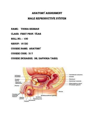
1. Anatomy ASSIGNMENT
MALE REPRODUCTIVE SYSTEM
NAME: TOOBA REHMAN
CLASS: FIRST PROF. YEAR
ROLL NO. : 100
Group: 16 (B)
COURSE NAME: ANATOMY
COURSE CODE: 317
COURSE INCHARGE: dr. safoora tariq
2. INTRODUCTION
REPRODUCTION
The process in which one or two parent organisms form a new individual is called
reproduction and the organs involve in reproduction are collectively called
reproductive system.
MALE REPRODUCTIVE SYSTEM
Genital organs of a male body include in reproductive system are collectively called
male reproductive system. Male reproductive system consist of f0llowing organs
named below.
1) Testes
2) Scrotum
3) Seminal vesical
4) Prostate gland
5) Cowper’s gland
6) Epididymis
7) Vas deferens
8) Spermatic cord
9) Urinogenital duct
10) Penis
3. EXPLANATION
TESTES
(singular=Testis)
Testes are r0ugh egg like in shape and about 3 cm long contain a mass of
semineferous tubules that manufacture the male sperm cell called
Spermatozoa. The testes (TES-te¯z), or testicles, are paired oval glands in
the scrotum measuring about 5 cm long and 2.5 cm in diameter . Each testis
(singular) has a mass of 10–15 grams. The testes develop near the kidneys, in
the posterior portion of the abdomen, and they usually begin their descent
into the scrotum through the inguinal canals (passage-ways in the anterior
abdominal wall;) during the latter half of the seventh month of fetal
development.A serous membrane called the tunica vaginalis (tunica
sheath), which is derived from the peritoneum and forms during
the descent of the testes, partially covers the testes. A collection
of serous fluid in the tunica vaginalis is called a hydrocele.
SCROTUM
The scrotum(SKRO-tum = bag), the supporting structure for the
testes, consists of loose skin and underlying subcutaneous layer that
hangs from the root (attached portion) of the penis .Externally, the scrotum
looks like a single pouch of skin separated into lateral portions by a median
ridge called the raphe (RA—fe= seam). Internally, the scrotal septumdivides
the scro-tum into two sacs, each containing a single testis .The septum is
made up of a subcutaneous layer and muscle tissue called the dartos muscle
(DAR-tos =skinned), which is composed of bundles of smooth muscle fibers.
The dartos muscle is also found in the subcutaneous layer of the
scrotum. Associated with each testis in the scrotum is the
cremaster muscle(kre-MAS-ter = suspender), a series of small
bands of skeletal muscle that descend as an extension of the internal
oblique muscle through the spermatic cord to surround the testes.
4. SEMINAL VESICAL
The paired seminal vesicles(VES-i-kuls) or seminal glandsare convoluted
pouchlike structures, about 5 cm in length, lying posterior to the base of the
urinary bladder and anterior to the rectum. Seminal vesical produces much
of the seminal fluid and lies lateral to the ductus on the posterior wall of
the bladder with its upper end just below the p0int of enterance of ureter
into the bladder. The very short duct leaves the l0wer end to join the
ductus deference at the edge of the prostate gland and form the
ejaculat0ry duct.
PROSTATE GLAND
The prostate (PROS-ta t) is a single, doughnut-shaped gland
about the size of a golf ball. It measures about 4 cm from
side to side, about 3 cm from top to bottom, and about 2 cm
from front to back. It is inferior to the urinary bladder and
surrounds the prostatic urethra . The prostate slowly increases
in size from birth to puberty. It then expands rapidly until about
age 30, after which time its size typically remains stable until about
age 45, when further enlargement may occur. Prostate consist of glands
embedded in a mass of connective tissues and smooth muscle. It secrets
about 30% of seminal fluid.
COWPER’s GLAND
The paired bulbourethral glands (bul-bo -u -RE--thral), or
Cowper’s glands are about the size of peas. They are located
inferior to the prostate on either side of the membranous urethra
within the deep muscles of the perineum, and their ducts open into the
spongy urethra .
5. EPIDIDYMIS
The epididymis(ep-i-DID-i-mis; epi- above or over; -didymis=testis)
is a comma-shaped organ about 4 cm long that lies along the posterior
border of each testis.The plural is epididymides (ep-i-did-IM-i-des).
Each epi-didymis consists mostly of the tightly coiled ductus epididymis.
The efferent ducts from the testis join the ductus epididymis at
the larger, superior portion of the epididymis called the head.
The bodyis the narrow midportion of the epididymis, and the
tail is the smaller, inferior portion. At its distal end, the tail of
the epididymis continues as the ductus (vas) deferens .The ductus
epididymis would measure about 6 m in length if it were uncoiled.
It is lined with pseudostratified columnar epithelium and encircled
by layers of smooth muscle.
The free surfaces of the columnar cells contain stereocilia,
which despite their name are long, branching microvilli (not
cilia) that increase the surface area for the reabsorption of degenerated
sperm. Connective tissue around the muscle layer attaches the loops of the
ductus epididymis and carries blood vessels and nerves.
VAS (DUCTUS) DEFERENS
Within the tail of the epididymis, the ductus epididymis becomes less
convoluted, and its diameter increases. Beyond this point, the duct is
known as the ductus deferens 0R vas deferens.The ductus deferens,
which is about 45 cm long, ascends along the posterior border of the
epididymis through the spermatic cord and then enters the pelvic
cavity. There it loops over the ureter and passes over the side and
down the posterior surface of the urinary bladder. The dilated
terminal portion of the ductus deferens is the ampulla (am-PUL-la
= little jar). The mucosa of the ductus deferens consists of
pseudostratified columnar epithelium and lamina propria (areolar
connective tissue). The muscularis is composed of three layers of
smooth muscle; the inner and outer layers are longitudinal, and the
middle layer is circular.
6. SPERMATIC CORD
The collective name f0r the ductus deferens, the testicular and 0ther
vessels and nerves and various connective tissue c0verings derived from the
abd0minal musculature that f0rm the inguinal canal.
The spermatic cordis a supporting
structure of the male repro-ductive system that ascends out of the
scrotum. It consists of the ductus (vas) deferens as it ascends through
the scrotum, the testicular artery, veins that drain the testes and carry
testosterone into circulation (the pampiniform plexus), autonomic nerves,
lymphatic vessels, and the cremaster muscle. The spermatic cord and
ilioinguinal nerve pass through the inguinal canal (IN-gwin-al =groin), an
oblique passageway in the anterior abdominal wall just superior and
parallel to the medial half of the inguinal ligament. The canal, which is
about 4–5 cm long, originates at the deep (abdominal) inguinal ring,a
slit like opening in the aponeurosis of the transversus ab-dominis muscle; the
canal ends at the superficial (subcutaneous) inguinal ring, a somewhat
triangular opening in the aponeurosis of the external oblique muscle. In
females, the round ligament of the uterus and ilioinguinal nerve
pass through the inguinal canal.
7. URINOGENITAL DUCT
In males, the urethrais the shared terminal duct of the reproductive and
urinary systems; so it is also kn0wn as urinogenital duct.
It serves as a passageway for both semen and urine. About 20 cm long, it
passes through the prostate, the deep muscles of the perineum, and the
penis, and is subdivided into three parts . The prostatic urethrais 2–3 cm
long and passes through the prostate. As this duct continues inferiorly, it
passes through the deep muscles of the perineum, where it is known as the
membranous urethra.The membranous urethra is about 1 cm in length. As
this duct passes through the corpus spongiosum of the penis, it is known as
the spongy (penile) urethra,which is about 15–20 cm long. The spongy urethra
ends at the external urethral orifice.
PENIS
The peniscontains the urethra and is a passageway for the ejaculation of
semen and the excretion of urine . It is cylindrical in shape and consists of a
body, glans penis, and a root. The body of the penisis composed of three
cylindrical masses of tissue, each surrounded by fibrous tissue called the
tunica albuginea . The two dorsolateral masses are called the corpora
cavernosa penis(corpora=main bod-ies; cavernosa =hollow). The smaller
midventral mass, the corpus spongiosum penis, contains the spongy urethra
and keeps it open during ejaculation. Skin and a subcutaneous layer enclose
all three masses, which consist of erectile tissue. Erectile tissue is
composed of numerous blood sinuses (vascular spaces) lined by endothelial
cells and surrounded by smooth muscle and elastic connective tissue.
8. BLOOD SUPPLY TO THE MALE REPRODUCTIVE
ORGANS :FOR TESTES AND EPIDIDYMIS: Internal to the tunica albuginea is the
tunica vasculosa, containing a plexus of blood vessels and connective tissue.
Bilateral testicular arteries originating from the aorta, just inferior to the
renal arteries, provide arterial supply to the testes. The testicular arteries
enter the scrotum in the spermatic cord via the inguinal canal and split into
two branches at the posterosuperior border of the testis. Additionally, the
testes receive blood from the cremasteric branch of the inferior epigastric
artery and the artery to the ductus deferens. The pampiniform plexus
drains both the testis and epididymis before coalescing to form the
testicular vein, usually above the spermatic cord formation at the deep
inguinal ring. The tenth and eleventh thoracic spinal nerves supply the testes
via the renal and aortic autonomic plexuses. Blood supply for epididymis is
same as for testes.
FOR PROSTATE GLAND: The arterial supply to the prostate gland is
derived from the inferior vesical artery and branches of the middle rectal
artery. Venous drainage of the prostate forms the prostatic plexus, which
eventually drains into the internal iliac vein
FOR SEMINAL VESICLES AND COWPER’s GLAND: Arterial blood supply
to the seminal vesicles includes branches from the inferior vesical and
middle rectal arteries, while venous accompanies these arteries. The inferior
division of the hypogastric plexus provides innervation to the seminal
vesicles. Blood supply for cowper’s gland is same as for seminal vesicles.
FOR DUCTUS DEFERENS AND URINOGENITAL DUCT: Each ductus
deferens has an artery usually derived from the superior vesical artery
(artery to the ductus), with venous drainage to the pelvic venous plexus.
Same blood supplying process for urinogenital duct.
9. FOR PENIS: The vasculature of the penis is extensive. The perineal artery (a
branch of the internal pudendal artery) together with the posterior scrotal artery
and the inferior rectal artery supply tissues from the bulb of the penis to the
anus. The artery of the bulb of the penis, from the internal pudendal, penetrates
the penile bulb and subsequently supplies the corpus spongiosum. The deep artery
of the penis is one of two terminal branches of the internal pudendal artery; it
enters the crus of the penis and continues through the length of the bilateral
corpus cavernosum. The other terminal branch of the internal pudendal artery is
the dorsal artery of the penis running along the dorsal surface of the penis
supplying the penile skin and the glans penis.
The venous drainage of the penis includes the veins draining the corpora cavernosa,
which subsequently drains into the circumflex veins. These veins receive venous
blood from the corpus spongiosum on the ventral aspect of the penis and wrap
around the penis to drain into the deep dorsal vein. The superficial dorsal vein
drains the penile skin and prepuce before draining via the superficial external
pudendal vein into the external pudendal veins. The deep dorsal vein further drains
blood from the glans penis and corpora cavernosa before joining the prostatic
venous plexus.