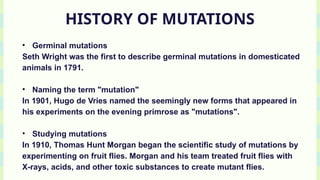
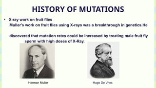
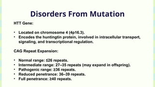
GURU GHASIDAS VISHWAVIDYALAYA
DEPARTMENT OF BIOTECHNOLOGY
INTERNAL ASSESMENT PRESENTATION
( SESSION 2024-25 )
PRESENTED BY :
YUGESHVAR YADAV
M.SC. IN MICROBIOLOGY
FIRST SEMESTER.
GUIDED BY :
DR. ASHISH KUMAR
ASSOCIATE PROFESSOR
DEPARTMENT OF
BIOTECHNOLOGY.
MUTATION IN
PROTEIN
CODING GENES .
HISTORY OF MUTATIONS
• Germinal mutations
Seth Wright was the first to describe germinal mutations in domesticated
animals in 1791.
• Naming the term "mutation"
In 1901, Hugo de Vries named the seemingly new forms that appeared in
his experiments on the evening primrose as "mutations".
• Studying mutations
In 1910, Thomas Hunt Morgan began the scientific study of mutations by
experimenting on fruit flies. Morgan and his team treated fruit flies with
X-rays, acids, and other toxic substances to create mutant flies.
HISTORY OF MUTATIONS
• X-ray work on fruit flies
Muller's work on fruit flies using X-rays was a breakthrough in genetics.He
discovered that mutation rates could be increased by treating male fruit fly
sperm with high doses of X-Ray.
Herman Muller Hugo De Vries
Introduction to Genetic
Mutations
• DEFINITION : A genetic mutation is a change in the DNA sequence of
an organism.
• OVERVIEW : Mutations can significantly impact protein structure and
function by altering the primary, secondary, tertiary, and quaternary
structure, leading to changes in enzyme activity, protein-protein
interactions, protein-ligand interactions, and cellular localization.
Point mutations, frameshift mutations, and chromosomal
rearrangements can introduce steric hindrance, electrostatic changes,
and hydrophobic interactions, disrupting protein function.
Introduction to Genetic
Mutations
• Techniques such as X-ray crystallography, NMR spectroscopy,
molecular dynamics simulations, site-directed mutagenesis, and
biochemical assays help study mutational effects.
• RELEVANCE : Understanding these effects informs personalized
medicine, protein engineering, and gene editing, with implications for
diseases like sickle cell anemia, cystic fibrosis, and cancer,
highlighting the importance of protein structure-function relationships
in therapeutic applications.
Types Of Mutations
• POINT MUTATIONS :
i.Missense
ii.Non Sense
iii.Silent
• FRAMESHIFT MUTATION :
i.Insertion
ii.Deletion
• SPLICE SITE MUTATION
• OTHERS : Expansions like Trinucleotide Repeats.
Point Mutations
• A point mutation is a genetic alteration involving a change to a single
nucleotide within the DNA sequence. It can lead to alterations in
protein structure and function, which may result in phenotypic
consequences or disease.
• SUB TYPES : Missense
Nonsense
Silent Mutations.
LET US DEAL WITH THREE OF THEM IN DETAILS....
Missense Mutations
• A single nucleotide change results in the substitution of one amino
acid for another in the protein sequence.
• Example: Sickle cell anemia (GAG → GTG in the β-globin gene causes
a glutamic acid-to-valine substitution).
• Implications: Can be conservative (minimal impact on protein
structure) or non-conservative (significant impact due to changes in
charge, polarity, or size).
Non Sense Mutations
• A nonsense mutation is a point mutation where a single nucleotide
change in the DNA sequence converts a codon that codes for an
amino acid into a premature stop codon (e.g., UAG, UAA, or UGA).
This results in the termination of translation and the production of a
truncated, often nonfunctional protein.
• A single base substitution, typically from a purine (A or G) to a
pyrimidine (T or C), leads to the creation of a stop codon.
Example: CAG (codes for glutamine) → TAG (a stop codon).
Nonsense Mutations
Nonsense-Mediated Decay (NMD):
• If the premature stop codon occurs far upstream of the last exon-exon
junction, the mRNA is typically recognized as defective and degraded
by the nonsense-mediated mRNA decay pathway.
• NMD prevents the accumulation of truncated proteins that might be
harmful to the cell.
Silent Mutations
• A silent mutation is a type of substitution mutation where a
change in the nucleotide sequence does not result in a change in
the amino acid sequence of the protein. This occurs because the
genetic code is degenerate—multiple codons can encode the
same amino acid.
• For example:
Codons GAA and GAG both code for glutamic acid. Changing the
DNA sequence from GAA to GAG is a silent mutation.
Frameshift Mutations
• A frameshift mutation occurs when nucleotides are inserted or
deleted from the DNA sequence in a number that is not a multiple
of three. This disrupts the triplet codon reading frame of mRNA
during translation, altering the downstream amino acid sequence
and often leading to the production of a truncated or
nonfunctional protein.
• Also Called InDel Mutation.
• TYPES : Insertion And Deletion.
Frameshift Mutation
• Insertion:
Addition of one or more nucleotides into the coding sequence.
Example: Adding a single adenine (A) in the sequence AUG-GCU →
AUG-AGC-U (disrupting the reading frame).
• Deletion:
Removal of one or more nucleotides from the coding sequence.
Example: Deleting one nucleotide from AUG-GCU → AUG-CU (shifts
the reading frame).
Frameshift Mutation
Resulting Effects:
• Alters all codons downstream of the mutation site.
• Frequently leads to the creation of a premature stop codon.
• Often activates nonsense-mediated mRNA decay (NMD) to
degrade the defective mRNA.
Frameshift Mutation
• Loss of Function: Truncated proteins often lose critical functional
domains, rendering them nonfunctional.
• Gain of Toxic Function: In some cases, altered proteins may
aggregate or interfere with normal cellular processes.
• Haploinsufficiency: If one allele is affected, the remaining allele
may not produce enough functional protein to maintain normal
cellular function.
• Frameshift mutations can extend the protein sequence into
noncoding regions, potentially disrupting downstream genes or
regulatory elements.
Splice Site Mutation
• A splice site mutation occurs when a nucleotide change affects
the conserved sequences at the intron-exon junctions or
regulatory regions critical for the removal of introns during pre-
mRNA splicing.
• These mutations can disrupt the normal recognition of splice
sites by the spliceosome, leading to aberrant mRNA and protein
products.
Trinucleotide Repeat Expansions
Trinucleotide repeat expansions are mutations where a sequence of
three nucleotides (e.g., CAG, CGG, GAA) is repeated more times than
normal in a specific region of a gene.
When the number of repeats exceeds a threshold, it can lead to
disease due to:
• Loss of function of the gene.
• Toxic gain of function at the RNA or protein level.
Trinucleotide Repeat Expansions
• Coding Region Expansions:
Typically encode polyglutamine (polyQ) tracts due to CAG repeats.
Diseases: Huntington’s disease, spinocerebellar ataxias.
Mechanism: Toxic gain of function at the protein level (misfolding
and aggregation).
• Non-Coding Region Expansions:
5' UTR: Fragile X syndrome (FMR1, CGG repeats).
Intron: Friedreich’s ataxia (FXN, GAA repeats).
3' UTR: Myotonic dystrophy type 1 (DMPK, CTG repeats).
Mechanism: RNA toxicity, epigenetic silencing, or sequestration of
RNA-binding proteins.
Disorders From Mutation
• Huntington's disease is an autosomal dominant
neurodegenerative disorder caused by a CAG trinucleotide repeat
expansion in the HTT gene, leading to an elongated polyglutamine
(polyQ) tract in the huntingtin protein.
• This results in toxic protein aggregation, neuronal dysfunction,
and cell death, primarily in the striatum and cortex.
Disorders From Mutation
HTT Gene:
• Located on chromosome 4 (4p16.3).
• Encodes the huntingtin protein, involved in intracellular transport,
signaling, and transcriptional regulation.
CAG Repeat Expansion:
• Normal range: ≤26 repeats.
• Intermediate range: 27–35 repeats (may expand in offspring).
• Pathogenic range: ≥36 repeats.
• Reduced penetrance: 36–39 repeats.
• Full penetrance: ≥40 repeats.
Disorders From Mutation
Motor Symptoms:
• Early: Subtle clumsiness, twitching, and fidgeting.
• Late: Chorea (involuntary jerky movements), dystonia(muscle
contract), and rigidity.
Cognitive Decline:
• Progressive impairment in memory, executive function, and
problem-solving.
• Eventually leads to dementia.
Disorders From Mutation
Psychiatric Symptoms:
• Depression, anxiety, irritability, and obsessive-compulsive
behaviors.
• High risk of suicide.
Disease Progression:
• Symptoms typically begin between ages 30–50.
• Duration from onset to death is approximately 15–20 years.
Case Study Of Sickle Cell Anemia
Gene:
Mutation in the HBB gene, which encodes the β-globin subunit of
hemoglobin, located on chromosome 11p15.5.
Mutation:
Single-nucleotide substitution (GAG → GTG) in codon 6 of the HBB
gene.
Results in the replacement of glutamic acid with valine in the β-
globin chain (p.Glu6Val).
Case Study Of Sickle Cell Anemia
Impact on Hemoglobin Structure:
Alters the structure of hemoglobin A (HbA), converting it to
hemoglobin S (HbS).
HbS polymerizes under low oxygen conditions, forming rigid fibers
that distort red blood cells into a "sickle" shape.
THANK YOU..!!

Mutations in Protein-Coding Genes: Mechanisms and Implications

  • 1.
    GURU GHASIDAS VISHWAVIDYALAYA DEPARTMENTOF BIOTECHNOLOGY INTERNAL ASSESMENT PRESENTATION ( SESSION 2024-25 ) PRESENTED BY : YUGESHVAR YADAV M.SC. IN MICROBIOLOGY FIRST SEMESTER. GUIDED BY : DR. ASHISH KUMAR ASSOCIATE PROFESSOR DEPARTMENT OF BIOTECHNOLOGY.
  • 2.
  • 3.
    HISTORY OF MUTATIONS •Germinal mutations Seth Wright was the first to describe germinal mutations in domesticated animals in 1791. • Naming the term "mutation" In 1901, Hugo de Vries named the seemingly new forms that appeared in his experiments on the evening primrose as "mutations". • Studying mutations In 1910, Thomas Hunt Morgan began the scientific study of mutations by experimenting on fruit flies. Morgan and his team treated fruit flies with X-rays, acids, and other toxic substances to create mutant flies.
  • 4.
    HISTORY OF MUTATIONS •X-ray work on fruit flies Muller's work on fruit flies using X-rays was a breakthrough in genetics.He discovered that mutation rates could be increased by treating male fruit fly sperm with high doses of X-Ray. Herman Muller Hugo De Vries
  • 5.
    Introduction to Genetic Mutations •DEFINITION : A genetic mutation is a change in the DNA sequence of an organism. • OVERVIEW : Mutations can significantly impact protein structure and function by altering the primary, secondary, tertiary, and quaternary structure, leading to changes in enzyme activity, protein-protein interactions, protein-ligand interactions, and cellular localization. Point mutations, frameshift mutations, and chromosomal rearrangements can introduce steric hindrance, electrostatic changes, and hydrophobic interactions, disrupting protein function.
  • 6.
    Introduction to Genetic Mutations •Techniques such as X-ray crystallography, NMR spectroscopy, molecular dynamics simulations, site-directed mutagenesis, and biochemical assays help study mutational effects. • RELEVANCE : Understanding these effects informs personalized medicine, protein engineering, and gene editing, with implications for diseases like sickle cell anemia, cystic fibrosis, and cancer, highlighting the importance of protein structure-function relationships in therapeutic applications.
  • 7.
    Types Of Mutations •POINT MUTATIONS : i.Missense ii.Non Sense iii.Silent • FRAMESHIFT MUTATION : i.Insertion ii.Deletion • SPLICE SITE MUTATION • OTHERS : Expansions like Trinucleotide Repeats.
  • 8.
    Point Mutations • Apoint mutation is a genetic alteration involving a change to a single nucleotide within the DNA sequence. It can lead to alterations in protein structure and function, which may result in phenotypic consequences or disease. • SUB TYPES : Missense Nonsense Silent Mutations. LET US DEAL WITH THREE OF THEM IN DETAILS....
  • 9.
    Missense Mutations • Asingle nucleotide change results in the substitution of one amino acid for another in the protein sequence. • Example: Sickle cell anemia (GAG → GTG in the β-globin gene causes a glutamic acid-to-valine substitution). • Implications: Can be conservative (minimal impact on protein structure) or non-conservative (significant impact due to changes in charge, polarity, or size).
  • 11.
    Non Sense Mutations •A nonsense mutation is a point mutation where a single nucleotide change in the DNA sequence converts a codon that codes for an amino acid into a premature stop codon (e.g., UAG, UAA, or UGA). This results in the termination of translation and the production of a truncated, often nonfunctional protein. • A single base substitution, typically from a purine (A or G) to a pyrimidine (T or C), leads to the creation of a stop codon. Example: CAG (codes for glutamine) → TAG (a stop codon).
  • 12.
    Nonsense Mutations Nonsense-Mediated Decay(NMD): • If the premature stop codon occurs far upstream of the last exon-exon junction, the mRNA is typically recognized as defective and degraded by the nonsense-mediated mRNA decay pathway. • NMD prevents the accumulation of truncated proteins that might be harmful to the cell.
  • 14.
    Silent Mutations • Asilent mutation is a type of substitution mutation where a change in the nucleotide sequence does not result in a change in the amino acid sequence of the protein. This occurs because the genetic code is degenerate—multiple codons can encode the same amino acid. • For example: Codons GAA and GAG both code for glutamic acid. Changing the DNA sequence from GAA to GAG is a silent mutation.
  • 16.
    Frameshift Mutations • Aframeshift mutation occurs when nucleotides are inserted or deleted from the DNA sequence in a number that is not a multiple of three. This disrupts the triplet codon reading frame of mRNA during translation, altering the downstream amino acid sequence and often leading to the production of a truncated or nonfunctional protein. • Also Called InDel Mutation. • TYPES : Insertion And Deletion.
  • 17.
    Frameshift Mutation • Insertion: Additionof one or more nucleotides into the coding sequence. Example: Adding a single adenine (A) in the sequence AUG-GCU → AUG-AGC-U (disrupting the reading frame). • Deletion: Removal of one or more nucleotides from the coding sequence. Example: Deleting one nucleotide from AUG-GCU → AUG-CU (shifts the reading frame).
  • 18.
    Frameshift Mutation Resulting Effects: •Alters all codons downstream of the mutation site. • Frequently leads to the creation of a premature stop codon. • Often activates nonsense-mediated mRNA decay (NMD) to degrade the defective mRNA.
  • 19.
    Frameshift Mutation • Lossof Function: Truncated proteins often lose critical functional domains, rendering them nonfunctional. • Gain of Toxic Function: In some cases, altered proteins may aggregate or interfere with normal cellular processes. • Haploinsufficiency: If one allele is affected, the remaining allele may not produce enough functional protein to maintain normal cellular function. • Frameshift mutations can extend the protein sequence into noncoding regions, potentially disrupting downstream genes or regulatory elements.
  • 21.
    Splice Site Mutation •A splice site mutation occurs when a nucleotide change affects the conserved sequences at the intron-exon junctions or regulatory regions critical for the removal of introns during pre- mRNA splicing. • These mutations can disrupt the normal recognition of splice sites by the spliceosome, leading to aberrant mRNA and protein products.
  • 23.
    Trinucleotide Repeat Expansions Trinucleotiderepeat expansions are mutations where a sequence of three nucleotides (e.g., CAG, CGG, GAA) is repeated more times than normal in a specific region of a gene. When the number of repeats exceeds a threshold, it can lead to disease due to: • Loss of function of the gene. • Toxic gain of function at the RNA or protein level.
  • 24.
    Trinucleotide Repeat Expansions •Coding Region Expansions: Typically encode polyglutamine (polyQ) tracts due to CAG repeats. Diseases: Huntington’s disease, spinocerebellar ataxias. Mechanism: Toxic gain of function at the protein level (misfolding and aggregation). • Non-Coding Region Expansions: 5' UTR: Fragile X syndrome (FMR1, CGG repeats). Intron: Friedreich’s ataxia (FXN, GAA repeats). 3' UTR: Myotonic dystrophy type 1 (DMPK, CTG repeats). Mechanism: RNA toxicity, epigenetic silencing, or sequestration of RNA-binding proteins.
  • 26.
    Disorders From Mutation •Huntington's disease is an autosomal dominant neurodegenerative disorder caused by a CAG trinucleotide repeat expansion in the HTT gene, leading to an elongated polyglutamine (polyQ) tract in the huntingtin protein. • This results in toxic protein aggregation, neuronal dysfunction, and cell death, primarily in the striatum and cortex.
  • 27.
    Disorders From Mutation HTTGene: • Located on chromosome 4 (4p16.3). • Encodes the huntingtin protein, involved in intracellular transport, signaling, and transcriptional regulation. CAG Repeat Expansion: • Normal range: ≤26 repeats. • Intermediate range: 27–35 repeats (may expand in offspring). • Pathogenic range: ≥36 repeats. • Reduced penetrance: 36–39 repeats. • Full penetrance: ≥40 repeats.
  • 28.
    Disorders From Mutation MotorSymptoms: • Early: Subtle clumsiness, twitching, and fidgeting. • Late: Chorea (involuntary jerky movements), dystonia(muscle contract), and rigidity. Cognitive Decline: • Progressive impairment in memory, executive function, and problem-solving. • Eventually leads to dementia.
  • 29.
    Disorders From Mutation PsychiatricSymptoms: • Depression, anxiety, irritability, and obsessive-compulsive behaviors. • High risk of suicide. Disease Progression: • Symptoms typically begin between ages 30–50. • Duration from onset to death is approximately 15–20 years.
  • 30.
    Case Study OfSickle Cell Anemia Gene: Mutation in the HBB gene, which encodes the β-globin subunit of hemoglobin, located on chromosome 11p15.5. Mutation: Single-nucleotide substitution (GAG → GTG) in codon 6 of the HBB gene. Results in the replacement of glutamic acid with valine in the β- globin chain (p.Glu6Val).
  • 31.
    Case Study OfSickle Cell Anemia Impact on Hemoglobin Structure: Alters the structure of hemoglobin A (HbA), converting it to hemoglobin S (HbS). HbS polymerizes under low oxygen conditions, forming rigid fibers that distort red blood cells into a "sickle" shape.
  • 34.