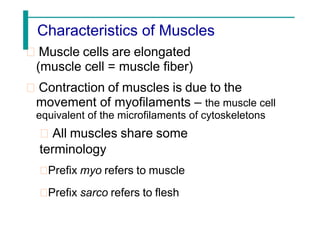
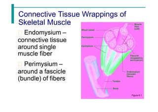
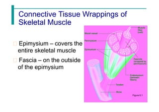
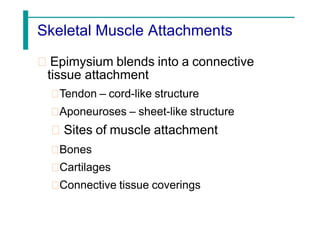
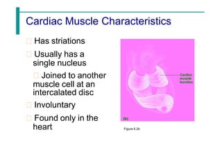
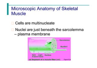
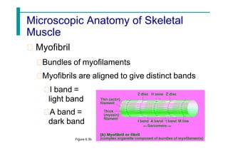
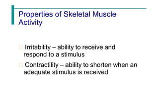
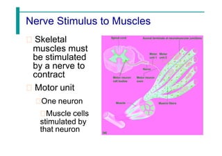
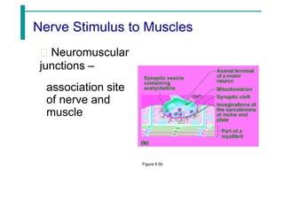
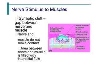
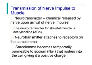
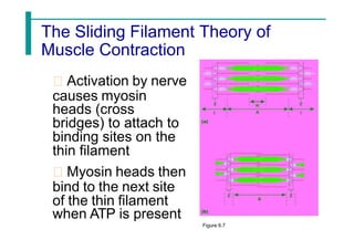
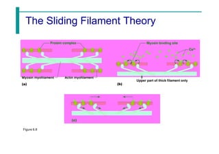
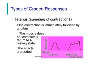
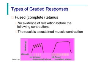
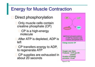
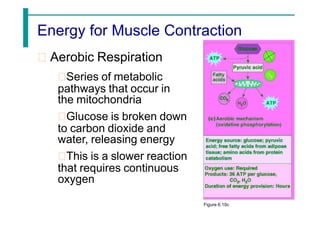
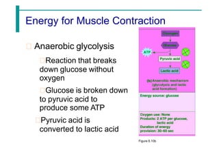
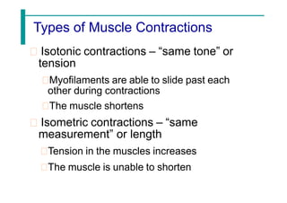
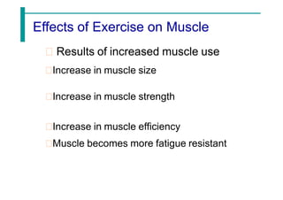
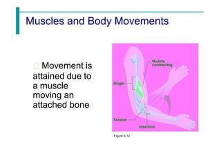
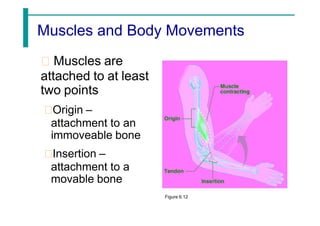
Essentials of Human Anatomy & Physiology
For. Romualdo B. De Guzman, Jr., MSA, Mbio
Associate Professor II
Chapter 5
The Muscular System
The Muscular System
Muscles are responsible for all types of
body movement
Three basic muscle types are found in
the body
Skeletal muscle
Cardiac muscle
Smooth muscle
Slide 6.2
Copyright © 2003 Pearson Education, Inc. publishing as Benjamin Cummings
Characteristics of Muscles
Muscle cells are elongated
(muscle cell = muscle fiber)
Contraction of muscles is due to the
movement of myofilaments – the muscle cell
equivalent of the microfilaments of cytoskeletons
All muscles share some
terminology
Prefix myo refers to muscle
Prefix sarco refers to flesh
Skeletal Muscle Characteristics
Most are attached by tendons to bones
Cells are multinucleate & cigar-shaped
Striated – have visible banding
Voluntary – subject to conscious control
Cells are surrounded and bundled by
connective tissue
Connective Tissue Wrappings of
Skeletal Muscle
Endomysium –
connective tissue
around single
muscle fiber
Perimysium –
around a fascicle
(bundle) of fibers
Figure 6.1
Connective Tissue Wrappings of
Skeletal Muscle
Epimysium – covers the
entire skeletal muscle
Fascia – on the outside
of the epimysium
Figure 6.1
Skeletal Muscle Attachments
Epimysium blends into a connective
tissue attachment
Tendon – cord-like structure
Aponeuroses – sheet-like structure
Sites of muscle attachment
Bones
Cartilages
Connective tissue coverings
Smooth Muscle Characteristics
Has no striations
Spindle-shaped cells
Single nucleus
Involuntary – no
conscious control
Found mainly in the
walls of hollow organs
– visceral
Arranged in two sheets
or layers Figure 6.2a
Cardiac Muscle Characteristics
Has striations
Usually has a
single nucleus
Joined to another
muscle cell at an
intercalated disc
Involuntary
Found only in the
heart Figure 6.2b
Function of Muscles
Produce movement
Maintain posture
Stabilize joints
Generate heat
Microscopic Anatomy of Skeletal
Muscle
Cells are multinucleate
Nuclei are just beneath the sarcolemma
– plasma membrane
Figure 6.3a
Microscopic Anatomy of Skeletal
Muscle
Sarcolemma – specialized plasma
membrane
Sarcoplasmic reticulum – specialized
smooth endoplasmic reticulum
Figure 6.3a
Microscopic Anatomy of Skeletal
Muscle
Myofibril
Bundles of myofilaments
Myofibrils are aligned to give distinct bands
I band =
light band
A band =
dark band
Figure 6.3b
Microscopic Anatomy of Skeletal
Muscle
Sarcomere
Contractile unit of a muscle fiber
Figure 6.3b
Microscopic Anatomy of
Skeletal
Muscle
Organization of the sarcomere
Thick filaments = myosin filaments
Composed of the protein myosin
Has ATPase enzymes
Figure 6.3c
Microscopic Anatomy of Skeletal
Muscle
Organization of the sarcomere
Thin filaments = actin filaments
Composed of the protein actin and
regulatory proteins
Figure 6.3c
Microscopic Anatomy of Skeletal
Muscle
Myosin filaments have heads
(extensions, or cross bridges)
Myosin
and
actin overlap
somewhat
Figure 6.3d
Microscopic Anatomy of Skeletal
Muscle
At rest, there is a bare zone that lacks
actin filaments – the H zone
Sarcoplasmic
reticulum
(SR) – for
storage of
calcium
Figure 6.3d
Properties of Skeletal Muscle
Activity
Irritability – ability to receive and
respond to a stimulus
Contractility – ability to shorten when an
adequate stimulus is received
Nerve Stimulus to Muscles
Skeletal
muscles must
be stimulated
by a nerve to
contract
Motor unit
One neuron
Muscle cells
stimulated by
that neuron
Nerve Stimulus to Muscles
Neuromuscular
junctions –
association site
of nerve and
muscle
Figure 6.5b
Nerve Stimulus to Muscles
Synaptic cleft –
gap between
nerve and
muscle
Nerve and
muscle do not
make contact
Area between
nerve and muscle
is filled with
interstitial fluid
Transmission of Nerve Impulse to
Muscle
Neurotransmitter – chemical released by
nerve upon arrival of nerve impulse
The neurotransmitter for skeletal muscle is
acetylcholine (ACh)
Neurotransmitter attaches to receptors on
the sarcolemma
Sarcolemma becomes temporarily
permeable to sodium (Na+) that rushes into
the cell giving it a positive charge
Transmission of Nerve Impulse to
Muscle
Sodium rushing into the cell generates an
action potential
Once started, muscle contraction cannot be
stopped
To return to resting state
Potassium ions (K+) diffuse out of the cell
Sodium-potassium pump pumps sodium and
potassium back to their original positions
The Sliding Filament Theory of
Muscle Contraction
Activation by nerve
causes myosin
heads (cross
bridges) to attach to
binding sites on the
thin filament
Myosin heads then
bind to the next site
of the thin filament
when ATP is present
Figure 6.7
The Sliding Filament Theory of
Muscle Contraction
This continued action
causes a sliding of the
myosin along the actin
The result is that the
muscle is shortened
(contracted)
Calcium ions are
required
for the attachment of
myosin cross bridges to
actin
Figure 6.7
The Sliding Filament Theory
Figure 6.8
Contraction of a Skeletal Muscle
Muscle fiber contraction is “all or none”
Within a skeletal muscle, not all fibers may
be stimulated during the same interval
Different combinations of muscle fiber
contractions may give differing responses
Graded responses – different degrees of
skeletal muscle shortening
Changing frequency of stimulation
Changing number of muscle cells stimulated
Types of Graded Responses
Twitch
Single, brief jerky contraction
Not a normal muscle function
Figure 6.9a, b
Types of Graded Responses
Tetanus (summing of contractions)
One contraction is immediately followed by
another
The muscle does
not completely
return to a
resting state
The effects
are added
Figure 6.9a, b
Types of Graded Responses
Unfused (incomplete) tetanus
Some relaxation occurs between
contractions
The results are summed
Figure 6.9c,d
Types of Graded Responses
Fused (complete) tetanus
No evidence of relaxation before the
following contractions
The result is a sustained muscle contraction
Figure 6.9c,d
Muscle Response to Strong Stimuli
Muscle force depends upon the number
of fibers stimulated
More fibers contracting results in
greater muscle tension
Muscles can continue to contract unless
they run out of energy
Energy for Muscle Contraction
Initially, muscles used stored ATP for
energy
Bonds of ATP are broken to release energy
Only 4-6 seconds worth of ATP is stored by
muscles
After this initial time, other pathways
must be utilized to produce ATP
Energy for Muscle Contraction
Direct phosphorylation
Only muscle cells contain
creatine phosphate (CP)
CP is a high-energy
molecule
After ATP is depleted, ADP is
left
CP transfers energy to ADP,
to regenerate ATP
CP supplies are exhausted in
about 20 seconds
Figure 6.10a
Energy for Muscle Contraction
Aerobic Respiration
Series of metabolic
pathways that occur in
the mitochondria
Glucose is broken down
to carbon dioxide and
water, releasing energy
This is a slower reaction
that requires continuous
oxygen
Figure 6.10c
Energy for Muscle Contraction
Anaerobic glycolysis
Reaction that breaks
down glucose without
oxygen
Glucose is broken down
to pyruvic acid to
produce some ATP
Pyruvic acid is
converted to lactic acid
Figure 6.10b
Energy for Muscle Contraction
Anaerobic glycolysis
(continued)
This reaction is not as
efficient, but is fast
Huge amounts of
glucose are needed
Lactic acid produces
muscle fatigue
Figure 6.10b
Muscle Fatigue and Oxygen Debt
When a muscle is fatigued, it is unable to
contract even when stimulated
The common reason for muscle fatigue is
oxygen debt
Oxygen must be “repaid” to tissue to remove
oxygen debt
Oxygen is required to get rid of accumulated
lactic acid
Increasing acidity (from lactic acid) and lack
of ATP causes the muscle to contract less
Types of Muscle Contractions
Isotonic contractions – “same tone” or
tension
Myofilaments are able to slide past each
other during contractions
The muscle shortens
Isometric contractions – “same
measurement” or length
Tension in the muscles increases
The muscle is unable to shorten
Muscle Tone
Some fibers are contracted even in a
relaxed muscle
Different fibers contract at different
times to provide muscle tone
The process of stimulating various
fibers is under involuntary control
Effects of Exercise on Muscle
Results of increased muscle use
Increase in muscle size
Increase in muscle strength
Increase in muscle efficiency
Muscle becomes more fatigue resistant
Muscles and Body Movements
Movement is
attained due to
a muscle
moving an
attached bone
Figure 6.12
Muscles and Body Movements
Muscles are
attached to at least
two points
Origin –
attachment to an
immoveable bone
Insertion –
attachment to a
movable bone
Figure 6.12
Types of Ordinary Body Movements
Flexion – brings 2 bones closer together
Extension – increases distance between 2
bones
Rotation
Abduction – moving a limb away from the
midline of the body
Adduction – moving a limb toward the midline
Circumduction – combination of all of the
above except rotation
Body Movements
Figure 6.13
Special Movements
Dorsiflexion – lifting the foot
Plantar flexion – depressing the foot
Inversion – turn foot inward
Eversion – turn foot outward
Supination – hand facing upward
Pronation – hand facing downward
Opposition – touching thumb to other fingers
Types of Muscles
Prime mover – muscle with the major
responsibility for a certain movement
Antagonist – muscle that opposes or
reverses a prime mover
Synergist – muscle that aids a prime
mover in the same movement and helps
prevent rotation or unwanted movement
Fixator – stabilizes the origin of a
prime
mover so all tension can be used to
move the insertion bone
Naming of Skeletal Muscles
Direction of muscle fibers
Example: rectus (straight) or oblique (slanted)
Relative size of the muscle
Examples: maximus (largest),
minimus
(smallest), longus (long)
Naming of Skeletal Muscles
Location of the muscle
Example: many muscles are named
for bones (e.g., temporalis, which is
near the temporal bone)
Number of origins
Example: biceps, triceps, quadriceps
(two, three, or four origins or heads)
Naming of Skeletal Muscles
Location of the muscle’s origin and
insertion
Example: sterno (on the sternum) cleido
(clavicle) mastoid (on the mastoid process)
Shape of the muscle
Example: deltoid (triangular)
Action of the muscle
Example: flexor and extensor (flexes or
extends a bone)
Head and Neck Muscles
Figure 6.14
Trunk Muscles
Figure 6.15
Deep Trunk and Arm Muscles
Figure 6.16
Muscles of the Pelvis, Hip, and Thigh
Figure 6.18c
Muscles of the Lower Leg
Figure 6.19
Superficial Muscles: Anterior
Figure 6.20
Superficial Muscles: Posterior
Figure 6.21

Chapter 5 A Muscular System.pdf123456789

  • 1.
    Essentials of HumanAnatomy & Physiology For. Romualdo B. De Guzman, Jr., MSA, Mbio Associate Professor II Chapter 5 The Muscular System
  • 2.
    The Muscular System Musclesare responsible for all types of body movement Three basic muscle types are found in the body Skeletal muscle Cardiac muscle Smooth muscle Slide 6.2 Copyright © 2003 Pearson Education, Inc. publishing as Benjamin Cummings
  • 3.
    Characteristics of Muscles Musclecells are elongated (muscle cell = muscle fiber) Contraction of muscles is due to the movement of myofilaments – the muscle cell equivalent of the microfilaments of cytoskeletons All muscles share some terminology Prefix myo refers to muscle Prefix sarco refers to flesh
  • 4.
    Skeletal Muscle Characteristics Mostare attached by tendons to bones Cells are multinucleate & cigar-shaped Striated – have visible banding Voluntary – subject to conscious control Cells are surrounded and bundled by connective tissue
  • 5.
    Connective Tissue Wrappingsof Skeletal Muscle Endomysium – connective tissue around single muscle fiber Perimysium – around a fascicle (bundle) of fibers Figure 6.1
  • 6.
    Connective Tissue Wrappingsof Skeletal Muscle Epimysium – covers the entire skeletal muscle Fascia – on the outside of the epimysium Figure 6.1
  • 7.
    Skeletal Muscle Attachments Epimysiumblends into a connective tissue attachment Tendon – cord-like structure Aponeuroses – sheet-like structure Sites of muscle attachment Bones Cartilages Connective tissue coverings
  • 8.
    Smooth Muscle Characteristics Hasno striations Spindle-shaped cells Single nucleus Involuntary – no conscious control Found mainly in the walls of hollow organs – visceral Arranged in two sheets or layers Figure 6.2a
  • 9.
    Cardiac Muscle Characteristics Hasstriations Usually has a single nucleus Joined to another muscle cell at an intercalated disc Involuntary Found only in the heart Figure 6.2b
  • 10.
    Function of Muscles Producemovement Maintain posture Stabilize joints Generate heat
  • 11.
    Microscopic Anatomy ofSkeletal Muscle Cells are multinucleate Nuclei are just beneath the sarcolemma – plasma membrane Figure 6.3a
  • 12.
    Microscopic Anatomy ofSkeletal Muscle Sarcolemma – specialized plasma membrane Sarcoplasmic reticulum – specialized smooth endoplasmic reticulum Figure 6.3a
  • 13.
    Microscopic Anatomy ofSkeletal Muscle Myofibril Bundles of myofilaments Myofibrils are aligned to give distinct bands I band = light band A band = dark band Figure 6.3b
  • 14.
    Microscopic Anatomy ofSkeletal Muscle Sarcomere Contractile unit of a muscle fiber Figure 6.3b
  • 15.
    Microscopic Anatomy of Skeletal Muscle Organizationof the sarcomere Thick filaments = myosin filaments Composed of the protein myosin Has ATPase enzymes Figure 6.3c
  • 16.
    Microscopic Anatomy ofSkeletal Muscle Organization of the sarcomere Thin filaments = actin filaments Composed of the protein actin and regulatory proteins Figure 6.3c
  • 17.
    Microscopic Anatomy ofSkeletal Muscle Myosin filaments have heads (extensions, or cross bridges) Myosin and actin overlap somewhat Figure 6.3d
  • 18.
    Microscopic Anatomy ofSkeletal Muscle At rest, there is a bare zone that lacks actin filaments – the H zone Sarcoplasmic reticulum (SR) – for storage of calcium Figure 6.3d
  • 19.
    Properties of SkeletalMuscle Activity Irritability – ability to receive and respond to a stimulus Contractility – ability to shorten when an adequate stimulus is received
  • 20.
    Nerve Stimulus toMuscles Skeletal muscles must be stimulated by a nerve to contract Motor unit One neuron Muscle cells stimulated by that neuron
  • 21.
    Nerve Stimulus toMuscles Neuromuscular junctions – association site of nerve and muscle Figure 6.5b
  • 22.
    Nerve Stimulus toMuscles Synaptic cleft – gap between nerve and muscle Nerve and muscle do not make contact Area between nerve and muscle is filled with interstitial fluid
  • 23.
    Transmission of NerveImpulse to Muscle Neurotransmitter – chemical released by nerve upon arrival of nerve impulse The neurotransmitter for skeletal muscle is acetylcholine (ACh) Neurotransmitter attaches to receptors on the sarcolemma Sarcolemma becomes temporarily permeable to sodium (Na+) that rushes into the cell giving it a positive charge
  • 24.
    Transmission of NerveImpulse to Muscle Sodium rushing into the cell generates an action potential Once started, muscle contraction cannot be stopped To return to resting state Potassium ions (K+) diffuse out of the cell Sodium-potassium pump pumps sodium and potassium back to their original positions
  • 25.
    The Sliding FilamentTheory of Muscle Contraction Activation by nerve causes myosin heads (cross bridges) to attach to binding sites on the thin filament Myosin heads then bind to the next site of the thin filament when ATP is present Figure 6.7
  • 26.
    The Sliding FilamentTheory of Muscle Contraction This continued action causes a sliding of the myosin along the actin The result is that the muscle is shortened (contracted) Calcium ions are required for the attachment of myosin cross bridges to actin Figure 6.7
  • 27.
    The Sliding FilamentTheory Figure 6.8
  • 28.
    Contraction of aSkeletal Muscle Muscle fiber contraction is “all or none” Within a skeletal muscle, not all fibers may be stimulated during the same interval Different combinations of muscle fiber contractions may give differing responses Graded responses – different degrees of skeletal muscle shortening Changing frequency of stimulation Changing number of muscle cells stimulated
  • 29.
    Types of GradedResponses Twitch Single, brief jerky contraction Not a normal muscle function Figure 6.9a, b
  • 30.
    Types of GradedResponses Tetanus (summing of contractions) One contraction is immediately followed by another The muscle does not completely return to a resting state The effects are added Figure 6.9a, b
  • 31.
    Types of GradedResponses Unfused (incomplete) tetanus Some relaxation occurs between contractions The results are summed Figure 6.9c,d
  • 32.
    Types of GradedResponses Fused (complete) tetanus No evidence of relaxation before the following contractions The result is a sustained muscle contraction Figure 6.9c,d
  • 33.
    Muscle Response toStrong Stimuli Muscle force depends upon the number of fibers stimulated More fibers contracting results in greater muscle tension Muscles can continue to contract unless they run out of energy
  • 34.
    Energy for MuscleContraction Initially, muscles used stored ATP for energy Bonds of ATP are broken to release energy Only 4-6 seconds worth of ATP is stored by muscles After this initial time, other pathways must be utilized to produce ATP
  • 35.
    Energy for MuscleContraction Direct phosphorylation Only muscle cells contain creatine phosphate (CP) CP is a high-energy molecule After ATP is depleted, ADP is left CP transfers energy to ADP, to regenerate ATP CP supplies are exhausted in about 20 seconds Figure 6.10a
  • 36.
    Energy for MuscleContraction Aerobic Respiration Series of metabolic pathways that occur in the mitochondria Glucose is broken down to carbon dioxide and water, releasing energy This is a slower reaction that requires continuous oxygen Figure 6.10c
  • 37.
    Energy for MuscleContraction Anaerobic glycolysis Reaction that breaks down glucose without oxygen Glucose is broken down to pyruvic acid to produce some ATP Pyruvic acid is converted to lactic acid Figure 6.10b
  • 38.
    Energy for MuscleContraction Anaerobic glycolysis (continued) This reaction is not as efficient, but is fast Huge amounts of glucose are needed Lactic acid produces muscle fatigue Figure 6.10b
  • 39.
    Muscle Fatigue andOxygen Debt When a muscle is fatigued, it is unable to contract even when stimulated The common reason for muscle fatigue is oxygen debt Oxygen must be “repaid” to tissue to remove oxygen debt Oxygen is required to get rid of accumulated lactic acid Increasing acidity (from lactic acid) and lack of ATP causes the muscle to contract less
  • 40.
    Types of MuscleContractions Isotonic contractions – “same tone” or tension Myofilaments are able to slide past each other during contractions The muscle shortens Isometric contractions – “same measurement” or length Tension in the muscles increases The muscle is unable to shorten
  • 41.
    Muscle Tone Some fibersare contracted even in a relaxed muscle Different fibers contract at different times to provide muscle tone The process of stimulating various fibers is under involuntary control
  • 42.
    Effects of Exerciseon Muscle Results of increased muscle use Increase in muscle size Increase in muscle strength Increase in muscle efficiency Muscle becomes more fatigue resistant
  • 43.
    Muscles and BodyMovements Movement is attained due to a muscle moving an attached bone Figure 6.12
  • 44.
    Muscles and BodyMovements Muscles are attached to at least two points Origin – attachment to an immoveable bone Insertion – attachment to a movable bone Figure 6.12
  • 45.
    Types of OrdinaryBody Movements Flexion – brings 2 bones closer together Extension – increases distance between 2 bones Rotation Abduction – moving a limb away from the midline of the body Adduction – moving a limb toward the midline Circumduction – combination of all of the above except rotation
  • 46.
  • 47.
    Special Movements Dorsiflexion –lifting the foot Plantar flexion – depressing the foot Inversion – turn foot inward Eversion – turn foot outward Supination – hand facing upward Pronation – hand facing downward Opposition – touching thumb to other fingers
  • 48.
    Types of Muscles Primemover – muscle with the major responsibility for a certain movement Antagonist – muscle that opposes or reverses a prime mover Synergist – muscle that aids a prime mover in the same movement and helps prevent rotation or unwanted movement Fixator – stabilizes the origin of a prime mover so all tension can be used to move the insertion bone
  • 49.
    Naming of SkeletalMuscles Direction of muscle fibers Example: rectus (straight) or oblique (slanted) Relative size of the muscle Examples: maximus (largest), minimus (smallest), longus (long)
  • 50.
    Naming of SkeletalMuscles Location of the muscle Example: many muscles are named for bones (e.g., temporalis, which is near the temporal bone) Number of origins Example: biceps, triceps, quadriceps (two, three, or four origins or heads)
  • 51.
    Naming of SkeletalMuscles Location of the muscle’s origin and insertion Example: sterno (on the sternum) cleido (clavicle) mastoid (on the mastoid process) Shape of the muscle Example: deltoid (triangular) Action of the muscle Example: flexor and extensor (flexes or extends a bone)
  • 52.
    Head and NeckMuscles Figure 6.14
  • 53.
  • 54.
    Deep Trunk andArm Muscles Figure 6.16
  • 55.
    Muscles of thePelvis, Hip, and Thigh Figure 6.18c
  • 56.
    Muscles of theLower Leg Figure 6.19
  • 57.
  • 58.