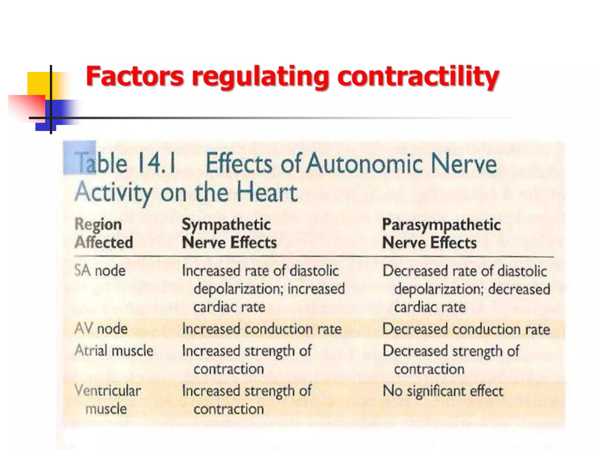
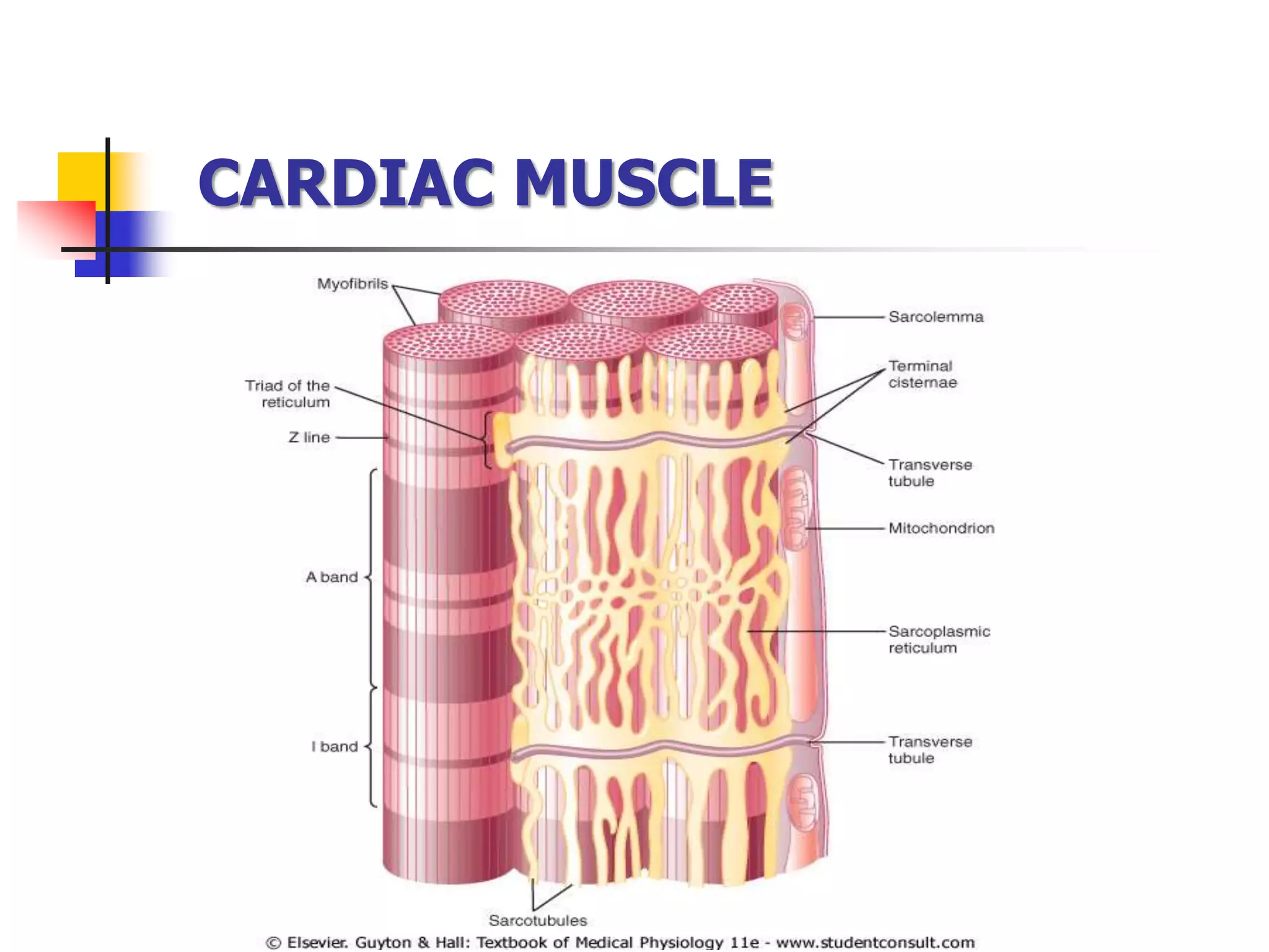
CARDIAC MUSCLE: CONTRACTILE
MECHANISM OF CARDIAC MUSCLE
Prof. Sultan Ayoub Meo
MBBS, M.Phil, Ph.D (Pak), M Med Ed (Dundee), FRCP (London),
FRCP (Dublin), FRCP (Glasgow), FRCP (Edinburgh)
Professor and Consultant, Department of Physiology,
College of Medicine, King Saud University, Riyadh, KSA
CARDIAC MUSCLE
 Atrial Muscle
 Ventricular Muscle
 Specialize Excitatory & Conductive Muscle
 The atrial and ventricular muscle contract in
much the same way as skeletal muscle, except
duration of contraction is much longer.
 The specialized excitatory and conductive fibers
contract weakly because they contain few
contractile fibrils Guyton pp 109
Heart is composed of three major types of cardiac muscle
• Cardiac muscle fibers are striated in appearance
• Functional unit is called Sarcomere
• Fibers are branched; connect to one another at
intercalated discs.
• The discs contain several Gap Junctions
• Nuclei are centrally located
• Abundant Mitochondria
• Sarcoplasmic Reticulum is less abundant than
in skeletal muscle, but greater in density than
smooth muscle
• Sarcolemma has specialized ion channels that
skeletal muscle does not – voltage-gated Ca2+
channels
• Fibers are not anchored at ends; allows for
greater sarcomere shortening and lengthening
CARDIAC MUSCLE
CARDIAC MUSCLE
The dark areas crossing the cardiac muscle
fibers are called intercalated discs; they
are actually cell membranes that
separate individual cardiac muscle cells
from one another. Guyton pp 109
At intercalated disc cell membranes fuse with one
another, form permeable “communicating” junctions
(gap junctions) allow free diffusion of ions.
Ions move with ease in the intracellular fluid
along the longitudinal axes of the cardiac muscle
fibers, so that action potentials travel easily from one
cardiac muscle cell to the next
Thus, cardiac muscle is a syncytium of many heart
muscle cells, action potential spreads to all of them.
CARDIAC MUSCLE
Guyton pp 104
isometric contraction: Muscle contraction
without significant shortening or change in
distance
isotonic contraction: Muscle contraction
without significant change in the force of
contraction
 Interconnected by intercalated discs
and form functional syncytia
 Within intercalated discs – two kinds of
membrane junctions
 Desmosomes
 Gap junctions
CARDIAC MUSCLE
 Heart beats rhythmically as result of action
potential, it generates by itself (Auto-
rhythmicity)
 Two specialized types of cardiac muscle cells
 Contractile cells
 99% of cardiac muscle cells
 Do mechanical work of pumping
 Autorhythmic cells
 Do not contract
 Specialized for initiating and conducting action
potentials responsible for contraction of working cells
CARDIAC MUSCLE
The cardiac muscle cells are responsible for the electrical
stimulation leads to mechanical function. The electro-
physiologic properties of cardiac muscles are:
Automaticity: Ability to spontaneously generate an
electrical impulse.
Excitability: Ability to respond to an electrical impulse.
Conductivity: Allows transmission of electrical impulse to
another cardiac cell.
Contractility: Ability to contract after electrical impulse
response
Rhythmicity: Ability to send electrical impulses in a
regularly manner.
CARDIAC MUSCLE PROPERTIES
Excitation-Contraction coupling
Excitation Contraction
[ Ca++
]i
(Action Potentials) (shortening)
Excitation-Contraction coupling
Excitation Contraction
[ Ca++
]i
(Action Potentials) (shortening)
Excitation of the heart is triggered by electrical impulse
rather than neural transmitters.
Contraction of the heart is triggered by elevation of
intracellular calcium influx.
Calcium ions regulate the contraction of cardiac muscle:
Entry of extracellular calcium ions causes the release of calcium from the sarcoplasmic
reticulum (calcium-induced calcium release), source of about 95% of calcium in cytosol.
Excitation-Contraction coupling
Excitation-
Contraction
Coupling in Cardiac
Contractile Cells
• Autonomic nervous system modulates the frequency of depolarization of pacemaker
• Sympathetic stimulation (neurotransmitter); binds to b1 receptors on the SA nodal
membranes
• Parasympathetic stimulation (neurotransmitter); binds to muscarinic receptors on
nodal membranes; increases conductivity of K+ and decreases conductivity of Ca2+
cardiac.ppt

cardiac.ppt

  • 1.
    CARDIAC MUSCLE: CONTRACTILE MECHANISMOF CARDIAC MUSCLE Prof. Sultan Ayoub Meo MBBS, M.Phil, Ph.D (Pak), M Med Ed (Dundee), FRCP (London), FRCP (Dublin), FRCP (Glasgow), FRCP (Edinburgh) Professor and Consultant, Department of Physiology, College of Medicine, King Saud University, Riyadh, KSA
  • 2.
    CARDIAC MUSCLE  AtrialMuscle  Ventricular Muscle  Specialize Excitatory & Conductive Muscle  The atrial and ventricular muscle contract in much the same way as skeletal muscle, except duration of contraction is much longer.  The specialized excitatory and conductive fibers contract weakly because they contain few contractile fibrils Guyton pp 109 Heart is composed of three major types of cardiac muscle
  • 3.
    • Cardiac musclefibers are striated in appearance • Functional unit is called Sarcomere • Fibers are branched; connect to one another at intercalated discs. • The discs contain several Gap Junctions • Nuclei are centrally located • Abundant Mitochondria • Sarcoplasmic Reticulum is less abundant than in skeletal muscle, but greater in density than smooth muscle • Sarcolemma has specialized ion channels that skeletal muscle does not – voltage-gated Ca2+ channels • Fibers are not anchored at ends; allows for greater sarcomere shortening and lengthening
  • 4.
  • 5.
    CARDIAC MUSCLE The darkareas crossing the cardiac muscle fibers are called intercalated discs; they are actually cell membranes that separate individual cardiac muscle cells from one another. Guyton pp 109
  • 6.
    At intercalated disccell membranes fuse with one another, form permeable “communicating” junctions (gap junctions) allow free diffusion of ions. Ions move with ease in the intracellular fluid along the longitudinal axes of the cardiac muscle fibers, so that action potentials travel easily from one cardiac muscle cell to the next Thus, cardiac muscle is a syncytium of many heart muscle cells, action potential spreads to all of them. CARDIAC MUSCLE Guyton pp 104
  • 7.
    isometric contraction: Musclecontraction without significant shortening or change in distance isotonic contraction: Muscle contraction without significant change in the force of contraction
  • 8.
     Interconnected byintercalated discs and form functional syncytia  Within intercalated discs – two kinds of membrane junctions  Desmosomes  Gap junctions
  • 9.
  • 10.
     Heart beatsrhythmically as result of action potential, it generates by itself (Auto- rhythmicity)  Two specialized types of cardiac muscle cells  Contractile cells  99% of cardiac muscle cells  Do mechanical work of pumping  Autorhythmic cells  Do not contract  Specialized for initiating and conducting action potentials responsible for contraction of working cells CARDIAC MUSCLE
  • 11.
    The cardiac musclecells are responsible for the electrical stimulation leads to mechanical function. The electro- physiologic properties of cardiac muscles are: Automaticity: Ability to spontaneously generate an electrical impulse. Excitability: Ability to respond to an electrical impulse. Conductivity: Allows transmission of electrical impulse to another cardiac cell. Contractility: Ability to contract after electrical impulse response Rhythmicity: Ability to send electrical impulses in a regularly manner. CARDIAC MUSCLE PROPERTIES
  • 13.
    Excitation-Contraction coupling Excitation Contraction [Ca++ ]i (Action Potentials) (shortening) Excitation-Contraction coupling
  • 14.
    Excitation Contraction [ Ca++ ]i (ActionPotentials) (shortening) Excitation of the heart is triggered by electrical impulse rather than neural transmitters. Contraction of the heart is triggered by elevation of intracellular calcium influx.
  • 16.
    Calcium ions regulatethe contraction of cardiac muscle: Entry of extracellular calcium ions causes the release of calcium from the sarcoplasmic reticulum (calcium-induced calcium release), source of about 95% of calcium in cytosol. Excitation-Contraction coupling
  • 17.
  • 20.
    • Autonomic nervoussystem modulates the frequency of depolarization of pacemaker • Sympathetic stimulation (neurotransmitter); binds to b1 receptors on the SA nodal membranes • Parasympathetic stimulation (neurotransmitter); binds to muscarinic receptors on nodal membranes; increases conductivity of K+ and decreases conductivity of Ca2+