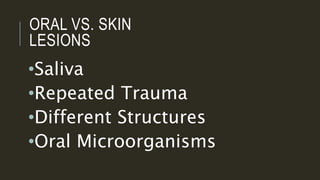
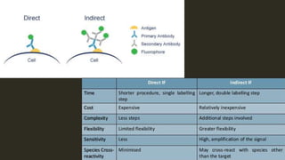
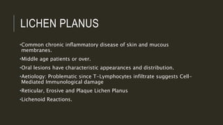
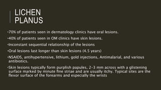
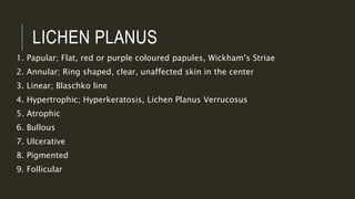
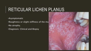
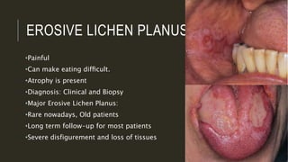
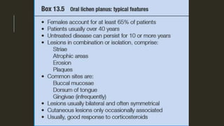
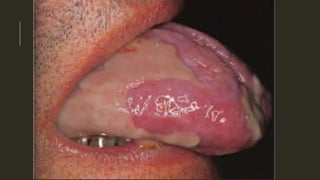
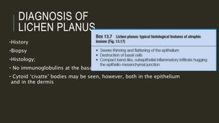
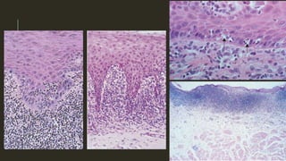
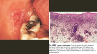
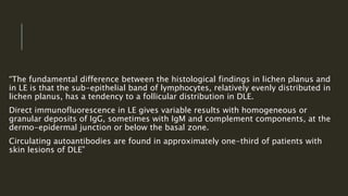
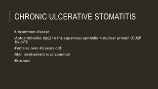
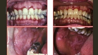
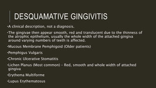
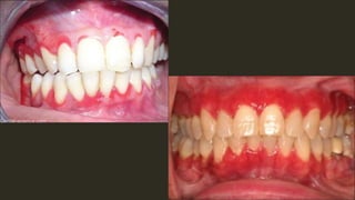
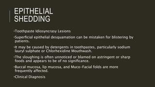
MUCOCUTANEOUS
DISEASES I
Dr. Hadi Munib
Oral and Maxillofacial
Surgery Resident
OUTLI
NE
•Differences between Oral and Skin lesions
•Immunofluorescence tests used in Mucocutaneous Diseases
•Lichen Planus
•Lupus Erythematosis
•Chronic Ulcerative Stomatitis
•Desquamative Gingivitis
•Epithelial Shedding
•Pemphigus Vulgaris
•Mucous Membrane Pemphigoid
•Erythema Multiforme
•References
ORAL VS. SKIN
LESIONS
•Saliva
•Repeated Trauma
•Different Structures
•Oral Microorganisms
IMMUNOFLUORESCENCE TESTS
•Direct Immunofluorescence
•Indirect Immunofluorescence
•Immunofluorescence: ​​use of antibodies to label a specific target antigen with a
fluorescent dye (also called fluorophores or fluorochromes) such
as fluorescein isothiocyanate (FITC).
•Direct IF uses a single antibody directed against the target of interest.
The primary antibody is directly conjugated to a fluorophore.
•Indirect IF uses two antibodies. The primary antibody is unconjugated and a
fluorophore-conjugated secondary antibody directed against the primary
antibody is used for detection.
LICHEN PLANUS
•Common chronic inflammatory disease of skin and mucous
membranes.
•Middle age patients or over.
•Oral lesions have characteristic appearances and distribution.
•Aetiology: Problematic since T-Lymphocytes infiltrate suggests Cell-
Mediated Immunological damage
•Reticular, Erosive and Plaque Lichen Planus
•Lichenoid Reactions.
LICHEN
PLANUS
•70% of patients seen in dermatology clinics have oral lesions.
•40% of patients seen in OM clinics have skin lesions.
•Inconstant sequential relationship of the lesions
•Oral lesions last longer than skin lesions (4.5 years)
•NSAIDS, antihypertensive, lithium, gold injections, Antimalarial, and various
antibiotics.
•Skin lesions typically form purplish papules, 2–3 mm across with a glistening
surface marked by minute fine striae and are usually itchy. Typical sites are the
flexor surface of the forearms and especially the wrists
LICHEN PLANUS
1. Papular; Flat, red or purple coloured papules, Wickham’s Striae
2. Annular; Ring shaped, clear, unaffected skin in the center
3. Linear; Blaschko line
4. Hypertrophic; Hyperkeratosis, Lichen Planus Verrucosus
5. Atrophic
6. Bullous
7. Ulcerative
8. Pigmented
9. Follicular
RETICULAR LICHEN PLANUS
•Asymptomatic
•Roughness or slight stiffness of the mucosa
•No atrophy.
•Diagnosis: Clinical and Biopsy
EROSIVE LICHEN PLANUS
•Painful
•Can make eating difficult.
•Atrophy is present
•Diagnosis: Clinical and Biopsy
•Major Erosive Lichen Planus:
•Rare nowadays, Old patients
•Long term follow-up for most patients
•Severe disfigurement and loss of tissues
DIAGNOSIS OF
LICHEN PLANUS
•History
•Biopsy
•Histology;
• No immunoglobulins at the basal zone.
• Cytoid ‘civatte’ bodies may be seen, however, both in the epithelium
and in the dermis
MANAGEMENT OF
LICHEN PLANUS
•Topical application of potent anti-inflammatory corticosteroids is
usually effective but monitoring is required
•Possible alternatives are to use similar corticosteroids
(beclomethasone) from aerosol inhalers.
•Thrush as a side effect
REPORTEDLY 1–4% OF PATIENTS
SUFFER MALIGNANT CHANGES
COMPLICATION AFTER 10 YEARS.
LICHENOID
REACTIONS
LUPUS ERYTHEMATOSUS
• Connective tissue disease which has two main forms, namely
systemic and cutaneous (‘discoid’).
•May be triggered by infections, elevated temperature, concurrent
systemic diseases, and exposure to ultraviolet light or drugs.
•Various body organs can be affected.
•Clinically, oral lesions appear in about 20% of cases of systemic
lupus.
CLINICAL FEATURES OF SYSTEMIC LUPUS
ERYTHEMATOSUS
•Typical lesions are white, often striate, areas with irregular atrophic
areas or shallow erosions, typically far less sharply defined than in
lichen planus.
•They are often patchy and unilateral and may be in the vault of the
palate.
•Lesions can form variable patterns of white and red areas.
•There may also be small slit-like ulcers just short of the gingival
margins.
•In about 30%, Sjögren’s syndrome develops and, rarely, cervical
lymphadenopathy is the first sign.
DISCOID LUPUS
ERYTHEMATOSUS
•Scaly red patches of the face
•Alopecia, follicular plugging of the skin
•Exposure to sunlight triggers exacerbations
•Lips are the most common site
•Oral lesions: irregular white keratotic plaques, or lesions with an
erythematous or ulcerated center surrounded by radiating white
striations
TREATMENT OF LUPUS
ERYTHEMATOSUS
I. Steroids
II. Antimalarials
III. Long-term observation of lip lesions (potentially premalignant)
“The fundamental difference between the histological findings in lichen planus and
in LE is that the sub-epithelial band of lymphocytes, relatively evenly distributed in
lichen planus, has a tendency to a follicular distribution in DLE.
Direct immunofluorescence in LE gives variable results with homogeneous or
granular deposits of IgG, sometimes with IgM and complement components, at the
dermo-epidermal junction or below the basal zone.
Circulating autoantibodies are found in approximately one-third of patients with
skin lesions of DLE”
CHRONIC ULCERATIVE STOMATITIS
•Uncommon disease
•Autoantibodies (IgG) to the squamous epithelium nuclear protein (CUSP
by p73)
•Females over 40 years old
•Skin involvement is uncommon
•Erosions
CHRONIC ULCERATIVE
STOMATITIS
•Immunofluorescence shows speckled antinuclear antibodies in the
perilesional mucosa and shaggy deposits of fibrinogen in the
basement membrane zone.
•Serum shows antinuclear antibody in high titer that reacts with
Guinea pig oesophagus substrate but the titer does not correspond
with clinical severity.
•The most effective treatment appears to be with chloroquine or
hydroxychloroquine, supplemented if necessary with prednisolone.
However complete clearance is not always attainable.
DESQUAMATIVE GINGIVITIS
•A clinical description, not a diagnosis.
•The gingivae then appear smooth, red and translucent due to the thinness of
the atrophic epithelium, usually the whole width of the attached gingiva
around varying numbers of teeth is affected.
•Mucous Membrane Pemphigoid (Older patients)
•Pemphigus Vulgaris
•Chronic Ulcerative Stomatitis
•Lichen Planus (Most common) – Red, smooth and whole width of attached
gingiva
•Erythema Multiforme
•Lupus Erythematosus
EPITHELIAL
SHEDDING
•Toothpaste Idiosyncrasy Lesions
•Superficial epithelial desquamation can be mistaken for blistering by
patients.
•It may be caused by detergents in toothpastes, particularly sodium
lauryl sulphate or Chlorhexidine Mouthwash.
•The sloughing is often unnoticed or blamed on astringent or sharp
foods and appears to be of no significance.
•Buccal mucosa, lip mucosa, and Muco-Facial folds are more
frequently affected.
•Clinical Diagnosis
REFERENCES
•Cawson’s Chapter 13: DISEASES OF THE ORAL MUCOSA: NON-INFECTIVE
STOMATITIS
•Tyldesley’s Chapter 11: Mucocutaneous disease and connective tissue
disorders
THANK YOU

Mucocutaneous Diseases I

  • 1.
    MUCOCUTANEOUS DISEASES I Dr. HadiMunib Oral and Maxillofacial Surgery Resident
  • 2.
    OUTLI NE •Differences between Oraland Skin lesions •Immunofluorescence tests used in Mucocutaneous Diseases •Lichen Planus •Lupus Erythematosis •Chronic Ulcerative Stomatitis •Desquamative Gingivitis •Epithelial Shedding •Pemphigus Vulgaris •Mucous Membrane Pemphigoid •Erythema Multiforme •References
  • 3.
    ORAL VS. SKIN LESIONS •Saliva •RepeatedTrauma •Different Structures •Oral Microorganisms
  • 4.
    IMMUNOFLUORESCENCE TESTS •Direct Immunofluorescence •IndirectImmunofluorescence •Immunofluorescence: ​​use of antibodies to label a specific target antigen with a fluorescent dye (also called fluorophores or fluorochromes) such as fluorescein isothiocyanate (FITC). •Direct IF uses a single antibody directed against the target of interest. The primary antibody is directly conjugated to a fluorophore. •Indirect IF uses two antibodies. The primary antibody is unconjugated and a fluorophore-conjugated secondary antibody directed against the primary antibody is used for detection.
  • 6.
    LICHEN PLANUS •Common chronicinflammatory disease of skin and mucous membranes. •Middle age patients or over. •Oral lesions have characteristic appearances and distribution. •Aetiology: Problematic since T-Lymphocytes infiltrate suggests Cell- Mediated Immunological damage •Reticular, Erosive and Plaque Lichen Planus •Lichenoid Reactions.
  • 7.
    LICHEN PLANUS •70% of patientsseen in dermatology clinics have oral lesions. •40% of patients seen in OM clinics have skin lesions. •Inconstant sequential relationship of the lesions •Oral lesions last longer than skin lesions (4.5 years) •NSAIDS, antihypertensive, lithium, gold injections, Antimalarial, and various antibiotics. •Skin lesions typically form purplish papules, 2–3 mm across with a glistening surface marked by minute fine striae and are usually itchy. Typical sites are the flexor surface of the forearms and especially the wrists
  • 8.
    LICHEN PLANUS 1. Papular;Flat, red or purple coloured papules, Wickham’s Striae 2. Annular; Ring shaped, clear, unaffected skin in the center 3. Linear; Blaschko line 4. Hypertrophic; Hyperkeratosis, Lichen Planus Verrucosus 5. Atrophic 6. Bullous 7. Ulcerative 8. Pigmented 9. Follicular
  • 9.
    RETICULAR LICHEN PLANUS •Asymptomatic •Roughnessor slight stiffness of the mucosa •No atrophy. •Diagnosis: Clinical and Biopsy
  • 10.
    EROSIVE LICHEN PLANUS •Painful •Canmake eating difficult. •Atrophy is present •Diagnosis: Clinical and Biopsy •Major Erosive Lichen Planus: •Rare nowadays, Old patients •Long term follow-up for most patients •Severe disfigurement and loss of tissues
  • 13.
    DIAGNOSIS OF LICHEN PLANUS •History •Biopsy •Histology; •No immunoglobulins at the basal zone. • Cytoid ‘civatte’ bodies may be seen, however, both in the epithelium and in the dermis
  • 15.
    MANAGEMENT OF LICHEN PLANUS •Topicalapplication of potent anti-inflammatory corticosteroids is usually effective but monitoring is required •Possible alternatives are to use similar corticosteroids (beclomethasone) from aerosol inhalers. •Thrush as a side effect
  • 16.
    REPORTEDLY 1–4% OFPATIENTS SUFFER MALIGNANT CHANGES COMPLICATION AFTER 10 YEARS.
  • 17.
  • 18.
    LUPUS ERYTHEMATOSUS • Connectivetissue disease which has two main forms, namely systemic and cutaneous (‘discoid’). •May be triggered by infections, elevated temperature, concurrent systemic diseases, and exposure to ultraviolet light or drugs. •Various body organs can be affected. •Clinically, oral lesions appear in about 20% of cases of systemic lupus.
  • 19.
    CLINICAL FEATURES OFSYSTEMIC LUPUS ERYTHEMATOSUS •Typical lesions are white, often striate, areas with irregular atrophic areas or shallow erosions, typically far less sharply defined than in lichen planus. •They are often patchy and unilateral and may be in the vault of the palate. •Lesions can form variable patterns of white and red areas. •There may also be small slit-like ulcers just short of the gingival margins. •In about 30%, Sjögren’s syndrome develops and, rarely, cervical lymphadenopathy is the first sign.
  • 20.
    DISCOID LUPUS ERYTHEMATOSUS •Scaly redpatches of the face •Alopecia, follicular plugging of the skin •Exposure to sunlight triggers exacerbations •Lips are the most common site •Oral lesions: irregular white keratotic plaques, or lesions with an erythematous or ulcerated center surrounded by radiating white striations
  • 21.
    TREATMENT OF LUPUS ERYTHEMATOSUS I.Steroids II. Antimalarials III. Long-term observation of lip lesions (potentially premalignant)
  • 23.
    “The fundamental differencebetween the histological findings in lichen planus and in LE is that the sub-epithelial band of lymphocytes, relatively evenly distributed in lichen planus, has a tendency to a follicular distribution in DLE. Direct immunofluorescence in LE gives variable results with homogeneous or granular deposits of IgG, sometimes with IgM and complement components, at the dermo-epidermal junction or below the basal zone. Circulating autoantibodies are found in approximately one-third of patients with skin lesions of DLE”
  • 24.
    CHRONIC ULCERATIVE STOMATITIS •Uncommondisease •Autoantibodies (IgG) to the squamous epithelium nuclear protein (CUSP by p73) •Females over 40 years old •Skin involvement is uncommon •Erosions
  • 25.
    CHRONIC ULCERATIVE STOMATITIS •Immunofluorescence showsspeckled antinuclear antibodies in the perilesional mucosa and shaggy deposits of fibrinogen in the basement membrane zone. •Serum shows antinuclear antibody in high titer that reacts with Guinea pig oesophagus substrate but the titer does not correspond with clinical severity. •The most effective treatment appears to be with chloroquine or hydroxychloroquine, supplemented if necessary with prednisolone. However complete clearance is not always attainable.
  • 27.
    DESQUAMATIVE GINGIVITIS •A clinicaldescription, not a diagnosis. •The gingivae then appear smooth, red and translucent due to the thinness of the atrophic epithelium, usually the whole width of the attached gingiva around varying numbers of teeth is affected. •Mucous Membrane Pemphigoid (Older patients) •Pemphigus Vulgaris •Chronic Ulcerative Stomatitis •Lichen Planus (Most common) – Red, smooth and whole width of attached gingiva •Erythema Multiforme •Lupus Erythematosus
  • 29.
    EPITHELIAL SHEDDING •Toothpaste Idiosyncrasy Lesions •Superficialepithelial desquamation can be mistaken for blistering by patients. •It may be caused by detergents in toothpastes, particularly sodium lauryl sulphate or Chlorhexidine Mouthwash. •The sloughing is often unnoticed or blamed on astringent or sharp foods and appears to be of no significance. •Buccal mucosa, lip mucosa, and Muco-Facial folds are more frequently affected. •Clinical Diagnosis
  • 31.
    REFERENCES •Cawson’s Chapter 13:DISEASES OF THE ORAL MUCOSA: NON-INFECTIVE STOMATITIS •Tyldesley’s Chapter 11: Mucocutaneous disease and connective tissue disorders
  • 32.