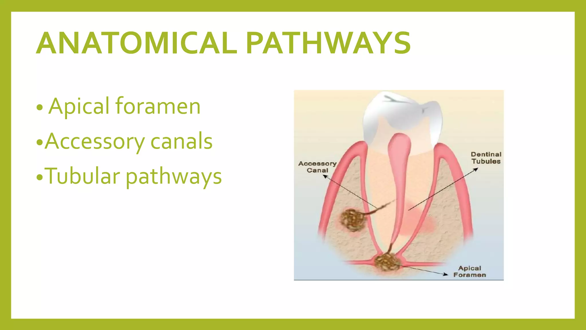
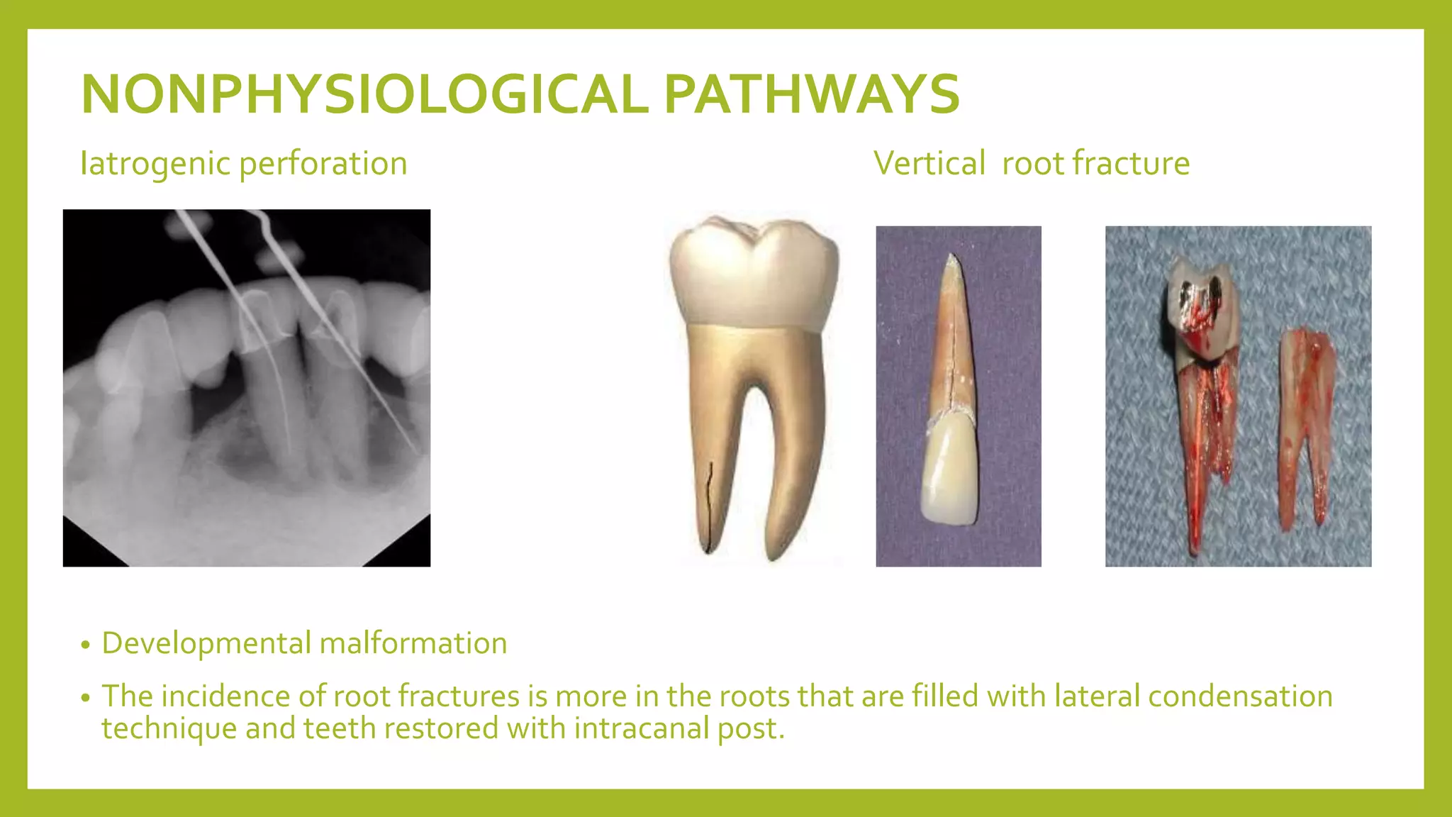
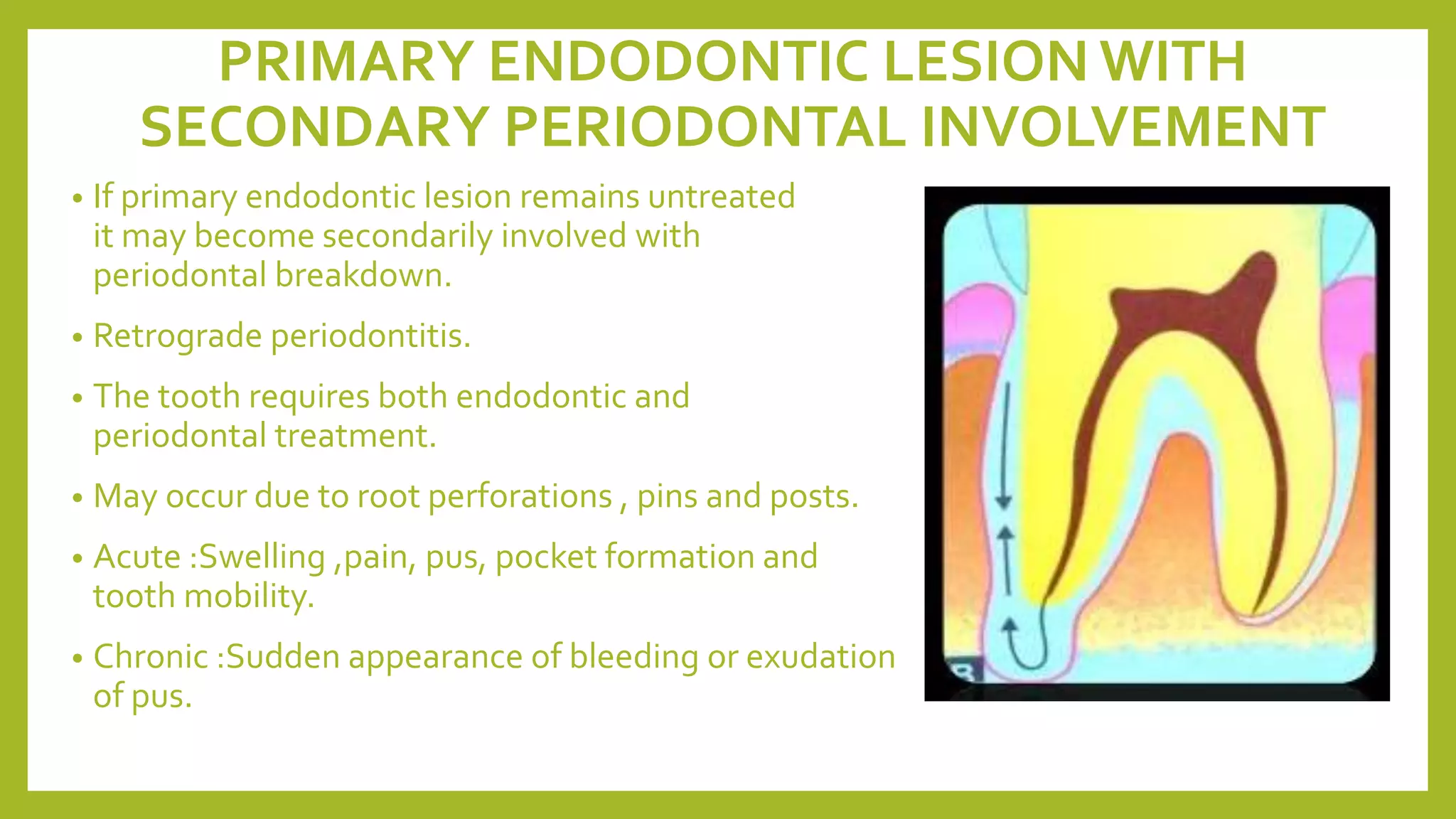
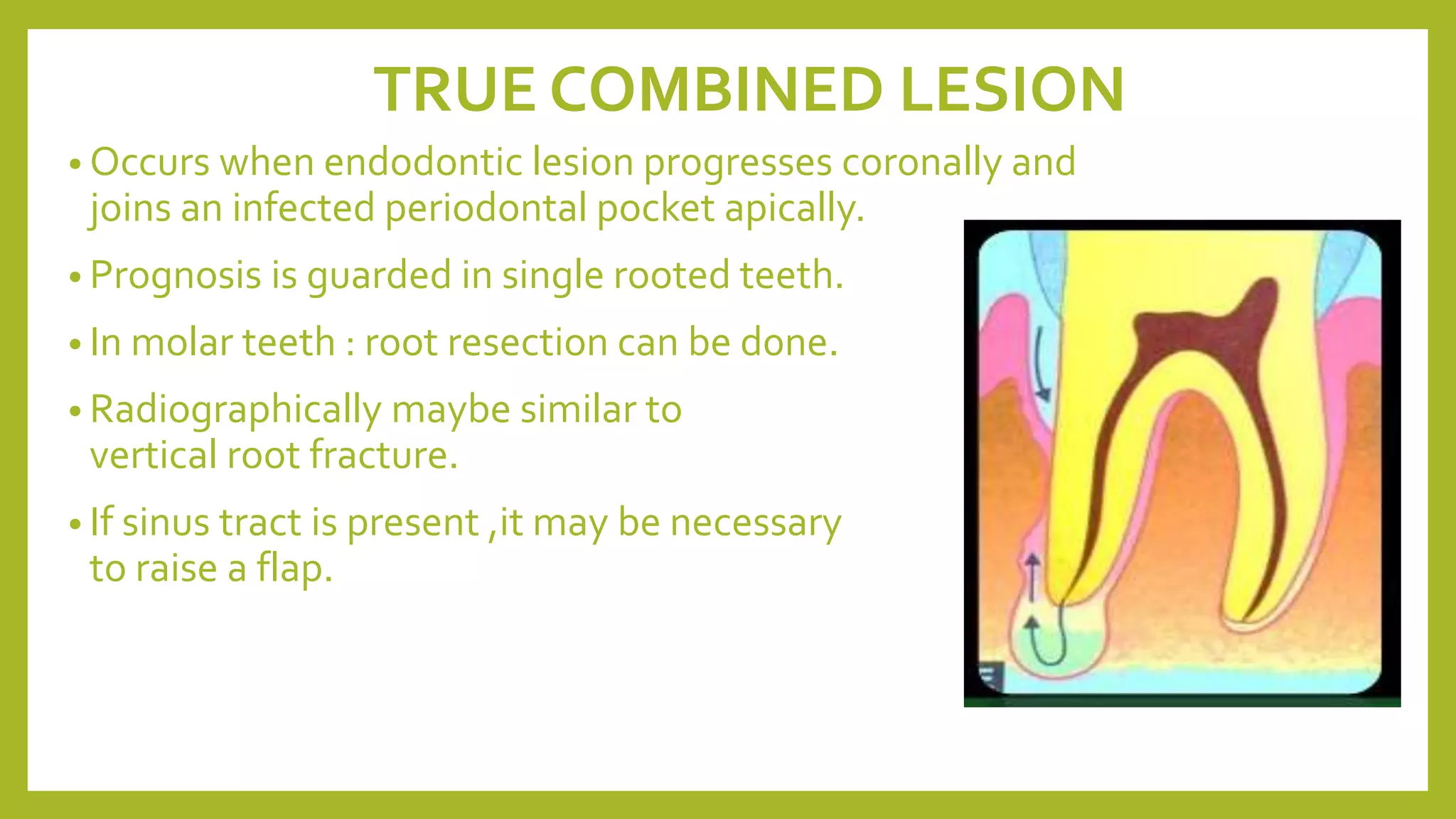
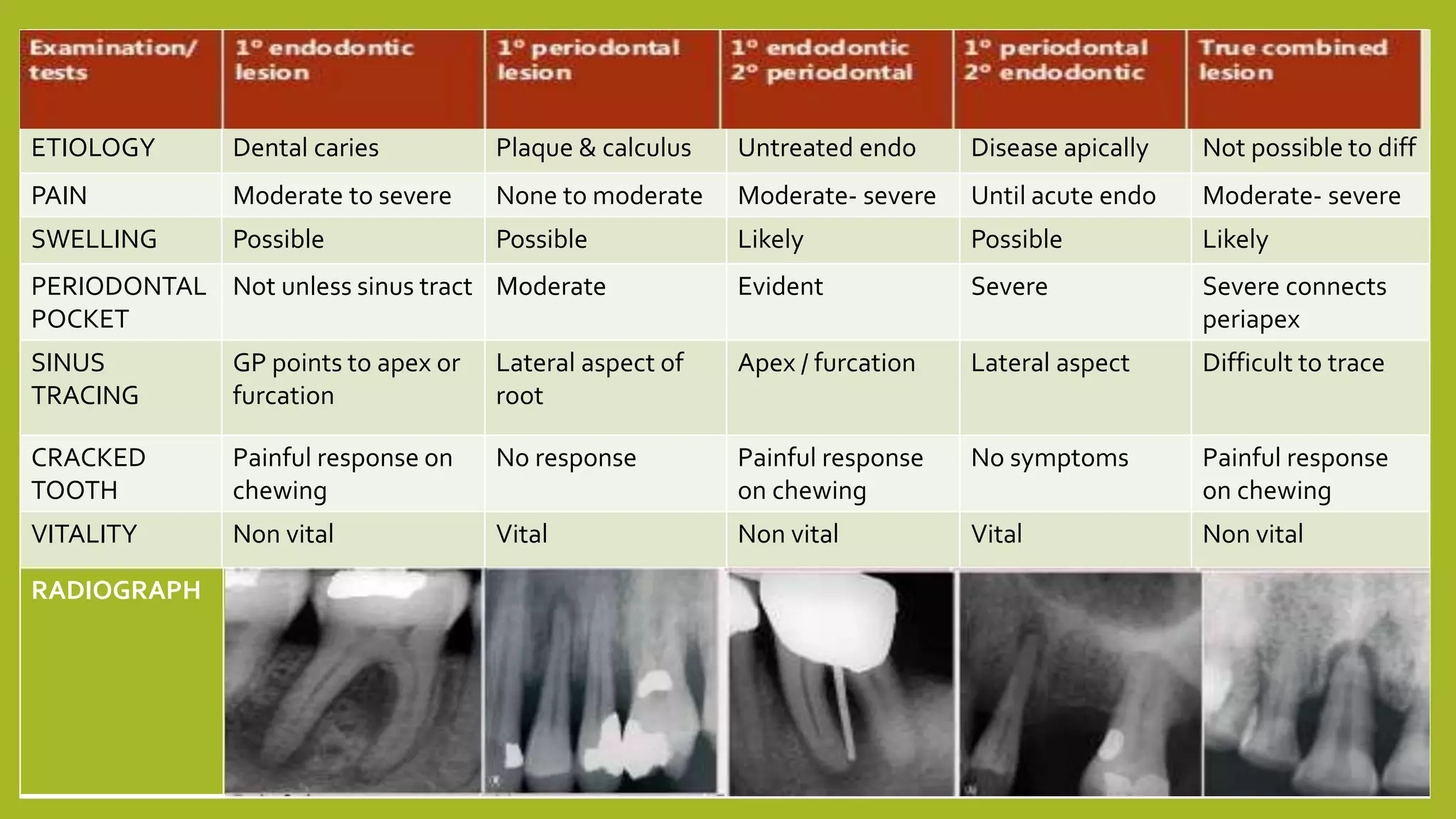
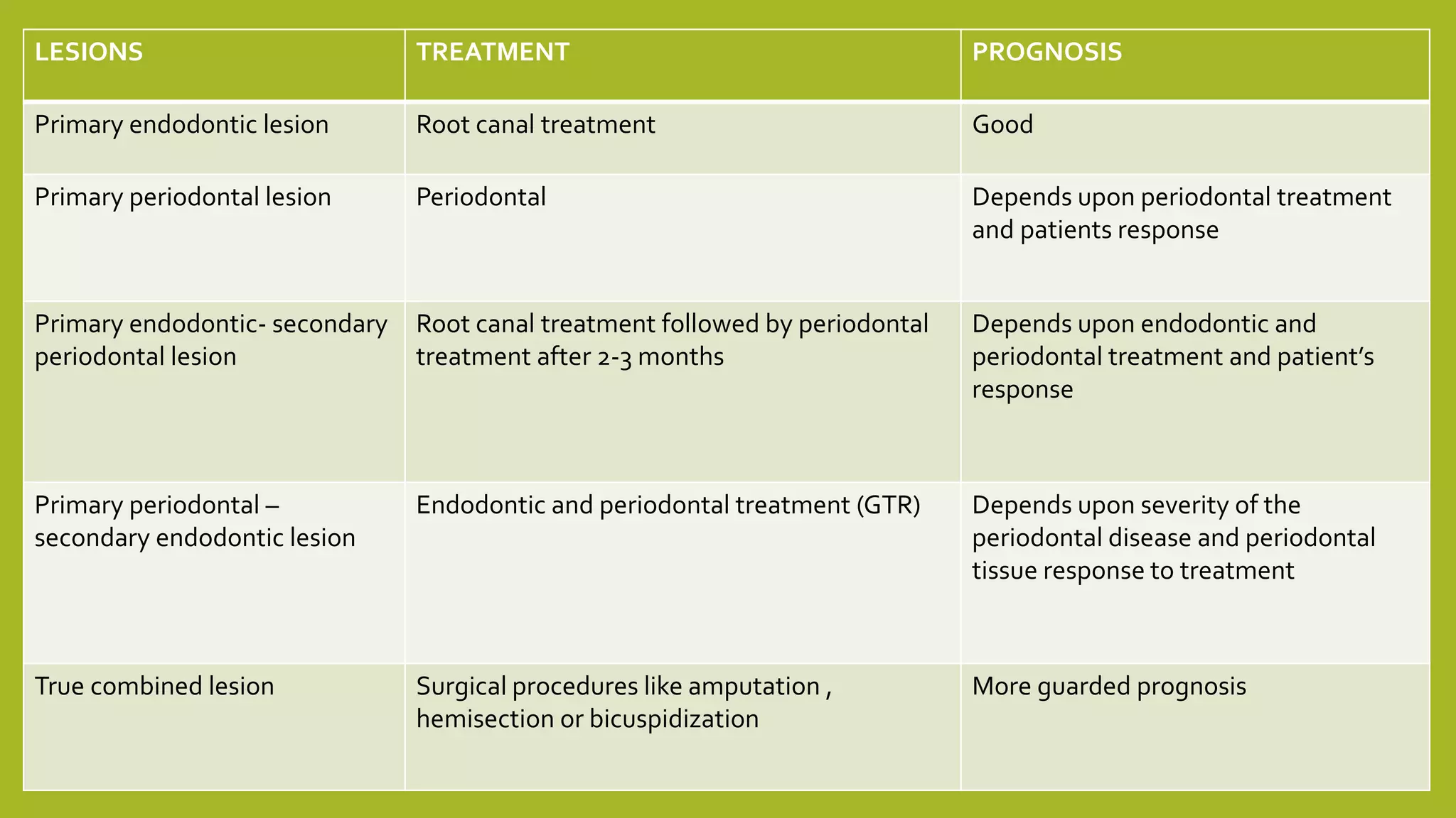
ENDODONTIC - PERIODONTAL
LESIONS
PRESENTED BY
Dr. Arsheya G.s
INTRODUCTION
•The term ENDO – PERIO has been an integral part of
dental vocabulary.
•The possible pathways for ingress of bacteria and
their products into these tissues can broadly be
divided into
•Anatomical pathways
•Non physiological pathways
ANATOMICAL PATHWAYS
• Apical foramen
•Accessory canals
•Tubular pathways
NONPHYSIOLOGICAL PATHWAYS
Iatrogenic perforation Vertical root fracture
• Developmental malformation
• The incidence of root fractures is more in the roots that are filled with lateral condensation
technique and teeth restored with intracanal post.
ETIOPATHOGENESIS OF PERIO-ENDO LESIONS
•Microbial agents
•Atrophic changes
•Inflammatory changes
•Resoprtion
CLASSIFICATION OF
ENDO-PERIO LESION
WEINE’S CLASSIFICATION (1982)
• Class 1:Tooth in which symptoms clinically and radiograohically
stimulate periodontal disease but are in fact due to pulpal
inflammation and /or necrosis.
• Class 2:Tooth that has both pulpal or periapical disease and
periodontal disease concomitantly.
• Class 3:Tooth that has no pulpal problem but requires Endodontic
therapy plus root amputation to gain periodontal healing.
• Class 4:Tooth that clinically and radiographically stimulates
pulpal or periapical diseases but in fact has periodontal disease.
SIMON,GLICK AND FRANK’S
CLASSIFICATION (1972)
•Primary endodontic lesion .
•Primary periodontal lesion .
•Primary endodontic lesion with secondary periodontal
involvement .
•Primary periodontal lesion with secondary endodontic
involvement .
•True combined lesion.
PRIMARY ENDODONTIC LESION
• Heals following root canal therapy.
• An acute exacerbation of chronic apical lesion on a tooth with
necrotic pulp may drain coronally through the periodontal
ligament into gingival sulcus.
• Usually mimic periodontal abscess (sinus tract).
• The sinus tract extending into gingival sulcus disappears
at an early stage if the necrotic plup is removed
and canal are sealed well.
PRIMARY PERIODONTAL LESION
• Caused primarily by periodontal pathogens.
• Progresses along root surface.
• Commonly due to accumulation of plaque
calculus and presence of deep pockets.
PRIMARY ENDODONTIC LESION WITH
SECONDARY PERIODONTAL INVOLVEMENT
• If primary endodontic lesion remains untreated
it may become secondarily involved with
periodontal breakdown.
• Retrograde periodontitis.
• The tooth requires both endodontic and
periodontal treatment.
• May occur due to root perforations , pins and posts.
• Acute :Swelling ,pain, pus, pocket formation and
tooth mobility.
• Chronic :Sudden appearance of bleeding or exudation
of pus.
PRIMARY PERIODONTAL LESION WITH
SECONDARY ENDODONTIC INVOLVEMENT
• Retrograde pulpitis: when bacteria and
inflammatory products could gain access
to the pulp via accessory canal
apical foramen and dentinal tubules.
• The apical progression of a periodontal pocket
continues until the apical tissues are involved.
• Pulp may become necrotic.
TRUE COMBINED LESION
• Occurs when endodontic lesion progresses coronally and
joins an infected periodontal pocket apically.
• Prognosis is guarded in single rooted teeth.
• In molar teeth : root resection can be d0ne.
• Radiographically maybe similar to
vertical root fracture.
• If sinus tract is present ,it may be necessary
to raise a flap.
DIAGNOSIS
•Vitality test
•Radiographic evaluation
•Pain and abscess formation
•Probing
•Mobility
•Percussion and palpation
EXAMINATION
ETIOLOGY Dental caries Plaque & calculus Untreated endo Disease apically Not possible to diff
PAIN Moderate to severe None to moderate Moderate- severe Until acute endo Moderate- severe
SWELLING Possible Possible Likely Possible Likely
PERIODONTAL
POCKET
Not unless sinus tract Moderate Evident Severe Severe connects
periapex
SINUS
TRACING
GP points to apex or
furcation
Lateral aspect of
root
Apex / furcation Lateral aspect Difficult to trace
CRACKED
TOOTH
Painful response on
chewing
No response Painful response
on chewing
No symptoms Painful response
on chewing
VITALITY Non vital Vital Non vital Vital Non vital
RADIOGRAPH
TREATMENT
AND
PROGNOSIS
OF PERIODONTAL –
ENDODONTIC LESION
LESIONS TREATMENT PROGNOSIS
Primary endodontic lesion Root canal treatment Good
Primary periodontal lesion Periodontal Depends upon periodontal treatment
and patients response
Primary endodontic- secondary
periodontal lesion
Root canal treatment followed by periodontal
treatment after 2-3 months
Depends upon endodontic and
periodontal treatment and patient’s
response
Primary periodontal –
secondary endodontic lesion
Endodontic and periodontal treatment (GTR) Depends upon severity of the
periodontal disease and periodontal
tissue response to treatment
True combined lesion Surgical procedures like amputation ,
hemisection or bicuspidization
More guarded prognosis
THANKYOU !

Endodontics periodontal lesions

  • 1.
  • 2.
    INTRODUCTION •The term ENDO– PERIO has been an integral part of dental vocabulary. •The possible pathways for ingress of bacteria and their products into these tissues can broadly be divided into •Anatomical pathways •Non physiological pathways
  • 3.
    ANATOMICAL PATHWAYS • Apicalforamen •Accessory canals •Tubular pathways
  • 4.
    NONPHYSIOLOGICAL PATHWAYS Iatrogenic perforationVertical root fracture • Developmental malformation • The incidence of root fractures is more in the roots that are filled with lateral condensation technique and teeth restored with intracanal post.
  • 5.
    ETIOPATHOGENESIS OF PERIO-ENDOLESIONS •Microbial agents •Atrophic changes •Inflammatory changes •Resoprtion
  • 6.
  • 7.
    WEINE’S CLASSIFICATION (1982) •Class 1:Tooth in which symptoms clinically and radiograohically stimulate periodontal disease but are in fact due to pulpal inflammation and /or necrosis. • Class 2:Tooth that has both pulpal or periapical disease and periodontal disease concomitantly. • Class 3:Tooth that has no pulpal problem but requires Endodontic therapy plus root amputation to gain periodontal healing. • Class 4:Tooth that clinically and radiographically stimulates pulpal or periapical diseases but in fact has periodontal disease.
  • 8.
    SIMON,GLICK AND FRANK’S CLASSIFICATION(1972) •Primary endodontic lesion . •Primary periodontal lesion . •Primary endodontic lesion with secondary periodontal involvement . •Primary periodontal lesion with secondary endodontic involvement . •True combined lesion.
  • 9.
    PRIMARY ENDODONTIC LESION •Heals following root canal therapy. • An acute exacerbation of chronic apical lesion on a tooth with necrotic pulp may drain coronally through the periodontal ligament into gingival sulcus. • Usually mimic periodontal abscess (sinus tract). • The sinus tract extending into gingival sulcus disappears at an early stage if the necrotic plup is removed and canal are sealed well.
  • 10.
    PRIMARY PERIODONTAL LESION •Caused primarily by periodontal pathogens. • Progresses along root surface. • Commonly due to accumulation of plaque calculus and presence of deep pockets.
  • 11.
    PRIMARY ENDODONTIC LESIONWITH SECONDARY PERIODONTAL INVOLVEMENT • If primary endodontic lesion remains untreated it may become secondarily involved with periodontal breakdown. • Retrograde periodontitis. • The tooth requires both endodontic and periodontal treatment. • May occur due to root perforations , pins and posts. • Acute :Swelling ,pain, pus, pocket formation and tooth mobility. • Chronic :Sudden appearance of bleeding or exudation of pus.
  • 12.
    PRIMARY PERIODONTAL LESIONWITH SECONDARY ENDODONTIC INVOLVEMENT • Retrograde pulpitis: when bacteria and inflammatory products could gain access to the pulp via accessory canal apical foramen and dentinal tubules. • The apical progression of a periodontal pocket continues until the apical tissues are involved. • Pulp may become necrotic.
  • 13.
    TRUE COMBINED LESION •Occurs when endodontic lesion progresses coronally and joins an infected periodontal pocket apically. • Prognosis is guarded in single rooted teeth. • In molar teeth : root resection can be d0ne. • Radiographically maybe similar to vertical root fracture. • If sinus tract is present ,it may be necessary to raise a flap.
  • 14.
  • 15.
    •Vitality test •Radiographic evaluation •Painand abscess formation •Probing •Mobility •Percussion and palpation
  • 16.
  • 17.
    ETIOLOGY Dental cariesPlaque & calculus Untreated endo Disease apically Not possible to diff PAIN Moderate to severe None to moderate Moderate- severe Until acute endo Moderate- severe SWELLING Possible Possible Likely Possible Likely PERIODONTAL POCKET Not unless sinus tract Moderate Evident Severe Severe connects periapex SINUS TRACING GP points to apex or furcation Lateral aspect of root Apex / furcation Lateral aspect Difficult to trace CRACKED TOOTH Painful response on chewing No response Painful response on chewing No symptoms Painful response on chewing VITALITY Non vital Vital Non vital Vital Non vital RADIOGRAPH
  • 18.
  • 19.
    LESIONS TREATMENT PROGNOSIS Primaryendodontic lesion Root canal treatment Good Primary periodontal lesion Periodontal Depends upon periodontal treatment and patients response Primary endodontic- secondary periodontal lesion Root canal treatment followed by periodontal treatment after 2-3 months Depends upon endodontic and periodontal treatment and patient’s response Primary periodontal – secondary endodontic lesion Endodontic and periodontal treatment (GTR) Depends upon severity of the periodontal disease and periodontal tissue response to treatment True combined lesion Surgical procedures like amputation , hemisection or bicuspidization More guarded prognosis
  • 20.