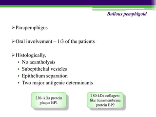
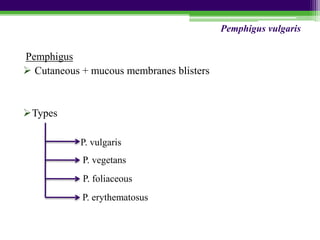
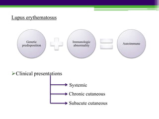
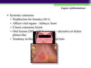
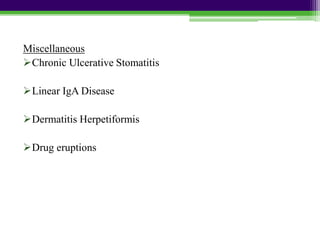
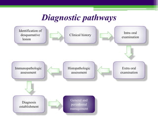
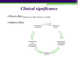
DESQUAMATIVE
GINGIVITIS
Contents
Introduction
History
Epidemiology
Classification
Disorders associated with desquamative gingivitis
• Lichen planus
• Pemphigoid
• Pemphigus
• Lupus erythematosus
• Erythema multiforme
• Miscellaneous
Conditions mimicking desquamative gingivitis
Diagnostic pathways
Clinical significance and implications
Conclusion
References
Introduction
 Current classification system
 Common characteristics
Immunomediated
pathogenesis
Desquamative
gingivitis
Introduction
Gingivosis
75% cases – dermatologic genesis
History
 1894
 Prinz, 1932
 McCarthy et al, 1960
 Glickman and Smulow, 1964
Epidemiologic features
 Highest prevalence - lichen planus and mucous membrane
pemphigoid
 Rarer conditions
Disorder Number of cases of
desquamative gingivitis
Mucous membrane pemphigoid 35-48%
Oral lichen planus 24- 45%
Pemphigus vulgaris 3-15%
Classification
1) Dermatoses
a) Cicatricial pemphigoid
b) Pemphigus
c) Lichen Planus
d) Erythema Multiforme
e) Lupus Erythematosis
f) Linear IgA disease
2) Hormonal influence
a) Estrogen deficiency following oophorectomy & post menopausal women
b) Testosterone imbalance
c) Hypothyroidism
3) Abnormal response to irritation
4) Chronic infection
a) Tuberculosis
b) Chronic candidiasis
c) Histoplasmosis
5) Aging
6) Idiopathic
Disorders associated with desquamative
gingivitis
General clinical features
• Predominantly affects women (Prinz 1932, Meritt 1993)
• Occurs in 4th – 5th decade
• Red, swollen & glossy gingiva
• Multiple vesicle and superficial
denuded areas
• Difficult to eat hot, spicy food
• Nikolsky’s sign may be positive
• Types
Disorders associated with desquamative gingivitis
• Scattered, irregularly shaped areas
• Denuded and red gingiva
• Extremely painful
• Elevation of epithelium → vesicle
• Patchy distribution
• Slight pitting with pressure
• Burning sensation
• Nikolsky’s sign
• Labial > Lingual
• Diffuse erythema
• Painless
Disorders associated with desquamative gingivitis
Lichen planus
Pemphigoid
Pemphigus
Lupus erythematosus
Erythema multiforme
Miscellaneous
Lichen planus
 Inflammatory mucocutaneous disorder
 Immunologically mediated - host T lymphocytes play a
central role (Ishii et al, 1987)
 0.1 - 4% of the population (Scully et al 1998)
 Female : Male – 2:1
 Etiology
Lichen
planus
Idiopathic
Stress
Traumatism
Malnutrition
and infection
Autoimmune
Hereditary
Grinspans
syndrome
 Oral lesions
• Relapsing –remitting course
• Papule arrangement
• Wickhams striae
• Various clinical forms
Atrophic
Erosive Reticular
Bullous
Lichen planus
• Painful erythematous areas
• Sensitive to heat, acid,
spicy foods
• Asymptomatic
• Bilateral interlacing
white strands
• Erythematous background
Reticular Erosive
Lichen planus
 Gingival lesions
• Keratotic lesions
• Erosive or ulcerative lesions
• Vesicular or bullous lesions
• Atrophic lesions
Lichen planus
Pathogenesis
CD8+ T cell TNF-α Apoptosis
CD4+ T cell IFN-γ, IL-2
↑ immune
response
Keratinocytes IL-1,8,10,12;
TNF-α
Lichen planus
Histopathology
•
 Pullon et al (1969)
• Irregularity of nuclear membrane
• Increased thickening and granularity of epithelial tonofibrils
Lichen planus
Immunopathology
• Direct immunofluorescence
• Indirect immunofluorescence
Lichen planus
Differential diagnosis
• Lupus erythematosus
• Chronic ulcerative stomatitis
• Cicatricial pemphigoid
• Pemphigus vulgaris
Lichen planus
Diagnosis of lichen planus
Erosive/UlcerativeAsymptomatic
No therapy
Follow up 6-12 months
Severe cases -
injections of
triamcinolone
acetonide
(10 to 20 mg)
Topical steroids
No resolution Resolution
Refer to
dermatologist
Retinoids, Dapsone,
Systemic steroids
Wean off and
monitor
Treatment of Lichen planus
Recalcitrant cases - 0.1% tacrolimus
Antifungal therapy
Risk for malignant transformation ~ 5% (Becker et al 2006)
Lichen planus
Pemphigoid
Cutaneous, immune-mediated, subepithelial bullous diseases
characterized by separation of the basement membrane zone
Bullous pemphigoid Cicatricial pemphigoid
Parapemphigus
Oral involvement – 1/3 of the patients
Histologically,
• No acantholysis
• Subepithelial vesicles
• Epithelium separation
• Two major antigenic determinants
Bullous pemphigoid
230- kDa protein
plaque BP1
180-kDa collagen-
like transmembrane
protein BP2
Immunopathology
Treatment
• Primary treatment - moderate dose of prednisone
• Steroid sparing strategies
• Localized lesions - high potency topical steroids or
tetracycline
Bullous pemphigoid
Mucous membrane pemphigoid
Oral cavity, conjunctiva, mucosa of the nose, vagina, rectum,
esophagus, urethra
 Female predilection
Five subtypes
Cicatricial pemphigoid
Oral pemphigoid
Multiple antigens
pemphigoid
Ocular pemphigoid
anti-BP antigen
mucosal
pemphigoid
Anti-epiligrin
pemphigoid
Oral lesions
• Areas of erythema, desquamation, ulceration
• Bullae - thick walled
• Healing - 3 weeks or longer
Ocular lesions
Cicatricial pemphigoid
Unilateral
conjunctivitis
Symblepharon Ankyloblepharon
Bilateral
conjunctivitis
BP 1, BP2,
laminin 5 and 6,
uncein,
α6β4integrin
Cytokine release
Sequestration of
leukocytes
Complement
activation
Production of
autoantibodies
Blister formation
Cicatricial pemphigoid
Release of
proteases,
collagenases,
elastases
Histopathology
• Non specific
• Subepithelial vesiculation
• Intact basal layer
Immunopathology
• Direct immunofluorescence
- IgG and C3
• Indirect immunofluorescence
- positive in <25%
Cicatricial pemphigoid
Differential diagnosis
Cicatricial
pemphigoid
Pemphigus
vulgaris
Erythema
multiforme
Epidermolysis
bullosa
acquisita
Bullous lichen
planus
Cicatricial pemphigoid
Diagnosis of cicatricial
pemphigoid
Asymptomatic
Plaque control
Mild to moderate
Topical steroids
Severe
Refer to dermatologist
Prednisolone
Azathioprine
Dapsone
Sulfonamide
Tetracycline
Treatment of Cicatricial pemphigoid
Pemphigus
 Cutaneous + mucous membranes blisters
Types
P. vulgaris
P. vegetans
P. foliaceous
P. erythematosus
Pemphigus vulgaris
0.1 - 0.5 cases /100,000 individuals/year
Higher predilection in women (Robinson et al 1997)
Potentially lethal: mortality rate < 10% (Black et al 2005)
 Etiology
Idiopathic
Drug induced
pemphigus
Paraneoplastic
pemphigus
Pemphigus vulgaris
Oral lesions
• Siegel et al 1994
• Painful small vesicles → large bullae
• Nikolsky’s sign
Pemphigus vulgaris
Soft palate > buccal mucosa > tongue > lower labial mucosa > gingiva
 Pathogenesis
Pemphigus vulgaris
Harman et al 2001
Histopathology
Intraepithelial vesiculation
↓
Fluid filled bulla
Pemphigus vulgaris
Acantholysis
↓
Intercellular bridges are lost
"Tombstone" appearance
 Cytology
• Tzanck cells
• Tzanck test
 Immunopathology
• Direct
• Indirect
Differential diagnosis
Pemphigus
vulgaris
Pemphigoid
Erythema
multiforme
Bullous
lichen
planus
Pemphigus vulgaris
Treatment
Pemphigus vulgaris
Diagnosis of
pemphigus vulgaris
Primary treatment
Secondary
treatment
Prednisone
Decrease dose
→maintenance dose
Azathioprine
Cyclosporine
Methyltrexate
Plasmaphoresis
Refer to
dermatologist
Lupus erythematosus
Clinical presentations
Genetic
predisposition
Immunologic
abnormality
Autoimmune
Systemic
Chronic cutaneous
Subacute cutaneous
 Systemic cutaneous
• Predilection for females (10:1)
• Affects vital organs – kidneys, heart
• Classic cutaneous lesion
• Oral lesions (36% of SLE patients) - ulcerative or lichen
planus-like
• Tendency to bleed, surrounded by red halo
Lupus erythematosus
Laboratory test
• LE cell inclusion phenomenon(Weiss and Swift et al 1955)
Immunopathology
• Direct - Perilesional and normal tissue: Ig and C3 deposits
at dermal-epidermal interface
Lupus erythematosus
 Chronic cutaneous
• Skin lesions – Discoid lupus erythematosus
• Produce scarring and atrophy
• Oral lesions - 9% of patients present lichen planus-like
plaques
Lupus erythematosus
Soft palate > buccal mucosa > tongue > lower labial mucosa > gingiva
Immunopathology
•
Subacute cutaneous lupus erythematosus
Lupus erythematosus
Differential diagnosis
Lupus
erythematosus
Pemphigus
vulgaris
Erythema
multiforme
Erosive lichen
planus
Lupus erythematosus
Treatment
Diagnosis of LE
Cutaneous lesion
Topical steroids
Sunscreens
Hydroxychloroquinine
Mucosal lesion
Topical steroids
Systemic involvement
Moderate- to-high
doses of prednisone
Lupus erythematosus
Erythema Multiforme
Acute bullous and/or macular inflammatory mucocutaneous
disease
Types
Etiology
Erythema multiforme minor
Erythema multiforme major/ Stevens-Johnson syndrome
Mycoplasma
infection
Herpes simplex
infection
Drug reactions
Male predilection
 Symmetric distribution of macules, papules or vesicles
“target”, “iris” or “bulls eye” appearance
Erythema multiforme
Oral lesions
• 70% of patients with skin involvement (McCarthy 1980)
• Multiple, large, painful ulcers with an erythematous border
• Hemorrhagic crusting of vermilion border of lips
Buccal mucosa > tongue > lower labial mucosa > floor of mouth > palate >gingiva
Erythema multiforme
Steven Johnsons Syndrome
• Severe bullous form
• Abrupt occurrence of fever, malaise, photophobia and
eruptions of oral mucosa, genitilia and skin
• Oral lesions → rupture → surfaces covered with thick white
or yellow exudate
• Lips - ulceration with bloody crusting
• ANUG
Erythema multiforme
Histopathology
• Epithelium
• Lamina propria
Immunopathology – negative
Erythema multiforme
 Treatment
Diagnosis of erythema
multiforme
Spontaneous
resolution
Mild cases
Systemic and local
antihistamines +
topical anesthetics +
debridement of lesions
Severe cases
Systemic
corticosteroids
Erythema multiforme
Miscellaneous
Chronic Ulcerative Stomatitis
Linear IgA Disease
Dermatitis Herpetiformis
Drug eruptions
Drug eruptions
• Stomatitis medicamentosa
• Stomatitis venenata or contact stomatitis
Etiology
• Mercurial compounds
• Toothpaste
Clinical presentation
Vesicular or bullous
Pigmented or nonpigmented macula
Erosions
Deep ulceration with purpura
Conditions Mimicking Desquamative
Gingivitis
Factitious lesions
Candidiasis
Graft versus host disease
Wegeners granulomatosis
Foreign body gingivitis
Diagnostic pathways
Identification of
desquamative
lesion
Immunopathologic
assessment
Histopathologic
assessment
Extra oral
examination
Intra oral
examination
Clinical history
Diagnosis
establishment
General and
periodontal
management
Clinical history
Onset
Chief complaint
Infections
Topical substances
Drug intake
Acute, subacute
Symptoms
Intraoral
examination
Location
Clinical presentation
Extraoral
examination
Other mucosae
Skin
Internal organsHistopathology
Epithelial changes
Basement membrane alterations
Lamina propria
Cell –cell attachment,
Acantholysis, epithelial
thickening
Inflammatory infiltrate
BM integrity and staining
Immunopathology
Serum
Involved tissue DIF
IIF
Clinical significance
Painful oral
lesions
Impede daily
oral hygiene
procedures
↑ plaque and
calculus
↑
inflammation
and
enhancement
of symptoms
Exacerbation
of lesions
Direct effect (Ramon et al 1999, Tricamo et al 2006)
Indirect effect
Koebner phenomenon
Clinical significance
Conclusion
Maintenance
and
supportive
periodontal
therapy
Monitoring
and
surveillance
References
 Newman, Takei, Klokkevold, Carranza. 10th edition. Carranza’s Clinical
Periodontology. W. B. Saunders Company.
 Lucio Lo Russo, Stefano Fedele, Rosario Guiglia,Domenico Ciavarella,
Lorenzo Lo Muzio, Pio Gallo, Chiara Di Liberto, and Giuseppina Campisi.
Diagnostic Pathways and Clinical Significance of Desquamative Gingivitis. J
Periodontol 2008;79:4-24.
 Shafer, Hine and Levy. 4th edition, A textbook of Oral Pathology.
W. B. Saunders Company.
 Burket L. Burket’s Oral Medicine – Diagnosis and Treatment. 9th edition.
Lippincott.
 Michele D. Mignogna, Giulio Fortuna, Stefania Leuci, Elvira Ruoppo, Flavio
Marasca and Sergio Matarasso. Nikolsky’s Sign on the Gingival Mucosa: A
Clinical Tool for Oral Health Practitioners.
J Periodontol 2008;79:2241-2246.
desquamative ginigivitis

desquamative ginigivitis

Editor's Notes

  • #10 Nikolskys sign – seperation attributed to prevesicular edema causing disruption of epidermal-dermal junction
  • #19 “Sawtooth” appearance of rete pegs. Hydropic degeneration of basal layer, with the appearance of a thin band of eosinophilic coagulum in this region. Epithelium becomes thin and atrophic or detaches off the underlying CT producing a Subepithelial vesicle. Colloid bodies or Civatte bodies are seen at the epithelium- CT interface.
  • #32 Dapsone (ocular lesions). Side effects – intolerance and hematological side effects
  • #41 Intravenoues human Ig has been shown to be successful in steroid resistant cases. (Yeh et al 2005)
  • #46 Igg
  • #56 Chronic Ulcerative Stomatitis – chronic oral ulcerations which present as painful solitary blisters and resemble erosive lichen planus Linear IgA disease – uncommon mucocutaneous disorder. Cud be trigerred by ACE inhibitors. Resemble erosive lichen planus clinically and histologically Dermatitis herpetiformis – cutaneous manifestaion of celiac disease with tissue transglutaminase as the predominant autoantigen. Characterised by painful ulceration preceeded by collapse of vesicles or bullae.