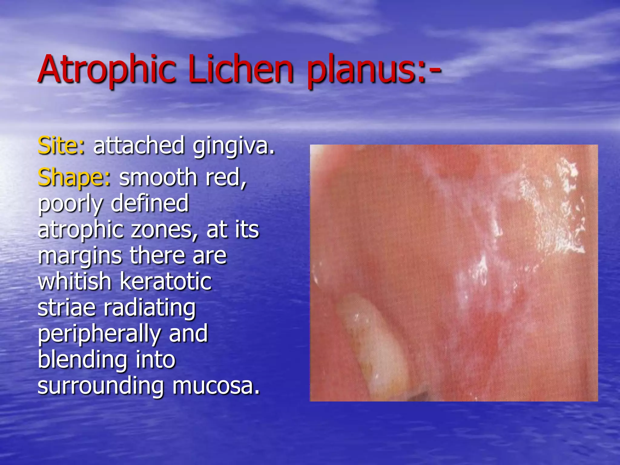
The presentation explain white lesions in oral cavity and the classification the demonstrate the etiology, histopathology, diagnosis and treatment for each one.
Odontogenic keratocyst (OKC) is the cyst arising from the cell rests of dental lamina. It can occur anywhere in the jaw, but commonly seen in the posterior part of the mandible. Radiographically, most OKCs are unilocular when presented at the periapex and can be mistaken for radicular or lateral periodontal cyst.
The Indian Dental Academy is the Leader in continuing dental education , training dentists in all aspects of dentistry and offering a wide range of dental certified courses in different formats.
Indian dental academy provides dental crown & Bridge,rotary endodontics,fixed orthodontics,
Dental implants courses.for details pls visit www.indiandentalacademy.com ,or call
00919248678078
A case presentation on a dermatological case,its investigations diagnosis and treatment regimen. Pemphigoid vulgaris is an auto immune disorder in which our own immune response and IgG antibodies acts against our own cells affecting the protein desmoglein 1 and 2 thus affecting the adhesion between cells and leading to fluid accumulation.
Common dermatologic disorders systemic lupus erythematosusDr. Faramarz Didar
SLE or lupus is a systemic autoimmune disease (or autoimmune connective tissue disease) that can affect any part of the body.
The immune system attacks the body's cells and tissue, resulting in inflammation and tissue damage.SLE most often harms the heart, joints, skin, lungs, blood vessels, liver, kidneys, and nervous system.
Characteristic facial rash of SLE is a butterfly rash which spread from one side of nose to other side.
It is very important to diagnose this Rash and SLE in patients who attend a cosmetic Clinic in order to solve their facial disfiguration. SLE butterfly facial rash is resistant to treatment by variety of cosmetic procedures like ablative and non-ablative laser, IPL , chemical peel and PRP. The diagnosis of SLE and systemic treatment od this disease is paramount to cosmetic approach. Cosmetic practitioner should have a broad knowledge of dermatological disorder and relevant approach to them.
Aim of the Presentation
1. Study the biodegradation process of pharmaceutical raw materials.
2. Purification of the biodegradation enzymes.
3. Identification of the biodegradation products.

Minimum intervention dentistry is a concept based on a better understanding of the caries process and development of the carious process and the development of new diagnostic technologies and adhesives, bioactive restorative materials.
The lecture presents skills and requirements of the initial interview in dental clinic, how could dentist gain patient rapport and patient's required information to reach diagnosis also identifying pits and errors of initial interview
Innervation of the face
The nervvous system
Nerve transmission
Definition of Pain
Pain Receptors
Pain nerve fibers
Reaction to pain
Pain Pathway
Control of Pain
Mode of action of local anesthesia
MANAGEMENT OF ATRIOVENTRICULAR CONDUCTION BLOCK.pdfJim Jacob Roy
Cardiac conduction defects can occur due to various causes.
Atrioventricular conduction blocks ( AV blocks ) are classified into 3 types.
This document describes the acute management of AV block.
micro teaching on communication m.sc nursing.pdfAnurag Sharma
Microteaching is a unique model of practice teaching. It is a viable instrument for the. desired change in the teaching behavior or the behavior potential which, in specified types of real. classroom situations, tends to facilitate the achievement of specified types of objectives.
Tom Selleck Health: A Comprehensive Look at the Iconic Actor’s Wellness Journeygreendigital
Tom Selleck, an enduring figure in Hollywood. has captivated audiences for decades with his rugged charm, iconic moustache. and memorable roles in television and film. From his breakout role as Thomas Magnum in Magnum P.I. to his current portrayal of Frank Reagan in Blue Bloods. Selleck's career has spanned over 50 years. But beyond his professional achievements. fans have often been curious about Tom Selleck Health. especially as he has aged in the public eye.
Follow us on: Pinterest
Introduction
Many have been interested in Tom Selleck health. not only because of his enduring presence on screen but also because of the challenges. and lifestyle choices he has faced and made over the years. This article delves into the various aspects of Tom Selleck health. exploring his fitness regimen, diet, mental health. and the challenges he has encountered as he ages. We'll look at how he maintains his well-being. the health issues he has faced, and his approach to ageing .
Early Life and Career
Childhood and Athletic Beginnings
Tom Selleck was born on January 29, 1945, in Detroit, Michigan, and grew up in Sherman Oaks, California. From an early age, he was involved in sports, particularly basketball. which played a significant role in his physical development. His athletic pursuits continued into college. where he attended the University of Southern California (USC) on a basketball scholarship. This early involvement in sports laid a strong foundation for his physical health and disciplined lifestyle.
Transition to Acting
Selleck's transition from an athlete to an actor came with its physical demands. His first significant role in "Magnum P.I." required him to perform various stunts and maintain a fit appearance. This role, which he played from 1980 to 1988. necessitated a rigorous fitness routine to meet the show's demands. setting the stage for his long-term commitment to health and wellness.
Fitness Regimen
Workout Routine
Tom Selleck health and fitness regimen has evolved. adapting to his changing roles and age. During his "Magnum, P.I." days. Selleck's workouts were intense and focused on building and maintaining muscle mass. His routine included weightlifting, cardiovascular exercises. and specific training for the stunts he performed on the show.
Selleck adjusted his fitness routine as he aged to suit his body's needs. Today, his workouts focus on maintaining flexibility, strength, and cardiovascular health. He incorporates low-impact exercises such as swimming, walking, and light weightlifting. This balanced approach helps him stay fit without putting undue strain on his joints and muscles.
Importance of Flexibility and Mobility
In recent years, Selleck has emphasized the importance of flexibility and mobility in his fitness regimen. Understanding the natural decline in muscle mass and joint flexibility with age. he includes stretching and yoga in his routine. These practices help prevent injuries, improve posture, and maintain mobilit
New Directions in Targeted Therapeutic Approaches for Older Adults With Mantl...i3 Health
i3 Health is pleased to make the speaker slides from this activity available for use as a non-accredited self-study or teaching resource.
This slide deck presented by Dr. Kami Maddocks, Professor-Clinical in the Division of Hematology and
Associate Division Director for Ambulatory Operations
The Ohio State University Comprehensive Cancer Center, will provide insight into new directions in targeted therapeutic approaches for older adults with mantle cell lymphoma.
STATEMENT OF NEED
Mantle cell lymphoma (MCL) is a rare, aggressive B-cell non-Hodgkin lymphoma (NHL) accounting for 5% to 7% of all lymphomas. Its prognosis ranges from indolent disease that does not require treatment for years to very aggressive disease, which is associated with poor survival (Silkenstedt et al, 2021). Typically, MCL is diagnosed at advanced stage and in older patients who cannot tolerate intensive therapy (NCCN, 2022). Although recent advances have slightly increased remission rates, recurrence and relapse remain very common, leading to a median overall survival between 3 and 6 years (LLS, 2021). Though there are several effective options, progress is still needed towards establishing an accepted frontline approach for MCL (Castellino et al, 2022). Treatment selection and management of MCL are complicated by the heterogeneity of prognosis, advanced age and comorbidities of patients, and lack of an established standard approach for treatment, making it vital that clinicians be familiar with the latest research and advances in this area. In this activity chaired by Michael Wang, MD, Professor in the Department of Lymphoma & Myeloma at MD Anderson Cancer Center, expert faculty will discuss prognostic factors informing treatment, the promising results of recent trials in new therapeutic approaches, and the implications of treatment resistance in therapeutic selection for MCL.
Target Audience
Hematology/oncology fellows, attending faculty, and other health care professionals involved in the treatment of patients with mantle cell lymphoma (MCL).
Learning Objectives
1.) Identify clinical and biological prognostic factors that can guide treatment decision making for older adults with MCL
2.) Evaluate emerging data on targeted therapeutic approaches for treatment-naive and relapsed/refractory MCL and their applicability to older adults
3.) Assess mechanisms of resistance to targeted therapies for MCL and their implications for treatment selection
The prostate is an exocrine gland of the male mammalian reproductive system
It is a walnut-sized gland that forms part of the male reproductive system and is located in front of the rectum and just below the urinary bladder
Function is to store and secrete a clear, slightly alkaline fluid that constitutes 10-30% of the volume of the seminal fluid that along with the spermatozoa, constitutes semen
A healthy human prostate measures (4cm-vertical, by 3cm-horizontal, 2cm ant-post ).
It surrounds the urethra just below the urinary bladder. It has anterior, median, posterior and two lateral lobes
It’s work is regulated by androgens which are responsible for male sex characteristics
Generalised disease of the prostate due to hormonal derangement which leads to non malignant enlargement of the gland (increase in the number of epithelial cells and stromal tissue)to cause compression of the urethra leading to symptoms (LUTS
Report Back from SGO 2024: What’s the Latest in Cervical Cancer?bkling
Are you curious about what’s new in cervical cancer research or unsure what the findings mean? Join Dr. Emily Ko, a gynecologic oncologist at Penn Medicine, to learn about the latest updates from the Society of Gynecologic Oncology (SGO) 2024 Annual Meeting on Women’s Cancer. Dr. Ko will discuss what the research presented at the conference means for you and answer your questions about the new developments.
These simplified slides by Dr. Sidra Arshad present an overview of the non-respiratory functions of the respiratory tract.
Learning objectives:
1. Enlist the non-respiratory functions of the respiratory tract
2. Briefly explain how these functions are carried out
3. Discuss the significance of dead space
4. Differentiate between minute ventilation and alveolar ventilation
5. Describe the cough and sneeze reflexes
Study Resources:
1. Chapter 39, Guyton and Hall Textbook of Medical Physiology, 14th edition
2. Chapter 34, Ganong’s Review of Medical Physiology, 26th edition
3. Chapter 17, Human Physiology by Lauralee Sherwood, 9th edition
4. Non-respiratory functions of the lungs https://academic.oup.com/bjaed/article/13/3/98/278874
Flu Vaccine Alert in Bangalore Karnatakaaddon Scans
As flu season approaches, health officials in Bangalore, Karnataka, are urging residents to get their flu vaccinations. The seasonal flu, while common, can lead to severe health complications, particularly for vulnerable populations such as young children, the elderly, and those with underlying health conditions.
Dr. Vidisha Kumari, a leading epidemiologist in Bangalore, emphasizes the importance of getting vaccinated. "The flu vaccine is our best defense against the influenza virus. It not only protects individuals but also helps prevent the spread of the virus in our communities," he says.
This year, the flu season is expected to coincide with a potential increase in other respiratory illnesses. The Karnataka Health Department has launched an awareness campaign highlighting the significance of flu vaccinations. They have set up multiple vaccination centers across Bangalore, making it convenient for residents to receive their shots.
To encourage widespread vaccination, the government is also collaborating with local schools, workplaces, and community centers to facilitate vaccination drives. Special attention is being given to ensuring that the vaccine is accessible to all, including marginalized communities who may have limited access to healthcare.
Residents are reminded that the flu vaccine is safe and effective. Common side effects are mild and may include soreness at the injection site, mild fever, or muscle aches. These side effects are generally short-lived and far less severe than the flu itself.
Healthcare providers are also stressing the importance of continuing COVID-19 precautions. Wearing masks, practicing good hand hygiene, and maintaining social distancing are still crucial, especially in crowded places.
Protect yourself and your loved ones by getting vaccinated. Together, we can help keep Bangalore healthy and safe this flu season. For more information on vaccination centers and schedules, residents can visit the Karnataka Health Department’s official website or follow their social media pages.
Stay informed, stay safe, and get your flu shot today!
2. Prof .Dr.Amal M. El Deeb
PROF.OF ORAL PATHOLOGY
FACULTY OF DENTISTRY,UMM AL
QURRA UNIVERSITY,MAKKA,SA
3. White lesions in oral cavity
Def.
lesions appear as white patches in oral
cavity.
Causes of white lesions:-
1-Increase in thickness of one or more of
epithelial layers.
2-Abnormal character of keratin.
3-Abnormal permeability of epithelium.
4. Classification:-
A) Keratotic White lesions:-
1.Focal (frictional) keratosis.
2.White sponge nevus.
3.Lichen planus.
4.Hairy leukoplakia.
5.Leukoplakia.
6.Candidal leukoplakia.
7.Discoid lupus erythromatosis.
B) Non keratotic white lesions:-
1.Leukodema.
2.Candidiasis.
3.Mucosal burns.
5.
6. 1) Focal (frictional) keratosis:-
Etiology:-
1-chronic rubbing of friction against an oral
mucosa.
2-It represents a protective action against
low grade, long term trauma as
habitual lip or cheek biting.
7. Clinically:-
Age: 5-6 decades.
Sex: male>female.
Site: mandibular mucosa, cheek,
palate, floor of the mouth,
maxillary mucosa, tongue,
buccal mucosa along occlusal
line, edentulous ridges.
Shape: focal keratosis clinically
show outlined white patches,
not indurated ,have no red
margin, painless.
9. Diagnosis:-
1. Careful history taking.
2. Careful examination.
3. Biopsy must be taken if no exact cause
is known.
Treatment:-
Removal of the cause, the lesion may
disappear in 2-3 weeks.
10. White sponge nevus
(familial white folded gingivostomatitis)
Etiology:-
It is a hereditary disease.
Site:-
Cheek mucosa along occlusal line bilaterally,
ventral surface of tongue, floor of mouth,
esophagus, rectum, vagina,larynx.
11. Shape:-
The mucosa appears
thickened, folded,
corrugated (velvety), with
a spongy texture and a
peculiar white opalescent
hue.
It is bilateral and
symmetrical
12. Histopathology:-
1- The epithelium is irregular,
thickened, showing both
hyperparakeratosis and
acanthosis.
2-The superficial epithelial cells fail
to take any stain (washed out
appearance).
3-These vaculated cells may show
pyknotic nuclei
4-The connective tissue show a mild
inflammatory cell infiltration.
5-Intra-cellular and inter-cellular
odema.
13. D.D.
1-hereditary bengin epithelial dyskeratosis.
2-Lichen planus (hypertrophic type).
3-Cheek biting.
4-Leukodema.
Treatment:-
No specific treatment.
Topical tetracycline.
14. Lichen Planus:-
Def:-
It is a chronic inflammatory disease, not
infectious.
It is one of the most common
dermatological disease to manifest itself in
oral cavity, in which oral lesions precede
the skin lesions.
15. Its importance relates to:-
1- its degree of frequency of occurrence.
2- its occasional similarity to other mucosal
diseases.
3- its occasional painful nature.
4- its possible connection to malignancy.
16. Etiology:-
Unknown, it may be:-
1)emotional stress, over work, trauma,
infection, malnutrition.
2)Psychosomatic in origin.
3)Auto-immune disease:
* the epithelial cells are the primary the
target cells.
* The mechanism of basal cell damage is
related to cell mediated immune process
involving Langerhans cells, T-lymphocytes,
Macrophages.
17. Stimulus activate langerhans cells,
macrophages Interleukin 1
attract T-
lymphocytes &
stimulate them to
produce Interleukin 2
18. Interleukin 2 T-cell proliferation.
T-cell activation.
Activated lymphocytes
toxic for basal cells.
Secrete Gamma
interferon
Gamma interferon induce keratinocytes to express
HLA-DR class 2 histocompatibility antigen.
Lymphocytes normally express HLA-DR.
Linkage of these HLA-DR occur which result in
inappropriate epithelial antigenic information
passed to lymphocytes.
So, self antigen may be recognized as foreign, by
host T-lymphocytes resulting in auto immune
response.
19. Clinically:-
Age: middle age, rare in children.
Sex: male=female.
Site:-
1) skin lesions: any where, bilateral, symmetrical
on flexor surface of wrist, inner aspect of
thighs, trunk, nails, vulvar mucosa.
2) Oral lesions: gingiva, cheek, lips, tongue,
palate.
21. Reticular Lichen planus:-
The most common type.
Site: posterior buccal
mucosa bilaterally, lips,
palate, gingiva.
Shape: radiating white,
velvety, thread like
papules in a linear,
annular or retiform
arrangement.
A tiny white elevated dots
is present at the
intersection of white lines
known as (striae of
wickham).
22. Erosive Lichen Planus:-
Site: posterior- inferior
aspect of buccal mucosa
adjacent to mandibular
molar teeth.
Shape: atrophic,
erythematous areas with
central ulceration, the
periphery of atrophic
regions is bordered by
fine, white radiating
striae.
23. Atrophic Lichen planus:-
Site: attached gingiva.
Shape: smooth red,
poorly defined
atrophic zones, at its
margins there are
whitish keratotic
striae radiating
peripherally and
blending into
surrounding mucosa.
24. Hypertrophic lichen planus:-
Site: dorsum of tongue
and buccal mucosa.
Shape: well
circumscribed white
lesions (plaque like)
which range from
slightly elevated and
smooth to slightly
irregular.
25. Histopathology:-
1-Hyperorthokeratosis or hyperparakeratosis.
2-Variable degree of acanthosis.
3- Destruction of basal cell layer of epithelium
(hydropic degeneration) with vacuolization of
basal cell layer.
4- Rete process may be absent, hyperplastic or
saw-toothed shape.
5- tearing between epithelium and C.T.
6-presence of colloid (civatte, hyaline, cytoid
bodies) as discrete eosinophilic ovoid bodies at
basal cell layer.
29. Treatment:-
No specific systemic or local therapy.
Corticosteroids (topical, intralesional ,systemic).
Antifungal therapy.
Retinoids.
30. Prognosis:-
It is a benign lesion , it was not considered
a premalignant condition But a large
number of cases of epidermoid carcinoma
developing in oral lesions of lichen planus.
The majority of cases of cancer have
occurred in erosive and atrophic types .
31. Hairy leukoplakia:-
Def.:-
It is an unusual white lesion with a hairy appearance or
corrugated surface that occurred on the lateral border or
dorsum of tongue.
Etiology:-
1-In male homosexuals.
2-An opportunistic infection relates to Epstein-Barr virus.
3-It is related to AIDS patients.
N.B. Viral particles are present and replicated within the epithelial
cells of tongue. Human papilloma virus present in co-existence
with EBV.
32. Clinically:-
Site: lateral surface of
tongue, dorsum of
tongue, floor of mouth,
palate.
Shape: unilateral or bilateral
surface which is folded or
corrugated or papillary(
hairy).
No associated symptoms
unless it is superimposed
by candidal infection.
33. Histopathology:-
1-Epithelial hyperplasia.
2-Marked hyperparakeratosis.
3- Formation of keratotic surface
irregularities and ridges.
4-Spinous cell layers show
koilocytosis.
5-Alterationas of nuclear chromatin in
form of viral inclusions.
6-Candidal albicans hyphae extend
into superficial epithelial layers.
7- No inflammatory cell infiltration in
C.T.
34. Diagnosis:-
1) Immunohistochemical staining technique
using anti-viral antibodies.
2) Ultrastructural study using electron
microscope.
3) Southern blot hybridization procedure .
38. Candidiasis (Moniliasis)
Def.
This is a term that encompasses a group of mucosal and
cutaneous conditions with a common etiological agent
from the Candida genus of fungi.
Etiology:-
The causative organism is Candida Albicans.
The predisposing factors are:-
1-topical corticosteroids.
2-malabsorption , malnutrition.
3-poor oral hygiene.
4-xerostomia.
5-systemic antibiotic therapy.
6-Cancer chemotherapy.
7- AIDS.
39. Classification of Oral Candidiasis:-
A) Acute Candidiasis:
1-pseudomembranous (Thrush).
2-atrophic (antibiotic sore mouth).
B) Chronic Candidiasis:
1-atrophic (denture sore mouth & angular chelitis)
2-hypertrophic (candidal leukoplakia &median
rhomboid glossitis & chronic multifocal
candidiasis).
C) Mucocutanous forms:
1-localized.
2-familial.
3-syndrome-associated.
41. Laboratory findings:-
1-remaval of a portion of
the candidal plaque.
2-It is smeared on a
microscopic slide,
macerated with 20%
potassium hydroxide.
3-Then examination for
typical hyphae.
4-Culture identification and
quantification of
organisms may be
performed with a variety
of media as blood agar or
cornmeal agar.
42. Histopathology:-
Histological section is stained with
periodic acid Schiff reagent PAS
will show presence of yeast cells
and hyphae in the superficial
and deeper layers of involved
epithelium give bright magenta
color.
Histological features include:-
1-hyperparakeratosis.
2- chronic inflammatory cell
infiltration in CT
3-collections of neutrophils (micro-
abscess) in parakeratin layer.
4-The candidal hyphae embedded
in parakeratin layer.
43. D.D.
1-slough associated with chemical burns.
2-traumatic ulcerations.
3-mucous patches of syphilis.
4-white keratotic lesions.
Treatment:-
1) Nystatin.
2)Imidazole agents.
3)Triazole agents.
44. Leukodema:-
Def.
It is an abnormality of the buccal mucosa of
unknown cause.
It may be considered as variation of the normal rather
than a disease.
Etiology:-
Causative factors as: smoking, alcohol, bacterial infection.
45. Site: buccal mucosa,
labial mucosa, floor of
the mouth.
Shape: bilateral,
symmetrical filmy
opalescence mucosa
become grayish white
in late stage with
corrugated surface.
Race: blacks > whites.
46. Histopathology:-
1-The epithelium is acanthotic, parakeratotic.
2-The enlarged cells in the superficial part of
stratum spinosum appear vacuolated because
they contain glycogen.
3-The rete ridges are broad and enlarged.
47. Diagnosis:-
1-gentle stroking with a gauze pad will not remove
it ( not rub off )
2-With stretching of buccal mucosa, the opaque
changes will dissipate.
D.D.
1-leukoplakia.
2-white sponge nevus.
3-response to chronic cheek biting.
Treatment:-
No treatment is necessary.
48. Mucosal burns:-
Chemical Burns:-
Topical applications of
chemicals as aspirin
tablets which is used in
self medication and held
locally against a painful
tooth and allowed to
dissolve slowly.
Thermal Burns:-
Common in hard palatal
mucosa caused by hot,
sticky food.
49.
50. Premalignant lesion:-
It is a benign , morphologically altered
tissue that has a greater than normal risk
of malignant transformation.
Ex. 1-leukoplakia.
2-erythroplakia.
3-sublingual keratosis.
4-candidal leukoplakia.
5-stomatitis nicotina.
51. Premalignant condition:-
It is a disease or patient habit that doesn`t
necessarily alter the clinical appearance of local
tissue but is associated with a greater than
normal risk of precancerous lesion or cancer
development in that tissue.
Ex. 1-Oral sub mucous fibrosis.
2-Paterson-Kelly syndrome.
3-lichen planus.
4-Discoid lupus erythematosis.
52. Leukoplakia:-
Def.
It is a white patch or plaque that cannot be characterized
clinically or pathologically as any other disease.
It is a clinical term.
Etiology:-
Exact etiology is unknown ,it may be:-
1-tobacco.
2-alcohol.
3-ultraviolet radiation.
4-trauma.
5-nutritional deficencies.
6-micro-organisms (treponema pallidum , candida albicans ,
human papilloma virus ).
53. Clinically:-
Age: middle age 4-6 decades.
Sex: male > female.
Site: Tongue, floor of mouth, buccal mucosa,
palate, lower lip, retro molar sites.
Shapes:-
1-mild ( thin ) leukoplakia.
2-Homogenous (thick) leukoplakia.
3-Granular or nodular leukoplakia.
4-Verrucous leukoplakia.
5-Proliferative verrucous leukoplakia.
6- Erythroleukoplakia or speckled leukoplakia.
57. Histopathology:-
It varies as follow:-
1-Hyperkeratosis: thickened keratin layer of
surface epithelium either hyperparakeratosis or
hyperorthokeratosis.
2-Acanthosis: thickened spinous layer.
3-Surface hyperkeratosis but show atrophy or
thinning of surface epithelium.
58. 4- epithelial dysplasia:-
It is a term to sum up various disturbances of epithelial
growth as:-
1-drop-shaped epithelial ridges.
2-basal layer hyperplasia.
3-loss of basal cell polarity.
4-loss of normal stratification.
5-cellular pleomorphism.
6-nucleal pleomorphism and hyperchromatism.
7-increased nuclear/cytoplasm ratio.
8-loss of intercellular adherence.
9-individual cell keratinization.
10-incrased normal and abnormal mitosis in shape, site,
number.
60. Epithelial dysplasia is classified according to the
severity as follow:-
Mild: when alterations limited to basal and
parabasal layers.
Moderate: when alterations involve from basal
layer to midportion of spinous layer.
Severe: when alterations involve from basal layer
to a level above midpoint of epithelium.
61. Carcinoma in situ:-
Def.
Dysplastic epithelial cells that extend from
basal layer to surface of mucosa (Top-to-
Bottom) changes.
It is called (intra-epithelial carcinoma).
63. Treatment:-
1-Identification of the etiological factor
discontinuation.
2-If no dysplastic changes are found, periodic
and careful follow-up is needed every 6
months.
3-Removal of dysplastic changes : surgically,
cryosurgery, electrodessication.
4- In case of extensive lesions, grafting is needed.
64. Candidal Leukoplakia:-
Age: adult
Histopathology:-
1-mitotic activity is 4 times higher than that of
idiopathic leukoplakia.
2-heavly infiltration of surface epithelium with hyphae
of Candida.
3-chronic inflammatory cells are more numerous.
Treatment: antifungal therapy may improve the
condition.
65. Nicotinic Stomatitis:-
Def.
It is the most frequently leukoplakic lesion
of the palate.
Etiology:-
1-pipe and cigar smoking.
2-long term use of extremely hot beverges.
3-reverse smoking.
66. Clinically:-
Age: more than 45 years.
Sex: male > female.
Site: palatal mucosa.
Shape:-
Erythematous patches over time
increase in keratinization
,opacification. Red dots are
seen in posterior portion of
hard palate.
These dots are surrounded by
white keratotic ring , these
dots represent inflammation of
ductal elements of underlying
minor salivary gland.
69. Erythroplakia:-
Def.
It is a clinical term represents a red patch that
can't be clinically or pathologically diagnosed as
any other condition.
Etiology:-
Unknown, Some etiological factors as : tobacco,
alcohol ,nutritional defects, chronic irritation.
N.B. Erythroplakia is less common but more
dangerous than leukoplakia.
70. Clinically:-
Age: 50-70 years.
Sex: male > female.
Site: floor of mouth,
retro molar area ,
tongue, soft palate.
Shape:-
1-homogenous: red
patch ,velvety ,well
demarcated, soft
macule or papule.
2-spkeled: associated
with focal white area.
71. Histopathology:-
90% of cases show
severe dysplasia.
50% of cases show
invasive squamous
cell carcinoma.
40% of cases show
carcinoma in situ.
75. Oral submucous fibrosis
Def.
It is a chronic , progressive, scarring high
precancerous condition of oral mucosa.
Etiology:-
1-chronic chewing of areca and betel nut.
2-general nutritional deficiency.
3-hypersensitivity to various dietary constituents
as spicy.
76. Clinically:-
Race: North America, Pakistanis.
Age: wide range, 20-40 years.
Site: buccal mucosa, retro molar area, soft
palate may extend into pharynx, esophagus.
Shape: White yellowish lesion, the oral mucosa
loses its resilience and elasticity, the process
progresses from lamina propria to underlying
musculature.
77. Histopathology:-
1-hyperkeratosis with epithelial atrophy.
2-variable degrees of dysplastic changes.
3-superficial portions of lamina propria are poorly
vascularized and hyalinized.
4-submucosal deposition of extremely dense and
a vascular collagenous C.T. with variable
numbers of chronic inflammatory.
78. D.D.
1-radiating related sub epithelial fibrosis.
2-mucosal scarring secondary to thermal or
chemical burn.
Treatment:-
1-stop the habit.
2-stretching exercises.
3-introlesional injection of corticosteroids.
4-surgical excision of fibrous bands and sub
mucosal placement of placental grafts.
81. Histopathology:-
1-atrophy of the epithelium.
2-complete absence of rete process.
3-hyalinization of lamina propria.
4-narrowing of blood vessels.
Treatment:
1-replacement nutritional therapy.
2-identification of the cause and treat it.
3-high protein diet.
82.
83. Nevus
Def.
It is a congenital or developmental malformation of skin
and mucosa leads to pigmented lesion composed of
nevus cells.
Nevus cell:-
Origin : melanoblasts originates from neural crest cells in
dorsal region of embryo and migrates to skin and
mucous membrane along the course of peripheral
nerves.
Shape:
*Oval, round or polygonal.
*Tend to make nests ( Theques).
*Produce melanin in superficial areas of lesion.
84. Clinically:-
Age: childhood.
Sex: female > male.
Race: whites > blackes.
Site:-
In skin : in any site most common above waist.
Intra-orally: palate, buccal mucosa, labial mucosa,
gingiva, alveolar mucosa, vermilion.
Colour: range from tan to black depending on the
amount of melanin produced and the depth of
the lesion.
85.
86. Histopathology:-
It is characterized by a benign unencapsulated
proliferation of nevus cells which has a
characteristic feature is that the superficial
nevus cells tend to be organized into round
aggregates (Theques).
There are 3 types:-
1-junctional nevus.
2-compound nevus.
3-intradermal nevus.
87. 1- junctional nevus:-
Theques of vevus cells are found only along the basal cell
layer of epithelium, especially at tips of rete ridges.
It presents at junctional zone between epithelium and
C.T.
88. 2-Compound nevus:-
Groups of nevus cells proliferate to drop off into
underlying dermis or lamina propria.
Nevus cells present both along junctional area and
within underlying C.T.
90. Oral Nevi:-
* The most common type is intramucosal
nevus.
* The lesion may or may not show some
degree of melanin pigmentation.
91. Malignant transformation of nevus
(dysplastic nevus):-
Clinically:-
1-varies pigmentation.
2-irregular margins.
3-distorted surface architecture.
Histopathology:-
1-disorded proliferation of nevus cells at dermal-
epidermal junction.
2-nuclear atypia as nuclear pleomorphism,
hyperchromatism.
92. Blue nevus:-
Def.
It is a benign proliferation of dermal
melanocytes usually deep within
subepithelial connective tissue.
Types:-
1-common blue nevus.
2-cellular blue nevus.
93. 1-Common blue nevus:-
Site: dorsa of hands, feet, palate.
Age: children.
Sex: female.
Shape: macular lesion, blue or black.
Size: < 1cm.
Histopathology:-
It is composed of collection of elongated,
slender melanocytes with branching
dendritic extensions located within dermis.
These cells align themselves parallel to
surface.
94. 2-Cellular blue nevus:-
Site: buttock region.
Age: 2-4 decades.
Size: > 2cm.
Shape: slow-growing blue-black papule or
nodule.
Histopathology:-
Wellcircumscribed ,highly cellular
aggregation of plump,melanin producing
spindle cells within dermis or submucosa ,
more typical pigmented dendritic spindle
cells are seen at the periphery of the
lesion.