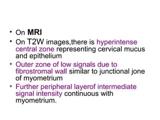
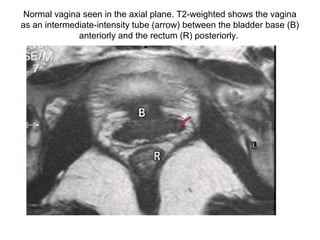
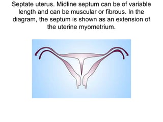
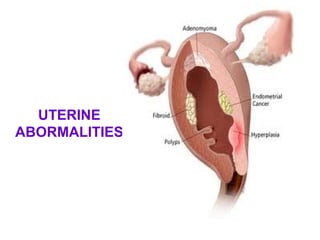
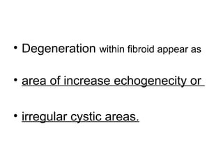
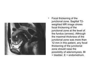
The document provides an overview of the normal radiological anatomy of the female pelvic organs including the uterus, endometrium, myometrium, cervix, vagina, ovaries and their appearance on ultrasound and MRI. It then discusses common uterine abnormalities such as congenital uterine anomalies, fibroids, adenomyosis and their imaging features. In adenomyosis, endometrial glands are present within the myometrium which can appear heterogeneous on ultrasound and cause diffuse or focal thickening of the junctional zone on MRI. Uterine fibroids appear as well-defined hypoechoic masses on ultrasound and may cause various signal changes on MRI depending on factors like degeneration. Congenital anomalies result from
In this presentation we will discuss
First trimester US especially TVS is an integral part for confirmation of intrauterine pregnancy and to rule out ectopic pregnancy.
First trimester US helps us in suggesting conceptus viability.
First trimester US especially TVS is very efficient in approaching and evaluating the cause of vaginal bleeding.
Sonographic evaluation of breast Dr. Muhammad Bin Zulfiqar
In this we will discuss role of high resolution Ultrasound in breast pathologies.
We will further discuss the role of Elastography in characterization of BIRADS.
Sonosalpingography. Dr. Sharda Jain, Dr. Jyoti Agarwal Lifecare Centre
Evaluation of fallopian tubes forms an essential part of evaluation
Tubal pathology is a cause of infertility in 30- 35% of infertile patients
Tubal Assessment
Fallopian tubes can be assessed by:
Hysterosalpingography
Hysterosalpingo-contrast-sonography (HycoSy)
Sonosalpingography
Laparoscopy & CHROMOTUBATION
ANATOMY OF UTERUS
ANATOMY OF OVARY
ANATOMY OF FALLOPIAN TUBES
ANATOMY OF UTERUS &ITS APPENDAGES
ANATOMY OF CERVIX
ANATOMY OF UTERUS PPT
BLOOD SUPPLY, NERVE SUPPLY, LYMPHATIC DRAINAGE
HISTOLOGY
basic anatomy and physiology of cervix to understand physiological changes in transformation zone during reproductive years, types of transformation zones and their importance
micro teaching on communication m.sc nursing.pdfAnurag Sharma
Microteaching is a unique model of practice teaching. It is a viable instrument for the. desired change in the teaching behavior or the behavior potential which, in specified types of real. classroom situations, tends to facilitate the achievement of specified types of objectives.
Pulmonary Thromboembolism - etilogy, types, medical- Surgical and nursing man...VarunMahajani
Disruption of blood supply to lung alveoli due to blockage of one or more pulmonary blood vessels is called as Pulmonary thromboembolism. In this presentation we will discuss its causes, types and its management in depth.
New Directions in Targeted Therapeutic Approaches for Older Adults With Mantl...i3 Health
i3 Health is pleased to make the speaker slides from this activity available for use as a non-accredited self-study or teaching resource.
This slide deck presented by Dr. Kami Maddocks, Professor-Clinical in the Division of Hematology and
Associate Division Director for Ambulatory Operations
The Ohio State University Comprehensive Cancer Center, will provide insight into new directions in targeted therapeutic approaches for older adults with mantle cell lymphoma.
STATEMENT OF NEED
Mantle cell lymphoma (MCL) is a rare, aggressive B-cell non-Hodgkin lymphoma (NHL) accounting for 5% to 7% of all lymphomas. Its prognosis ranges from indolent disease that does not require treatment for years to very aggressive disease, which is associated with poor survival (Silkenstedt et al, 2021). Typically, MCL is diagnosed at advanced stage and in older patients who cannot tolerate intensive therapy (NCCN, 2022). Although recent advances have slightly increased remission rates, recurrence and relapse remain very common, leading to a median overall survival between 3 and 6 years (LLS, 2021). Though there are several effective options, progress is still needed towards establishing an accepted frontline approach for MCL (Castellino et al, 2022). Treatment selection and management of MCL are complicated by the heterogeneity of prognosis, advanced age and comorbidities of patients, and lack of an established standard approach for treatment, making it vital that clinicians be familiar with the latest research and advances in this area. In this activity chaired by Michael Wang, MD, Professor in the Department of Lymphoma & Myeloma at MD Anderson Cancer Center, expert faculty will discuss prognostic factors informing treatment, the promising results of recent trials in new therapeutic approaches, and the implications of treatment resistance in therapeutic selection for MCL.
Target Audience
Hematology/oncology fellows, attending faculty, and other health care professionals involved in the treatment of patients with mantle cell lymphoma (MCL).
Learning Objectives
1.) Identify clinical and biological prognostic factors that can guide treatment decision making for older adults with MCL
2.) Evaluate emerging data on targeted therapeutic approaches for treatment-naive and relapsed/refractory MCL and their applicability to older adults
3.) Assess mechanisms of resistance to targeted therapies for MCL and their implications for treatment selection
Acute scrotum is a general term referring to an emergency condition affecting the contents or the wall of the scrotum.
There are a number of conditions that present acutely, predominantly with pain and/or swelling
A careful and detailed history and examination, and in some cases, investigations allow differentiation between these diagnoses. A prompt diagnosis is essential as the patient may require urgent surgical intervention
Testicular torsion refers to twisting of the spermatic cord, causing ischaemia of the testicle.
Testicular torsion results from inadequate fixation of the testis to the tunica vaginalis producing ischemia from reduced arterial inflow and venous outflow obstruction.
The prevalence of testicular torsion in adult patients hospitalized with acute scrotal pain is approximately 25 to 50 percent
Lung Cancer: Artificial Intelligence, Synergetics, Complex System Analysis, S...Oleg Kshivets
RESULTS: Overall life span (LS) was 2252.1±1742.5 days and cumulative 5-year survival (5YS) reached 73.2%, 10 years – 64.8%, 20 years – 42.5%. 513 LCP lived more than 5 years (LS=3124.6±1525.6 days), 148 LCP – more than 10 years (LS=5054.4±1504.1 days).199 LCP died because of LC (LS=562.7±374.5 days). 5YS of LCP after bi/lobectomies was significantly superior in comparison with LCP after pneumonectomies (78.1% vs.63.7%, P=0.00001 by log-rank test). AT significantly improved 5YS (66.3% vs. 34.8%) (P=0.00000 by log-rank test) only for LCP with N1-2. Cox modeling displayed that 5YS of LCP significantly depended on: phase transition (PT) early-invasive LC in terms of synergetics, PT N0—N12, cell ratio factors (ratio between cancer cells- CC and blood cells subpopulations), G1-3, histology, glucose, AT, blood cell circuit, prothrombin index, heparin tolerance, recalcification time (P=0.000-0.038). Neural networks, genetic algorithm selection and bootstrap simulation revealed relationships between 5YS and PT early-invasive LC (rank=1), PT N0—N12 (rank=2), thrombocytes/CC (3), erythrocytes/CC (4), eosinophils/CC (5), healthy cells/CC (6), lymphocytes/CC (7), segmented neutrophils/CC (8), stick neutrophils/CC (9), monocytes/CC (10); leucocytes/CC (11). Correct prediction of 5YS was 100% by neural networks computing (area under ROC curve=1.0; error=0.0).
CONCLUSIONS: 5YS of LCP after radical procedures significantly depended on: 1) PT early-invasive cancer; 2) PT N0--N12; 3) cell ratio factors; 4) blood cell circuit; 5) biochemical factors; 6) hemostasis system; 7) AT; 8) LC characteristics; 9) LC cell dynamics; 10) surgery type: lobectomy/pneumonectomy; 11) anthropometric data. Optimal diagnosis and treatment strategies for LC are: 1) screening and early detection of LC; 2) availability of experienced thoracic surgeons because of complexity of radical procedures; 3) aggressive en block surgery and adequate lymph node dissection for completeness; 4) precise prediction; 5) adjuvant chemoimmunoradiotherapy for LCP with unfavorable prognosis.
These lecture slides, by Dr Sidra Arshad, offer a quick overview of physiological basis of a normal electrocardiogram.
Learning objectives:
1. Define an electrocardiogram (ECG) and electrocardiography
2. Describe how dipoles generated by the heart produce the waveforms of the ECG
3. Describe the components of a normal electrocardiogram of a typical bipolar leads (limb II)
4. Differentiate between intervals and segments
5. Enlist some common indications for obtaining an ECG
Study Resources:
1. Chapter 11, Guyton and Hall Textbook of Medical Physiology, 14th edition
2. Chapter 9, Human Physiology - From Cells to Systems, Lauralee Sherwood, 9th edition
3. Chapter 29, Ganong’s Review of Medical Physiology, 26th edition
4. Electrocardiogram, StatPearls - https://www.ncbi.nlm.nih.gov/books/NBK549803/
5. ECG in Medical Practice by ABM Abdullah, 4th edition
6. ECG Basics, http://www.nataliescasebook.com/tag/e-c-g-basics
HOT NEW PRODUCT! BIG SALES FAST SHIPPING NOW FROM CHINA!! EU KU DB BK substit...GL Anaacs
Contact us if you are interested:
Email / Skype : kefaya1771@gmail.com
Threema: PXHY5PDH
New BATCH Ku !!! MUCH IN DEMAND FAST SALE EVERY BATCH HAPPY GOOD EFFECT BIG BATCH !
Contact me on Threema or skype to start big business!!
Hot-sale products:
NEW HOT EUTYLONE WHITE CRYSTAL!!
5cl-adba precursor (semi finished )
5cl-adba raw materials
ADBB precursor (semi finished )
ADBB raw materials
APVP powder
5fadb/4f-adb
Jwh018 / Jwh210
Eutylone crystal
Protonitazene (hydrochloride) CAS: 119276-01-6
Flubrotizolam CAS: 57801-95-3
Metonitazene CAS: 14680-51-4
Payment terms: Western Union,MoneyGram,Bitcoin or USDT.
Deliver Time: Usually 7-15days
Shipping method: FedEx, TNT, DHL,UPS etc.Our deliveries are 100% safe, fast, reliable and discreet.
Samples will be sent for your evaluation!If you are interested in, please contact me, let's talk details.
We specializes in exporting high quality Research chemical, medical intermediate, Pharmaceutical chemicals and so on. Products are exported to USA, Canada, France, Korea, Japan,Russia, Southeast Asia and other countries.
Couples presenting to the infertility clinic- Do they really have infertility...Sujoy Dasgupta
Dr Sujoy Dasgupta presented the study on "Couples presenting to the infertility clinic- Do they really have infertility? – The unexplored stories of non-consummation" in the 13th Congress of the Asia Pacific Initiative on Reproduction (ASPIRE 2024) at Manila on 24 May, 2024.
Ozempic: Preoperative Management of Patients on GLP-1 Receptor Agonists Saeid Safari
Preoperative Management of Patients on GLP-1 Receptor Agonists like Ozempic and Semiglutide
ASA GUIDELINE
NYSORA Guideline
2 Case Reports of Gastric Ultrasound
Report Back from SGO 2024: What’s the Latest in Cervical Cancer?bkling
Are you curious about what’s new in cervical cancer research or unsure what the findings mean? Join Dr. Emily Ko, a gynecologic oncologist at Penn Medicine, to learn about the latest updates from the Society of Gynecologic Oncology (SGO) 2024 Annual Meeting on Women’s Cancer. Dr. Ko will discuss what the research presented at the conference means for you and answer your questions about the new developments.
Ethanol (CH3CH2OH), or beverage alcohol, is a two-carbon alcohol
that is rapidly distributed in the body and brain. Ethanol alters many
neurochemical systems and has rewarding and addictive properties. It
is the oldest recreational drug and likely contributes to more morbidity,
mortality, and public health costs than all illicit drugs combined. The
5th edition of the Diagnostic and Statistical Manual of Mental Disorders
(DSM-5) integrates alcohol abuse and alcohol dependence into a single
disorder called alcohol use disorder (AUD), with mild, moderate,
and severe subclassifications (American Psychiatric Association, 2013).
In the DSM-5, all types of substance abuse and dependence have been
combined into a single substance use disorder (SUD) on a continuum
from mild to severe. A diagnosis of AUD requires that at least two of
the 11 DSM-5 behaviors be present within a 12-month period (mild
AUD: 2–3 criteria; moderate AUD: 4–5 criteria; severe AUD: 6–11 criteria).
The four main behavioral effects of AUD are impaired control over
drinking, negative social consequences, risky use, and altered physiological
effects (tolerance, withdrawal). This chapter presents an overview
of the prevalence and harmful consequences of AUD in the U.S.,
the systemic nature of the disease, neurocircuitry and stages of AUD,
comorbidities, fetal alcohol spectrum disorders, genetic risk factors, and
pharmacotherapies for AUD.
These simplified slides by Dr. Sidra Arshad present an overview of the non-respiratory functions of the respiratory tract.
Learning objectives:
1. Enlist the non-respiratory functions of the respiratory tract
2. Briefly explain how these functions are carried out
3. Discuss the significance of dead space
4. Differentiate between minute ventilation and alveolar ventilation
5. Describe the cough and sneeze reflexes
Study Resources:
1. Chapter 39, Guyton and Hall Textbook of Medical Physiology, 14th edition
2. Chapter 34, Ganong’s Review of Medical Physiology, 26th edition
3. Chapter 17, Human Physiology by Lauralee Sherwood, 9th edition
4. Non-respiratory functions of the lungs https://academic.oup.com/bjaed/article/13/3/98/278874
2. NORMAL RADIOLOGICAL
ANATOMY
• UTERUS
• It is pear shaped structure
• varies in size
• Before puberty cervix large and body of
uterus small.
• After puberty uterus size increases to 7 to
8 cm.
• After menopause involute to 5 to 6 cm
3.
4. • May be retrovertes or anteverted
• Retroflexed or anteflexed
• There is difficulty in seeing the fundus if
retroverted retroflexed
5. On MRI T2W images
endometrium is of
High signal intensity
7. Sagittal US image of the uterus
obtained during menstruation
shows a thin endometrial lining
8. Sagittal US image of the uterus obtained during
the late proliferative phase of the menstrual cycle
demonstrates the endometrium with a multilayered
appearance
9. Sagittal US image of the uterus obtained during
the secretory phase of the menstrual cycle shows
a thickened, echogenic endometrium
10. • ENDOMETRIUM IS CONSIDERED
ABMORMAL IF IT MEASURES MORE
THAN 1.4cm IN PREMENOPAUAL
AND MORE THAN 4mm IN POST
MENOPAUSAL.
11. • On USG endometrium is of high
echogenecity .
• At the start of follicular phase of menstrual cycle
it is thin echogenic line.
• From day 8 to 10 of menstrual cycle
endometrium thickens and become five layered
at midcycle.
• In the luteal phase endometrium loses its five
layered appearance and becomes progressively
more echogenic with normal thickness of 1.2 to
1.4cm.
12. • MYOMETRIUM IS HYPOECHOIC ON
USG
• ON MRI T2W The layer of myometrium
just below endometrium is of low signal
intensity and the remaining myometrium is
of intermediate signal intensity.
13. The zonal anatomy, with differentiation between the high-
intensity endometrium (E), low-intensity junctional zone
(arrow), and intermediate-intensity outer myometrium, is
well seen.
14. T2-weighted MR image shows the normal
endometrium (straight arrow) and junctional
zone (curved arrow).
15. • Parametrium is hypoechoic on USG
• and on MRI T1W intermediate signals and
increase in signals on T2W.
16. CERVIX
• Cervix is cylinderical shape structure 2 to
4cm.
• connecting with body of uterus at internal
os.
17. Normal Uterus–Transabdominal. Longitudinal scan through the urine-filled
bladder (B) demonstrates a normal adult uterus with smooth contours, pear
shape, and well-defined bright endometrial echo (open arrow). The cervix (arrow)
is recognized at the junction of imaginary lines drawn through the long axis of the
uterus and the long axis of the vagina (between arrowheads). This uterus is
anteverted.
18. Transvaginal sonogram in early pregnancy
showing a normal cervix. Arrows point to the
internal and external os.
20. • On MRI
• On T2W images,there is hyperintense
central zone representing cervical mucus
and epithelium
• Outer zone of low signals due to
fibrostromal wall similar to junctional jone
of myometrium
• Further peripheral layerof intermediate
signal intensity continuous with
myometrium.
21. VAGINA RADIOLOGICAL
ANATOMY
• On USG Vagina is visualizes as two
linear hypoechoic muscular walls around
and echogenic mucosa.
• On MRI T2W images vagina has a high
signal central zone of mucus and
epithelium surrounded by low signal
muscular wall.
22.
23. Normal vagina seen in the axial plane. T2-weighted shows the vagina
as an intermediate-intensity tube (arrow) between the bladder base (B)
anteriorly and the rectum (R) posteriorly.
24. OVARY
• These are oval structures lying in relation
with internal iliac vessels on the pelvic
side walls.
• They are less echogenic than the
uterus.
• may contain variable number of
follicles/cysts.
25.
26. • IN MENSTRUATING WOMEN its
VOLUME SHOULD BE NORMALLY
LESS THAN 7.5ml
• AND IN POSTMENOPAUSLA WOMEN
NOT MORE THAN 3ml.
27. OVULATORY CYCLE
• At start of FOLLICULAR PHASE few
number of follicles start to develop
• From day 8 to 10 one follicle become
dominant nad continues to grow at a rate
of 2 to 3 mm/day.
• About midcycle it measures 18 to 25mm.
• after ovulation corpus luteum can be seen
seen as irregular cystcontaining internal
echoes due to blood or a hypoechoic
area.
28. Doppler studies
• During menstruation and early follicular
phase high impedance flow RI appro 0.7
• Following neovascularization and corpus
luteum development ,diastolic flow
increases leading to low impedance flow
and RI less than 0.6
• Doppler indices in postmenopausal are of
high resistive index.
29. Color Doppler US scan shows
abundant flow in the wall of the
corpus luteum.
30. • Ovaries are best seen in transverse
or coronal planes.
• They measure about 1.5 to 3 cm in
diameter.
• Ovary show low to intermediate
signal intensity similar to muscle
on T1W and low signals on T2W.
31.
32. Axial T2-weighted magnetic resonance image of
the pelvis. This image reveals multiple subcapsular
follicles in both ovaries
33. • Follicles are seen which are of Low to
intermediate signals on T1W and high
signals on T2W
• On T1W images difficulty arise in
differentiating ovaries from bowel and
uterus.After IV gadolinium normal
ovaries enhance
34. CONGENITAL UTERINE
ANOMALIES
• The upper two third of vagina uterus
and fallopian tube develop from fusion
and descent of paired mullerian
ducts.
• The lower third of vagina is derived from
urogenital sinus.
• Partial or complete failure of the ducts to
fuse result in spectrum of complex
abnormalities.
37. Axial T2-weighted FSE image (2,000/120) shows
a laterally deviated banana-shaped uterus (arrow).
No rudimentary horn could be detected.
38.
39. BICORNUATE UTERUS
• Bicornuate uterus results from partial
nonfusion of the müllerian ducts . Double
uterine bodies and a single cervix are
present. An arcuate uterus is considered a
milder form of bicornuate uterus; it has a
convex or flat external fundal contour and
mild impression on the endometrial cavity.
Bicornuate bicollis is a variant of
bicornuate uterus in which the anomaly is
combined with a muscular uterine septum
that extends to the external os
42. SEPTATE UTERUS
• In it uterine septum
fails to resorb
resulting in failure of
correct placental
implantation and
subsequent
miscarriage.
Septate uterus results from complete
fusion of the müllerian ducts with failure
of resorption of the central septum
44. the outer fundal contour (superior border) is flat
or slightly concave, which is sufficient to make the
diagnosis of septate uterus
45.
46. UTERUS DIDELPHYS
• .Didelphic uterus
results from complete
nonfusion of both
müllerian ducts Two
uterine bodies and
two cervices are
present
47. Didelphys uterus. Complete separation and full
development of both müllerian ducts is noted. (a) Two
vaginas and 2 cervices; (b) 2 distinct cervices; (c) 2 uterine
horns are widely splayed; (d) cross section of uterine
bodies and cervices.
48. Bicornuate uterus. The midline uterine external fundal cleft
(superior border) has a depression greater than 1 cm,
excluding septate uterus from the differential diagnosis.
This image is of bicornuate bicollis, since 2 cervices are
present
49.
50. Normal uterus. Note a single
uterine cavity and flat or convex
outer fundal contour.
51. Septate uterus. Midline septum can be of variable
length and can be muscular or fibrous. In the
diagram, the septum is shown as an extension of
the uterine myometrium.
52. Bicornuate uterus. Note the partial fusion of the lower
uterine segment and persistently separated upper uterine
segments. Of key importance is the prominent fundal cleft
(>1 cm), which distinguishes the anomaly from septate
uterus.
54. Didelphys uterus. Note the
complete separation but full
development of each müllerian
duct.
55. Arcuate uterus. Mild thickening of the
midline fundal myometrium resulting in
fundal cavity indentation but normal outer
fundal contour.
56. Diethylstilbestrol-exposed uterus. Myometrial hypertrophy
results in a T-shaped uterine cavity and cavity irregularity,
which is pathognomonic for the anomaly. Typically, the
uteri are hypoplastic.
57. HEMATOMETRIUM AND
HEMATOCOLPOS
• Primary amenorrhea
• cyclical abdominal pain
• pelvic pain
• severe dysmenorrhea
• USG reveals a thick walled cystic
mass either due to obstructed
vagina or obstructed uterus with
low level internal echoes due to
blood.
58. Transverse vaginal septum (class 2). Sagittal T2-weighted shows a
transverse septum in the middle of the vagina (arrow), causing
dilatation of the proximal vagina (V) and uterus (U) (hematocolpos and
hetmatometria).
59. Obstructed hymen (class 2). Sagittal T1-weighted
spin-echo image (500/8) shows a dilated
hematometrocolpos. The obstruction is at the level
of perineum
60. Hematometrocolpos in a 12-year-old girl with
abdominal pain. Sagittal US image demonstrates a
markedly distended vagina (straight arrow) and
uterine cavity (curved arrow)
67. UTERINE FIBROIDS
• SUBMUCOUS arise within cavity and
distort it
• MURAL may or maynot distort cavity
depends on precise location
• SUBSEROSAL cause bulge on surface of
uterus
•
68.
69.
70. • On USG most fibroids are
• ROUND WELL DEFINED
• HYPOECHOIC MASSES WITH
CHARACTERISTIC INTERNAL
ARCHITECTURE SHOWING
RECURRENT SHADOWING
71. • Nondegenerating fibroids
• have a uniform signal intensity
indistinguishable from myometrium on
T1W images and with lower signals
on T2W images.
72. • Degenerating fibroids show variable
and nonspecific signal
appearanceswith an intermediate to high
signals on T1W and high signals on T2W
images.
• Malignant transformation cant be
differentiated from degenerating
fibroids
90. • They can be hyperechoic and may
be calcified particularly in
postmenopausal women.
91. • Degeneration within fibroid appear as
• area of increase echogenecity or
• irregular cystic areas.
92. Fibroids can undergo various degenerative changes,
especially when large. This fibroid of the uterus measures
11.2 cms. and shows multiple hypoechoic and hyperechoic
patchy areas
95. • It is the presence of endometrial
glands within myometrium
associated with adjacent
myometrial hyperplasia.
96. • It is usually a diffuse process but may form
a localazised mass or adenomyoma.
• Clinical findings are dysmenorrhea and
menorrhagia with a tender bulky uterus.
97. USG features
• On USG poorly defined areas of
decreased echogenecity and
heterogeneity in myometriumassociated
with small 2to 5mm cystic spaces in
myometrium in 50% cases.
• Focal adenomyoma cause focal bulge in
myometrium and may be hypo or
hyperechoic but less well defined than
fibroids.
98. Normal uterus. Sagittal endovaginal US scan shows a normal
myometrium (M), which is moderately echogenic and has a
homogeneous echotexture. The subendometrial halo, which represents
the innermost layer of the myometrium, is visualized subjacent to the
endometrium (E) as a thin hypoechoic band (arrows). The endometrium
is uniformly echogenic in this patient, who was in the secretory phase of
the menstrual cycle.
99. • sagittal oblique endovaginal US
scan shows that the myometrium
is thickened ventrally and has a
heterogeneous echotexture
(straight arrows). The echogenicity
of the ventral myometrium is
decreased relative to that of the
dorsal myometrium. Additional
features of adenomyosis seen in
this image include poor definition
of the endomyometrial junction
and a myometrial cyst (curved
arrow).
101. MRI features
• Maximum thickness of junctional zone is
12mm
• And in adenomyosis there is diffuse or
focal thickening of junctional zone,with or
without focal areas of high signal on T2W.
• On T2W images focal adenomyosis
appear as poorly marginated low signal
mass within the myometrium.
102. • sagittal T2-weighted MR image
shows marked thickening of
the junctional zone. The result
is a poorly defined low-signal-
intensity mass that replaces
the ventral myometrium
(arrows). The numerous bright
foci, some of which have a
rounded appearance whereas
others have a linear or
fingerlike appearance,
represent the heterotopic
endometrium. Bl = bladder.
103. • Focal thickening of the
junctional zone. Sagittal T2-
weighted MR image shows
focal thickening of the
junctional zone at the level of
the fundus (arrows). Although
the maximal thickness of the
junctional zone was more than
12 mm in this patient, any focal
thickening of the junctional
zone should raise the
possibility of adenomyosis. Bl
= bladder, E = endometrium.
106. Endometrial hyperplasia. US image shows an
endometrium with diffuse thickening (maximum
thickness, 1.74 cm) due to hyperplasia
107. ENDOMETRIAL CARCINOMA
• Occurs mainly in postmenopausal
women
• Most common presenting symptom
abnormal uterine bleeding
108. Ultrasound features
• vary from endometrial thickening
to an irregular hypoechoic
intracavitatory mass to an enlarged
diffusely infiltrated uterus.
109. Endometrial adenocarcinoma. (a) US image reveals a
heterogeneous endometrial mass (arrows) that is difficult to
distinguish from the myometrium. Cursors indicate the
entire transverse width of the uterus.
110. CT features
• Hypodense irregular mass expanding the
uterine cavity sometimes associated with
blood,fluid or pus within cavity.
• CT is good at determining the extent of
extrauterine disease but cant easily
differnentiate stage1 from stage 2 disease.
112. A 57-year-old woman with stage IVB poorly
differentiated endometrial carcinoma. CT image
through the uterus. The cervix is markedly
enlarged and replaced by tumor.
113. CT scan also reveals a
heterogeneous tumor
(arrowheads).
114. endometrial adenocarcinoma. CT image through
the uterus shows fluid-filled cavity marginated by
tumor involving most of the endometrial
andcervical regionenlarged lymph node right pelvic
115. T2-weighted MR image shows a large,
heterogeneous tumor distending the
endometrial canal (arrows).
116. • Endometrial
carcinoma in a 58-
year-old patient. (a)
Sagittal fast SE T2-
weighted (4,000/119
[effective]) MR image
shows nodular,
discretely irregular
foci of low signal
intensity (arrowheads)
in the endometrial
cavity.
117.
118. • THE MOST RELIABLE CRITERIA FOR
THE DIAGNOSIS OF MYOMETRIAL
INVASION IS DISRUPTION OF
JUNCTIONAL ZONE.
119. • FOLLOWING IV CONTRAST
ENDOMETRIAL CARCINOMA
ENHANCES .
122. POLYCYSTIC OVARY
• LARGE OVARIES VOLUME >7.5ml
• MORE THAN 2to 5mm follicles MAINLY
PERIPHERAL DISTRIBUTION
INCREASED STROMA
LARGE /NORMAL UTERUS THICK
ENDOMETRIUM
130. • 3.5-cm simple ovarian
cyst (calipers).
Normal-appearing
ovarian tissue
(arrows) with a few
follicles around the
periphery confirms
the ovarian origin of
the cyst.
131.
132. • corpus luteum within
the ovary. It has a
slightly thick,
crenulated wall
(arrows) and a small
cystic center
135. HEMORRHAGIC CYST
• a retracting clot
(asterisk) with
concave margins
along the wall of a
hemorrhagic cyst
136. • hemarragic cyst
complex cystic mass
within the periphery of
the ovary. The
seemingly solid area
within the cystic mass
has concave margins
and no demonstrable
flow, both typical
features of a clot.
137. • hemorrhagic cyst
complex ovarian cyst
with a seemingly solid
area due to a clot (C).
This could be
mistaken for the solid
area of a neoplasm.
No flow was evident
138. • hemorrhagic cyst
complex ovarian cyst
with internal echoes.
There is a reticular or
fishnet pattern to the
internal echoes due to
fibrin strands
(arrows). Note how
the fibrin strands are
thin
139.
140. RUPTURED CYST
• right adnexa shows a
thick-walled ovarian
cyst (corpus luteum)
with surrounding
anechoic free fluid, a
finding indicative of
rupture
141. TORSION OF CYST
• Twisted vascular
pedicle showing the
circular string-of-
beads appearance of
dilated veins (arrows).
BL-indicates urinary
bladder; and CYST,
ovarian cyst.
144. A, Snail shell appearance of the twisted pedicle (arrows). BL
indicates urinary bladder; and CYST, ovarian cyst. B, Color
Doppler study of the twisted artery and vein.
145. • A, Twisted pedicle
appearing as a large
echogenic mass
(arrows). B, Color
Doppler study of the
pedicle showing
absent flow.
148. • endometrioma
complex ovarian cyst
with homogeneous
internal echoes. It
contains a small solid-
appearing area
(arrow). Color
Doppler US (not
shown) did not
demonstrate flow in
the solid area.
160. Endovaginal ultrasound scan. This image shows a
relatively enlarged right ovary, increased flow, and
a small amount of adjacent free fluid. These
findings are compatible with oophoritis.
163. This sonogram shows a markedly
heterogeneous and thickened endometrium,
a finding that is compatible with
endometritis.
164. This sonogram demonstrates a markedly heterogeneous and thickened
endometrium. On closer evaluation, a fluid-fluid level in the endometrial cavity is
revealed. In the appropriate clinical setting, this finding is compatible with
pyometrium, as it was in this case.
165. This sonogram reveals bilateral complex masses
in a patient who had pyometrium. The finding is
compatible with tubo-ovarian abscesses.
166. This sonogram reveals bilateral complex masses
in a patient who had pyometrium, a finding that is
compatible with tubo-ovarian abscess.
167. Power Doppler sonogram. This image shows increased
flow to the wall of a tubo-ovarian abscess. The inner
hypoechoic regions are due to the presence of purulent
material
168. Endometritis with air in the endometrial
cavity and bilateral tubo-ovarian abscesses
are shown.
170. Ovarian fibroma
• Ovarian fibroma in a
24-year-old woman.
Sagittal transvaginal
US scan reveals a
slightly hypoechoic
solid mass (M) within
and replacing most of
the ovary (calipers).
No distal acoustic
shadowing is present
171. • Ovarian fibromas are composed of
spindle cells that form collagen and
usually display low signal intensity
on both T1- and T2-weighted MRI.
High signal intensity on T2-
weighted images corresponded to
regions of hyalinization and
myxomatous changes [5].
Intratumoral edema is also
common in larger fibromas.
176. EPITHELIAL OVARIAN TUMORS
• serous
cystadenocarcinoma
complex ovarian cyst
(calipers) with several
thick septa (arrows)
and solid areas.
177. • Benign serous
cystadenoma in a 49-
year-old woman.
Contrast material-
enhanced CT scan
shows a unilocular
cystic mass in the
right lower quadrant
(arrows). The wall of
the mass is not
delineated, and there
is no evidence of any
excrescence within it.
178.
179.
180.
181. • 21-year-old woman
with ovarian serous
cystadenoma.
Sonogram shows
echogenic mural
nodule (arrow) in
cystic mass.
184. • 21-year-old woman
with ovarian serous
cystadenoma. Sagittal
contrast-enhanced
T1-weighted MR
image shows papillary
projection (arrows) in
cystic mass.
185. • 38-year-old woman
with ovarian
mucinous
cystadenofibroma. CT
scan shows large
cystic mass with
enhancing solid
portion
186. • Benign mucinous
cystadenoma in a 26-
year-old woman.
Contrast-enhanced
CT scan shows a
large, multilocular
cystic mass (arrows)
with a smooth
contour, honeycomb
appearance, and
heterogeneous
attenuation in the
locules.
187. • . Serous
cystadenocarcinoma
of the ovary with
peritoneal
carcinomatosis in a
60-year-old woman.
Contrast-enhanced
CT scans obtained at
the level of the liver
188. Brenner tumour
• They are mainly hypoechoic solid masses.
Calcifications have been reported in 50% of
Brenner tumours on ultrasound.
• CT
• calcifications have been reported in ~ 83% of
Brenner tumours on CT.
• solid component may show mild to moderate
enhancement post contrast.
• Pelvic MRI
• due to its predominantly fibrous content content
they appear hypointense on T2 weighted
sequences
189. Axial contrast-enhanced CT shows a solid mass
(arrows) with 8 cm diameter arising from the right
ovary. Extensive amorphous calcification within the
mass is seen
190. Axial T1-weighted image showing a large
hypointense adnexal mass (arrows) arising
from the right ovary.
191. Sagittal T2-weighted image demonstrating large right
adnexal mass to be uniformly hypointense (asterisk). Also
seen is anteverted and anteflexed uterus (arrow).
199. • The test uses “Levovist”, a non-iodine
contrast agent. Levovist is a microparticle
suspension consisting of 99.9% galactose
and 0.1% palmitic acid.
• The HyCoSy procedure is a safe and
reliable alternative to the conventional
hysterosalpingogram (HSG) which uses X-
rays. No radiation or iodinated contrast
material is used for a HyCoSy test.
200.
201.
202. • —Endometrial polyp
in 33-year-old
woman.
• A, Sonohysterogram
in transverse plane
shows solitary,
smooth, well-defined,
uniformly echogenic
intracavitary lesion.
Angle of lesion with
underlying
endometrium (arrows)
is acute.
203. • Endometrial polyp in
33-year-old woman.
• B, Color Doppler
image shows single
feeding vessel at
base of polyp (arrow).
204. • Endometrial cancer in 57-year-
old woman. Sonohysterogram
in sagittal plane with color
Doppler reveals that diffuse,
irregular frondlike thickening of
posterior endometrium shows
internal vascularity.
Endometrial-myometrial
interface has been lost, which
suggests invasion into
myometrium.
205. • Submucosal fibroid in
42-year-old woman at
sonohysterography.
• B, Transverse image
with application of
color Doppler
sonography shows
typical arborizing
vascular pattern of
fibroid lesion.