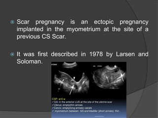
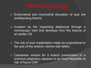
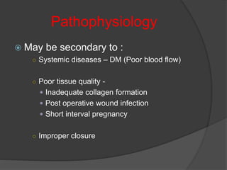
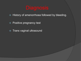
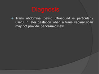
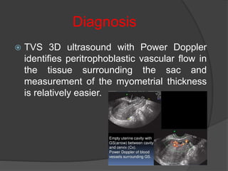
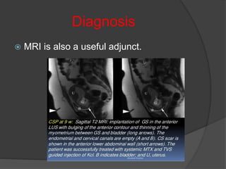
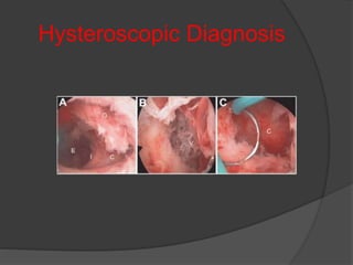
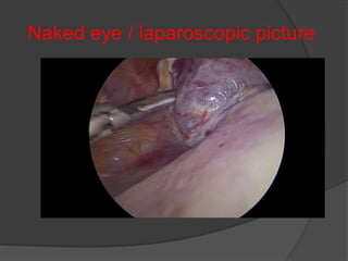
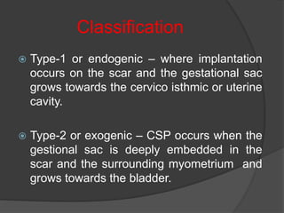
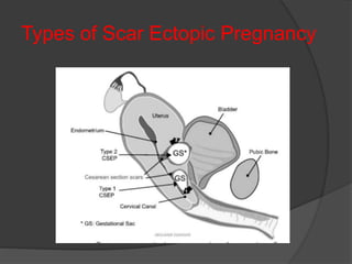
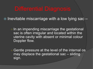
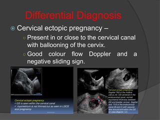
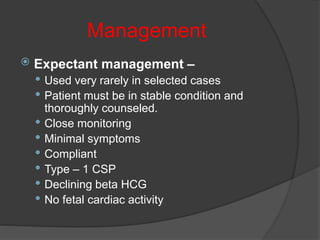
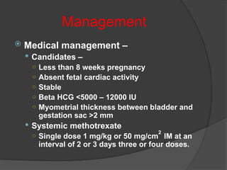
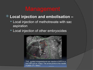
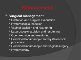
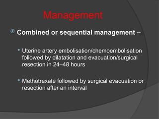
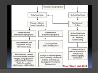
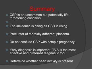
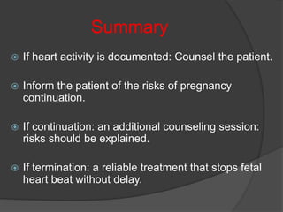
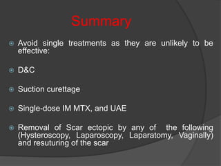
Dr. Alka Pandey
MD, Ph.D.,
Associate Professor, P.M.C.H., Patna
 Scar pregnancy is an ectopic pregnancy
implanted in the myometrium at the site of a
previous CS Scar.
 It was first described in 1978 by Larsen and
Soloman.
Prevalence
 Prevalence of CSP is increasing and to date
more than 1000 cases have been reported.
 This may be attributed to :
○ Increasing Number of CS
○ Increasing awareness
○ Better diagnostic technique
Risk Factors
 May occur after any myometrial trauma -
 Caesarean Section
 Manual Removal of placenta
 Myomectomy
 D & C
Pathophysiology
 Endometrial and myometrial disruption of scar are
predisposing factors.
 Invasion by the implanting blastocyst through a
microscopic tract that develops from the trauma of
an earlier CS.
 The risk of scar implantation might be proportional to
the size of the anterior uterine wall defect.
 Caesarean section for a breech presentation in a
previous pregnancy appears to be most frequently at
risk of future CSP.
Pathophysiology
 May be secondary to :
○ Systemic diseases – DM (Poor blood flow)
○ Poor tissue quality -
 Inadequate collagen formation
 Post operative wound infection
 Short interval pregnancy
○ Improper closure
Diagnosis
 History of amenorrhoea followed by bleeding.
 Positive pregnancy test
 Trans vaginal ultrasound
Diagnosis
○ Ultrasound criteria : -
 Empty uterine cavity , closed and empty cervical canal
 Placenta and/or a gestational sac embedded in the scar of a
previous caesarean section
 A triangular/round or oval‐shaped gestational sac that fills the
niche of the scar
 A thin or absent myometrial layer between the gestational sac
and the bladder
 Yolk sac, embryo and cardiac activity may or may not be
present
 Evidence of functional trophoblastic/placental circulation on
colour flow Doppler examination, characterised by high
velocity and low impedance blood flow
 Negative ‘sliding organs’ sign
Diagnosis
 Trans abdominal pelvic ultrasound is particularly
useful in later gestation when a trans vaginal scan
may not provide panoramic view.
Diagnosis
 TVS 3D ultrasound with Power Doppler
identifies peritrophoblastic vascular flow in
the tissue surrounding the sac and
measurement of the myometrial thickness
is relatively easier.
Diagnosis
 MRI is also a useful adjunct.
Hysteroscopic Diagnosis
Naked eye / laparoscopic picture
Classification
 Type-1 or endogenic – where implantation
occurs on the scar and the gestational sac
grows towards the cervico isthmic or uterine
cavity.
 Type-2 or exogenic – CSP occurs when the
gestional sac is deeply embedded in the
scar and the surrounding myometrium and
grows towards the bladder.
Types of Scar Ectopic Pregnancy
Classification
 The myometrial layer between the
gestational sac and the bladder becomes
very thin and may disappear with bulging of
the gestational sac through the gap as the
pregnancy advances.
 In two – third of the cases the thickness of
the scar may be less than 5 mm.
Clinical Presentation
 Asymptomatic – 37%
 Painless vaginal bleeding – 39%
 Generalized abdominal pain – 25%
Differential Diagnosis
 Inevitable miscarriage with a low lying sac –
○ In an impending miscarriage the gestational
sac is often irregular and located within the
uterine cavity with absent or minimal colour
Doppler flow.
○ Gentle pressure at the level of the internal os
may displace the gestational sac – sliding
sign.
Differential Diagnosis
 Cervical ectopic pregnancy –
○ Present in or close to the cervical canal
with ballooning of the cervix.
○ Good colour flow Doppler and a
negative sliding sign.
Risk factors influencing management
choices
Management
 Patient factors
 Symptoms
 Fertility wishes
 Acceptability of prolonged follow up
 Associated lesions
 Surgical risk factors
 Response to initial treatment
Management
 Caesarean scar pregnancy (CSP)
 Gestational age
 Human chorionic gonadotropin (hCG) levels
 Size of CSP mass
 Type of CSP
 Myometrial thickness
 Viability
 Facilities
 Interventional radiology
 Surgical expertise/facilities
 Monitoring facilities
Management
 No consensus on the preferred mode of treatment.
 All treatment options carry a risk of haemorrhage
and subsequent hysterectomy.
 Treatment should be individualized based on full
pre-treatment evaluation.
 Pregnancy should be ended as soon as possible
after confirming the diagnosis.
Management
 Expectant management –
 Used very rarely in selected cases
 Patient must be in stable condition and
thoroughly counseled.
 Close monitoring
 Minimal symptoms
 Compliant
 Type – 1 CSP
 Declining beta HCG
 No fetal cardiac activity
Management
 Medical management –
 Candidates –
○ Less than 8 weeks pregnancy
○ Absent fetal cardiac activity
○ Stable
○ Beta HCG <5000 – 12000 IU
○ Myometrial thickness between bladder and
gestation sac >2 mm
 Systemic methotrexate
○ Single dose 1 mg/kg or 50 mg/cm
2
IM at an
interval of 2 or 3 days three or four doses.
Management
 Local injection and embolisation –
 Local injection of methotrexate with sac
aspiration
 Local injection of other embryocides
Management
 Surgical management
 Dilatation and surgical evacuation
 Hysteroscopic resection
 Vaginal excision and resuturing
 Laparoscopic excision and resuturing
 Open excision and resuturing
 Combined laparoscopic and hysteroscopic
procedure
 Combined laparoscopic and vaginal surgery
 Hysterectomy
Management
 Combined or sequential management –
 Uterine artery embolisation/chemoembolisation
followed by dilatation and evacuation/surgical
resection in 24–48 hours
 Methotrexate followed by surgical evacuation or
resection after an interval
 Intrauterine Balloon Catheter – can be
used successfully to compress the
gestational sac.
 Catheter is left for 48 hours
 Antibiotics given
 Outer end of the catheter is fastened to
the thigh.
Prevention
 Avoid pregnancy 12 to 24 months.
 Surgical repair of uterine scar defects
with single or double layer closure.
Summary
 CSP is an uncommon but potentially life-
threatening condition.

 The incidence is rising as CSR is rising.
 Precursor of morbidly adherent placenta.
 Do not confuse CSP with ectopic pregnancy.
 Early diagnosis is important. TVS is the most
effective and preferred diagnostic tool.
 Determine whether heart activity is present.
Summary
 If heart activity is documented: Counsel the patient.
 Inform the patient of the risks of pregnancy
continuation.
 If continuation: an additional counseling session:
risks should be explained.
 If termination: a reliable treatment that stops fetal
heart beat without delay.
Summary
 Avoid single treatments as they are unlikely to be
effective:
 D&C
 Suction curettage
 Single-dose IM MTX, and UAE
 Removal of Scar ectopic by any of the following
(Hysteroscopy, Laparoscopy, Laparatomy, Vaginally)
and resuturing of the scar
Summary
 Consider combination treatments: best results.
direct injection of MTX or Kcl into GS with TVS
guidance.
 In a future pregnancy, an early visit for TVS is
important.
Complications
 Placenta previa/accreta
 Uterine rupture
 Massive Haemorrhage : increased
maternal morbidity and mortality.
Thank You

Scar ectopic pregnancy

  • 1.
    Dr. Alka Pandey MD,Ph.D., Associate Professor, P.M.C.H., Patna
  • 2.
     Scar pregnancyis an ectopic pregnancy implanted in the myometrium at the site of a previous CS Scar.  It was first described in 1978 by Larsen and Soloman.
  • 3.
    Prevalence  Prevalence ofCSP is increasing and to date more than 1000 cases have been reported.  This may be attributed to : ○ Increasing Number of CS ○ Increasing awareness ○ Better diagnostic technique
  • 4.
    Risk Factors  Mayoccur after any myometrial trauma -  Caesarean Section  Manual Removal of placenta  Myomectomy  D & C
  • 5.
    Pathophysiology  Endometrial andmyometrial disruption of scar are predisposing factors.  Invasion by the implanting blastocyst through a microscopic tract that develops from the trauma of an earlier CS.  The risk of scar implantation might be proportional to the size of the anterior uterine wall defect.  Caesarean section for a breech presentation in a previous pregnancy appears to be most frequently at risk of future CSP.
  • 6.
    Pathophysiology  May besecondary to : ○ Systemic diseases – DM (Poor blood flow) ○ Poor tissue quality -  Inadequate collagen formation  Post operative wound infection  Short interval pregnancy ○ Improper closure
  • 7.
    Diagnosis  History ofamenorrhoea followed by bleeding.  Positive pregnancy test  Trans vaginal ultrasound
  • 8.
    Diagnosis ○ Ultrasound criteria: -  Empty uterine cavity , closed and empty cervical canal  Placenta and/or a gestational sac embedded in the scar of a previous caesarean section  A triangular/round or oval‐shaped gestational sac that fills the niche of the scar  A thin or absent myometrial layer between the gestational sac and the bladder  Yolk sac, embryo and cardiac activity may or may not be present  Evidence of functional trophoblastic/placental circulation on colour flow Doppler examination, characterised by high velocity and low impedance blood flow  Negative ‘sliding organs’ sign
  • 9.
    Diagnosis  Trans abdominalpelvic ultrasound is particularly useful in later gestation when a trans vaginal scan may not provide panoramic view.
  • 10.
    Diagnosis  TVS 3Dultrasound with Power Doppler identifies peritrophoblastic vascular flow in the tissue surrounding the sac and measurement of the myometrial thickness is relatively easier.
  • 11.
    Diagnosis  MRI isalso a useful adjunct.
  • 12.
  • 13.
    Naked eye /laparoscopic picture
  • 14.
    Classification  Type-1 orendogenic – where implantation occurs on the scar and the gestational sac grows towards the cervico isthmic or uterine cavity.  Type-2 or exogenic – CSP occurs when the gestional sac is deeply embedded in the scar and the surrounding myometrium and grows towards the bladder.
  • 15.
    Types of ScarEctopic Pregnancy
  • 16.
    Classification  The myometriallayer between the gestational sac and the bladder becomes very thin and may disappear with bulging of the gestational sac through the gap as the pregnancy advances.  In two – third of the cases the thickness of the scar may be less than 5 mm.
  • 17.
    Clinical Presentation  Asymptomatic– 37%  Painless vaginal bleeding – 39%  Generalized abdominal pain – 25%
  • 18.
    Differential Diagnosis  Inevitablemiscarriage with a low lying sac – ○ In an impending miscarriage the gestational sac is often irregular and located within the uterine cavity with absent or minimal colour Doppler flow. ○ Gentle pressure at the level of the internal os may displace the gestational sac – sliding sign.
  • 19.
    Differential Diagnosis  Cervicalectopic pregnancy – ○ Present in or close to the cervical canal with ballooning of the cervix. ○ Good colour flow Doppler and a negative sliding sign.
  • 20.
    Risk factors influencingmanagement choices Management  Patient factors  Symptoms  Fertility wishes  Acceptability of prolonged follow up  Associated lesions  Surgical risk factors  Response to initial treatment
  • 21.
    Management  Caesarean scarpregnancy (CSP)  Gestational age  Human chorionic gonadotropin (hCG) levels  Size of CSP mass  Type of CSP  Myometrial thickness  Viability  Facilities  Interventional radiology  Surgical expertise/facilities  Monitoring facilities
  • 22.
    Management  No consensuson the preferred mode of treatment.  All treatment options carry a risk of haemorrhage and subsequent hysterectomy.  Treatment should be individualized based on full pre-treatment evaluation.  Pregnancy should be ended as soon as possible after confirming the diagnosis.
  • 23.
    Management  Expectant management–  Used very rarely in selected cases  Patient must be in stable condition and thoroughly counseled.  Close monitoring  Minimal symptoms  Compliant  Type – 1 CSP  Declining beta HCG  No fetal cardiac activity
  • 24.
    Management  Medical management–  Candidates – ○ Less than 8 weeks pregnancy ○ Absent fetal cardiac activity ○ Stable ○ Beta HCG <5000 – 12000 IU ○ Myometrial thickness between bladder and gestation sac >2 mm  Systemic methotrexate ○ Single dose 1 mg/kg or 50 mg/cm 2 IM at an interval of 2 or 3 days three or four doses.
  • 25.
    Management  Local injectionand embolisation –  Local injection of methotrexate with sac aspiration  Local injection of other embryocides
  • 26.
    Management  Surgical management Dilatation and surgical evacuation  Hysteroscopic resection  Vaginal excision and resuturing  Laparoscopic excision and resuturing  Open excision and resuturing  Combined laparoscopic and hysteroscopic procedure  Combined laparoscopic and vaginal surgery  Hysterectomy
  • 27.
    Management  Combined orsequential management –  Uterine artery embolisation/chemoembolisation followed by dilatation and evacuation/surgical resection in 24–48 hours  Methotrexate followed by surgical evacuation or resection after an interval
  • 28.
     Intrauterine BalloonCatheter – can be used successfully to compress the gestational sac.  Catheter is left for 48 hours  Antibiotics given  Outer end of the catheter is fastened to the thigh.
  • 30.
    Prevention  Avoid pregnancy12 to 24 months.  Surgical repair of uterine scar defects with single or double layer closure.
  • 31.
    Summary  CSP isan uncommon but potentially life- threatening condition.   The incidence is rising as CSR is rising.  Precursor of morbidly adherent placenta.  Do not confuse CSP with ectopic pregnancy.  Early diagnosis is important. TVS is the most effective and preferred diagnostic tool.  Determine whether heart activity is present.
  • 32.
    Summary  If heartactivity is documented: Counsel the patient.  Inform the patient of the risks of pregnancy continuation.  If continuation: an additional counseling session: risks should be explained.  If termination: a reliable treatment that stops fetal heart beat without delay.
  • 33.
    Summary  Avoid singletreatments as they are unlikely to be effective:  D&C  Suction curettage  Single-dose IM MTX, and UAE  Removal of Scar ectopic by any of the following (Hysteroscopy, Laparoscopy, Laparatomy, Vaginally) and resuturing of the scar
  • 34.
    Summary  Consider combinationtreatments: best results. direct injection of MTX or Kcl into GS with TVS guidance.  In a future pregnancy, an early visit for TVS is important.
  • 35.
    Complications  Placenta previa/accreta Uterine rupture  Massive Haemorrhage : increased maternal morbidity and mortality.
  • 36.