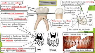
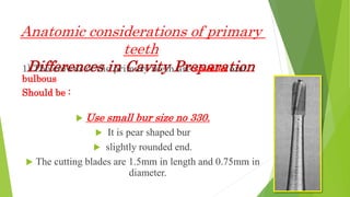
The primary teeth have several anatomical differences compared to permanent teeth that are important to consider for cavity preparation:
1) The crowns are smaller and more bulbous so smaller bur sizes should be used and preparations should be more conservative.
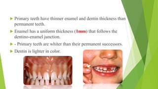
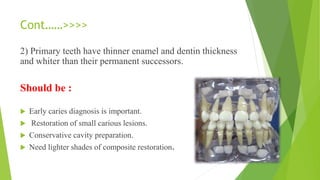
2) Enamel and dentin are thinner so early caries diagnosis and small restorations are important to avoid pulpal involvement.
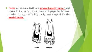
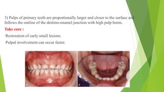
3) Pulps are larger and closer to the surface so care must be taken to not expose the pulp during preparation.
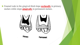
4) Enamel rods slope differently so beveling is not needed for class II preparations.