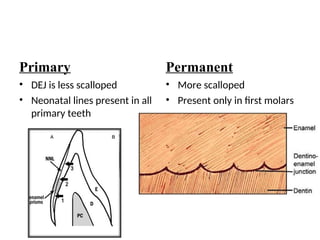
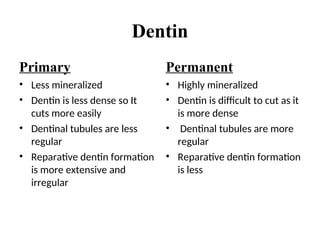
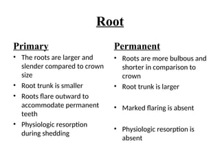
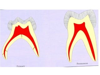
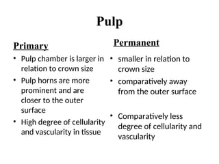
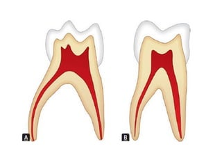
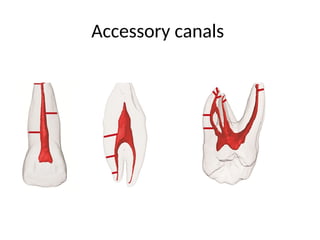
Primary vs Permanent Teeth
Prof. Dr. Deepthi K
Dept of Oral and Maxillofacial Pathology
Primary
• Lighter in colour: bluish
white –milky white
(MILK TEETH)
• 6 months to 5-6 years
• 20 teeth
• Smaller in dimension
Permanent
• Darker in colour, grayish
or yellowish white
• 6 years onwards
• 32 teeth
• Larger in dimension
Primary Permanent
• First molar is smaller in
dimension than second
molar
• Mamelons absent
• More supplemental grooves
• Contact areas are
broader ,flatter and gingival
• First molar is larger in
dimension than second
molar
• Mamelons are present on
newly erupted teeth
• Less supplemental grooves
• Contact points are more
occlusally placed
Primary Permanent
Contact points
Mamelons
Contact areas
Primary Permanent
wider mesiodistally wider cervico incisally
Primary
• Cervical ridges are
prominent especially on
buccal aspect
• Narrow occlusal tables
• Sharp cervical constriction
Permanent
• Cervical ridges are less
prominent
• Less convergence of buccal
and lingual surfaces hence
wider occlusal table
• Less constriction of the neck
Enamel
Primary
• Thinner and uniformly
thick(1mm)
• Less number of striae
• Less mineralized
Permanent
• About 2-3 mm thick
• More number of striae
• Highly mineralized
Primary
Enamel rods at the
cervical slopes
occlusally from DEJ
Permanent
Enamel rods are
oriented gingivally
Primary
• DEJ is less scalloped
• Neonatal lines present in all
primary teeth
Permanent
• More scalloped
• Present only in first molars
Dentin
Primary
• Less mineralized
• Dentin is less dense so It
cuts more easily
• Dentinal tubules are less
regular
• Reparative dentin formation
is more extensive and
irregular
Permanent
• Highly mineralized
• Dentin is difficult to cut as it
is more dense
• Dentinal tubules are more
regular
• Reparative dentin formation
is less
Root
Primary
• The roots are larger and
slender compared to crown
size
• Root trunk is smaller
• Roots flare outward to
accommodate permanent
teeth
• Physiologic resorption
during shedding
Permanent
• Roots are more bulbous and
shorter in comparison to
crown
• Root trunk is larger
• Marked flaring is absent
• Physiologic resorption is
absent
Physiologic resorption
Pulp
Primary
• Pulp chamber is larger in
relation to crown size
• Pulp horns are more
prominent and are
closer to the outer
surface
• High degree of cellularity
and vascularity in tissue
Permanent
• smaller in relation to
crown size
• comparatively away
from the outer surface
• Comparatively less
degree of cellularity and
vascularity
Pulp
Primary
• High potential for repair
• Root canals are ribbon like.
Radicular pulp is thin,
tortuous and branch
• Accessory canals are
present in good number
• Density of innervation is
less of which primary teeth
are less sensitive
Permanent
• Less potential for repair
• Root canals are well defined
with less branching
• Floor of pulp chamber does
not have any accessory
canals
• Density of innervation is
more
Accessory canals
Pulp
Primary
• Enlarged apical foramen,
abundant blood supply,
demonstrates an
inflammatory response
• Localisation of infection and
inflammation is poorer in
pulp
Permanent
• Foramens are restricted
Reduced blood supply
favours calcific response
• Inflammation and infection
in pulp is localised
Periodontium
Primary
• Cementum is very thin
• Secondary cementum is
absent
• Majority of CEJs are knife
edge
• Alveolar atrophy is rare
• Gingivitis is generally absent
in a healthy child
Permanent
• Thick
• Present
• Majority of CEJs are over
lapping type
• Alveolar atrophy occurs
• Gingivitis is common in
adults
Difference between primary and permanent.ppt

Difference between primary and permanent.ppt

  • 1.
    Primary vs PermanentTeeth Prof. Dr. Deepthi K Dept of Oral and Maxillofacial Pathology
  • 2.
    Primary • Lighter incolour: bluish white –milky white (MILK TEETH) • 6 months to 5-6 years • 20 teeth • Smaller in dimension Permanent • Darker in colour, grayish or yellowish white • 6 years onwards • 32 teeth • Larger in dimension
  • 4.
    Primary Permanent • Firstmolar is smaller in dimension than second molar • Mamelons absent • More supplemental grooves • Contact areas are broader ,flatter and gingival • First molar is larger in dimension than second molar • Mamelons are present on newly erupted teeth • Less supplemental grooves • Contact points are more occlusally placed
  • 5.
  • 6.
  • 7.
  • 8.
    Primary • Cervical ridgesare prominent especially on buccal aspect • Narrow occlusal tables • Sharp cervical constriction Permanent • Cervical ridges are less prominent • Less convergence of buccal and lingual surfaces hence wider occlusal table • Less constriction of the neck
  • 10.
    Enamel Primary • Thinner anduniformly thick(1mm) • Less number of striae • Less mineralized Permanent • About 2-3 mm thick • More number of striae • Highly mineralized
  • 11.
    Primary Enamel rods atthe cervical slopes occlusally from DEJ Permanent Enamel rods are oriented gingivally
  • 12.
    Primary • DEJ isless scalloped • Neonatal lines present in all primary teeth Permanent • More scalloped • Present only in first molars
  • 13.
    Dentin Primary • Less mineralized •Dentin is less dense so It cuts more easily • Dentinal tubules are less regular • Reparative dentin formation is more extensive and irregular Permanent • Highly mineralized • Dentin is difficult to cut as it is more dense • Dentinal tubules are more regular • Reparative dentin formation is less
  • 15.
    Root Primary • The rootsare larger and slender compared to crown size • Root trunk is smaller • Roots flare outward to accommodate permanent teeth • Physiologic resorption during shedding Permanent • Roots are more bulbous and shorter in comparison to crown • Root trunk is larger • Marked flaring is absent • Physiologic resorption is absent
  • 17.
  • 19.
    Pulp Primary • Pulp chamberis larger in relation to crown size • Pulp horns are more prominent and are closer to the outer surface • High degree of cellularity and vascularity in tissue Permanent • smaller in relation to crown size • comparatively away from the outer surface • Comparatively less degree of cellularity and vascularity
  • 21.
    Pulp Primary • High potentialfor repair • Root canals are ribbon like. Radicular pulp is thin, tortuous and branch • Accessory canals are present in good number • Density of innervation is less of which primary teeth are less sensitive Permanent • Less potential for repair • Root canals are well defined with less branching • Floor of pulp chamber does not have any accessory canals • Density of innervation is more
  • 22.
  • 23.
    Pulp Primary • Enlarged apicalforamen, abundant blood supply, demonstrates an inflammatory response • Localisation of infection and inflammation is poorer in pulp Permanent • Foramens are restricted Reduced blood supply favours calcific response • Inflammation and infection in pulp is localised
  • 25.
    Periodontium Primary • Cementum isvery thin • Secondary cementum is absent • Majority of CEJs are knife edge • Alveolar atrophy is rare • Gingivitis is generally absent in a healthy child Permanent • Thick • Present • Majority of CEJs are over lapping type • Alveolar atrophy occurs • Gingivitis is common in adults